Microdiscectomy Surgery
Microdiscectomy
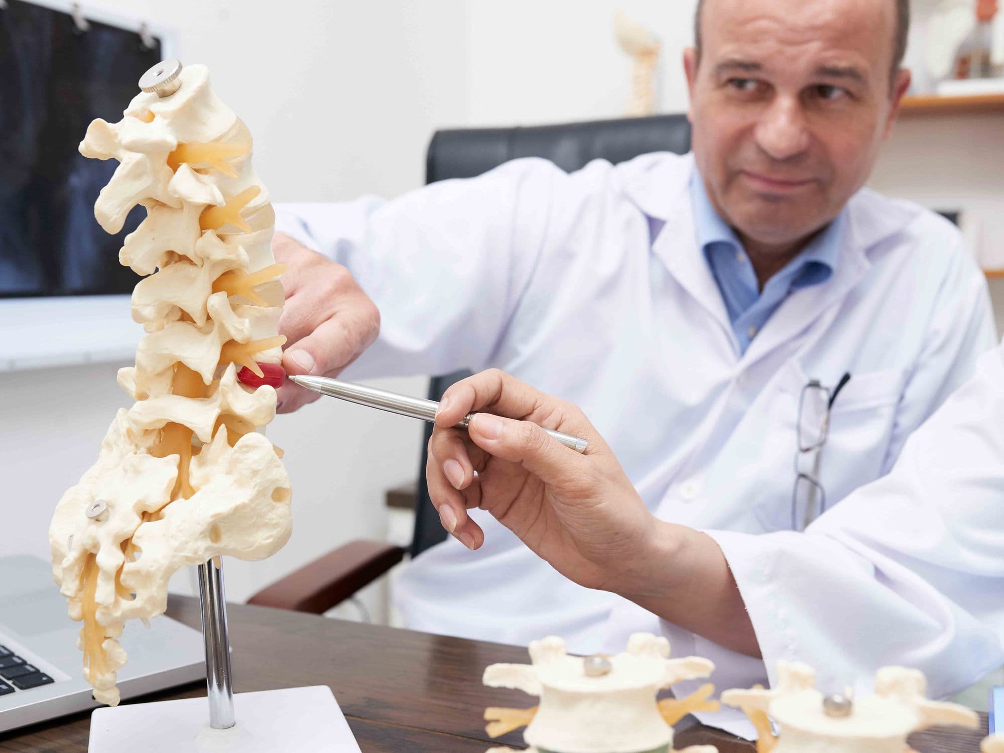
In 2026, a Microdiscectomy (also called microdecompression) is the gold-standard surgical procedure for treating a herniated lumbar disc that is pressing on a spinal nerve. Unlike a traditional discectomy, this version uses high-powered magnification—either a microscope or an endoscope—to allow the surgeon to work through a very small incision.
When You Should Consider Microdiscectomy
Sciatica: Sharp, "electric" radiating leg pain caused by nerve root compression.
Herniated Lumbar Disc: When the inner "jelly-like" material of a disc leaks out and pinches a spinal nerve.
Neurological Deficits: Numbness, tingling, or weakness in the legs or feet.
Failure of Conservative Treatment: When physical therapy, epidural injections, and medications fail to provide relief after 6–12 weeks.
Severe Nerve Impingement: Evidence of significant pressure on the nerve root as confirmed by advanced imaging.
Methods of Disc Decompression
Micro-Decompression: Using high-powered microscopes to visualize and treat the spine through a 1–2 cm incision.
Endoscopic Discectomy: A ultra-minimally invasive approach using a camera-equipped tube to reach the herniated fragment.
Muscle Preservation: Utilizing tubular dilators to stretch back muscles apart rather than cutting them away from the bone.
Disc Annular Repair: Using specialized biological glues or closure devices to "plug" the hole in the outer disc rim.
Laminotomy: The removal of a tiny piece of the overlying bone to safely reach the spinal canal and nerve root.
How Microdiscectomy Is Performed
Anesthesia: The procedure is performed under general anesthesia to ensure the patient remains perfectly still and comfortable.
Precision Access: A 1 to 2-centimeter incision is made directly over the affected disc level.
Nerve Retraction: The surgeon carefully moves the nerve root aside to access the disc space.
Fragment Removal: Only the "damaged" protruding part of the disc is removed, leaving the healthy portion to provide cushioning.
Annular Closure: Modern 2026 techniques may include sealing the disc wall to significantly reduce the risk of re-herniation.
Pre-Procedure Preparation
Fasting: Patients must follow strict fasting protocols for 8–12 hours prior to surgery.
Imaging Review: A final review of high-resolution MRI scans to confirm the exact location of the herniation.
Medical Clearance: Blood tests and an ECG are conducted to ensure the patient is a safe candidate for anesthesia.
Medication Adjustment: Pausing anti-inflammatory or blood-thinning medications as directed by the surgical team.
Recovery Support: Arranging for a support person to drive the patient home after the same-day procedure.
Tests Before Microdiscectomy
Lumbar MRI: The definitive imaging tool to visualize the disc herniation and nerve compression.
CT Scan: Occasionally used to assess the bone structure surrounding the herniated disc.
Electromyography (EMG): To measure the electrical activity of muscles and the extent of nerve damage.
ECG: To monitor the heart's electrical rhythm as part of the standard pre-surgical screening.
Physical Examination: Assessing muscle strength, reflexes, and sensation in the lower extremities.
Life After Microdiscectomy
Hospital Stay: Almost always an outpatient procedure in 2026, with most patients returning home within 3–5 hours.
Immediate Relief: Radiating leg pain often disappears immediately upon waking from surgery.
The "No BLT" Rule: For six weeks, patients must strictly avoid Bending, Lifting (over 2kg), or Twisting.
Activity Resumption: Light walking is encouraged immediately; sedentary work can typically be resumed in 1–2 weeks.
Long-term Care: Post-operative physical therapy often focuses on core strengthening to protect the spine.
Benefits of Microdiscectomy
High Success Rate: Offers a 90% to 95% success rate for the immediate relief of radiating leg pain.
Minimally Invasive: The tiny 1–2 cm incision results in minimal scarring and reduced surgical trauma.
Rapid Recovery: Outpatient nature allows patients to recover in the comfort of their own homes.
Nerve Protection: Prevents further decline and permanent damage to the compressed nerve roots.
Innovative Sealing: 2026 annular repair technologies significantly lower the risk of future re-herniation.