Shoulder Arthroscopy
Shoulder Arthroscopy
Shoulder Arthroscopy is a minimally invasive surgical procedure used to diagnose and treat various joint problems, such as rotator cuff tears and labral injuries. Using a tiny camera called an arthroscope, surgeons can see inside the joint and perform high-precision repairs through small, buttonhole-sized incisions.
When You Should Consider Shoulder Arthroscopy
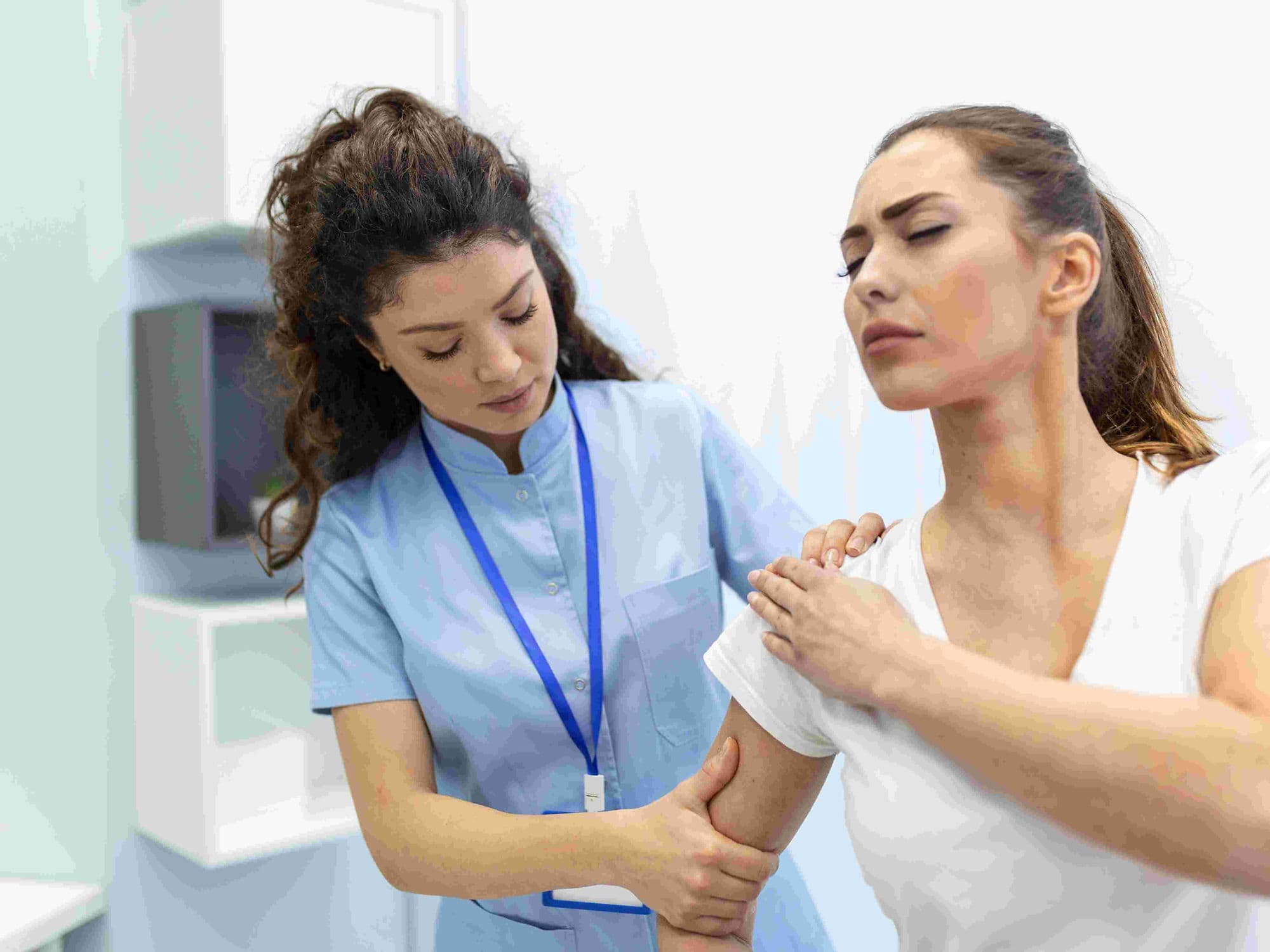
Persistent shoulder pain that has not improved with physical therapy, rest, or injections.
Loss of range of motion or weakness, often associated with a torn rotator cuff.
Recurrent shoulder dislocations or a feeling of "looseness" in the joint (instability).
Mechanical symptoms such as painful catching, clicking, or "locking" of the shoulder.
Presence of bone spurs that cause "impingement," where tendons are pinched during overhead movement.
Methods of Shoulder Arthroscopy
Rotator Cuff Repair: Reattaching torn tendons to the humerus (arm bone) using specialized suture anchors.
Labral Repair (Bankart or SLAP Repair): Stitching the cartilage ring (labrum) back to the socket to restore stability.
Subacromial Decompression: Shaving down bone spurs and inflamed tissue to create more space for the tendons to move.
Biceps Tenodesis: Relocating a damaged biceps tendon to a new attachment point to relieve pain.
Capsular Release: Surgically stretching or cutting tight tissue to treat severe "frozen shoulder."
How Shoulder Arthroscopy Is Performed
Fluid Insufflation: The surgeon injects sterile saline into the shoulder to inflate the joint capsule, providing a clear workspace and view.
Portal Placement: Two to three small incisions (0.5–1 cm) are made around the shoulder to serve as entry points for the camera and tools.
Joint Inspection: The arthroscope is inserted to project high-definition images of the tendons, ligaments, and cartilage onto a monitor.
Specialized Repair: Miniature tools, such as shavers or suture passers, are used to trim damaged tissue or anchor tendons back to the bone.
Portal Closure: Once the repair is complete, the saline is drained and the tiny incisions are closed with a single stitch or sterile tape.
Pre-Procedure Preparation
Diagnostic confirmation through MRI or X-rays to map the internal damage and plan the surgical approach.
Fasting (NPO) for 6–12 hours prior to the procedure to ensure safety during anesthesia.
Coordination of a regional nerve block, which numbs the entire arm for up to 24 hours to assist with immediate pain control.
Pausing certain medications, such as blood thinners or anti-inflammatories, as directed by the surgical team.
Tests Before Shoulder Arthroscopy
Shoulder MRI: The gold standard for visualizing soft tissue injuries like rotator cuff tears and labral damage.
MRI Arthrogram: An MRI where dye is injected into the joint to highlight small tears that might be missed on a standard scan.
X-rays: Used to identify bone spurs, arthritis, or fractures that may contribute to shoulder pain.
Physical Strength Testing: Assessing the deltoid and rotator cuff muscles to determine the functional impact of the injury.
Life After Shoulder Arthroscopy
This is typically an outpatient procedure, allowing most patients to return home the same day.
A shoulder sling is mandatory for 1 to 6 weeks, depending on the complexity of the repair (e.g., longer for a rotator cuff repair).
Pendulum exercises, involving gently swinging the arm, are often started within days to prevent "frozen shoulder" stiffness.
Driving is generally restricted for at least 1 week, or until the patient has stopped taking narcotic pain medications.
Heavy lifting and overhead sports are avoided for 3 to 6 months while the repaired tendons fully bond to the bone.
Benefits of Shoulder Arthroscopy
Significantly less post-operative pain and swelling compared to traditional "open" shoulder surgery.
Smaller incisions result in minimal scarring and a lower risk of wound-related complications.
High success rate for restoring shoulder stability and relieving chronic pain.
Allows for a more precise diagnosis by giving the surgeon a dynamic, 360-degree view of the internal joint structures.