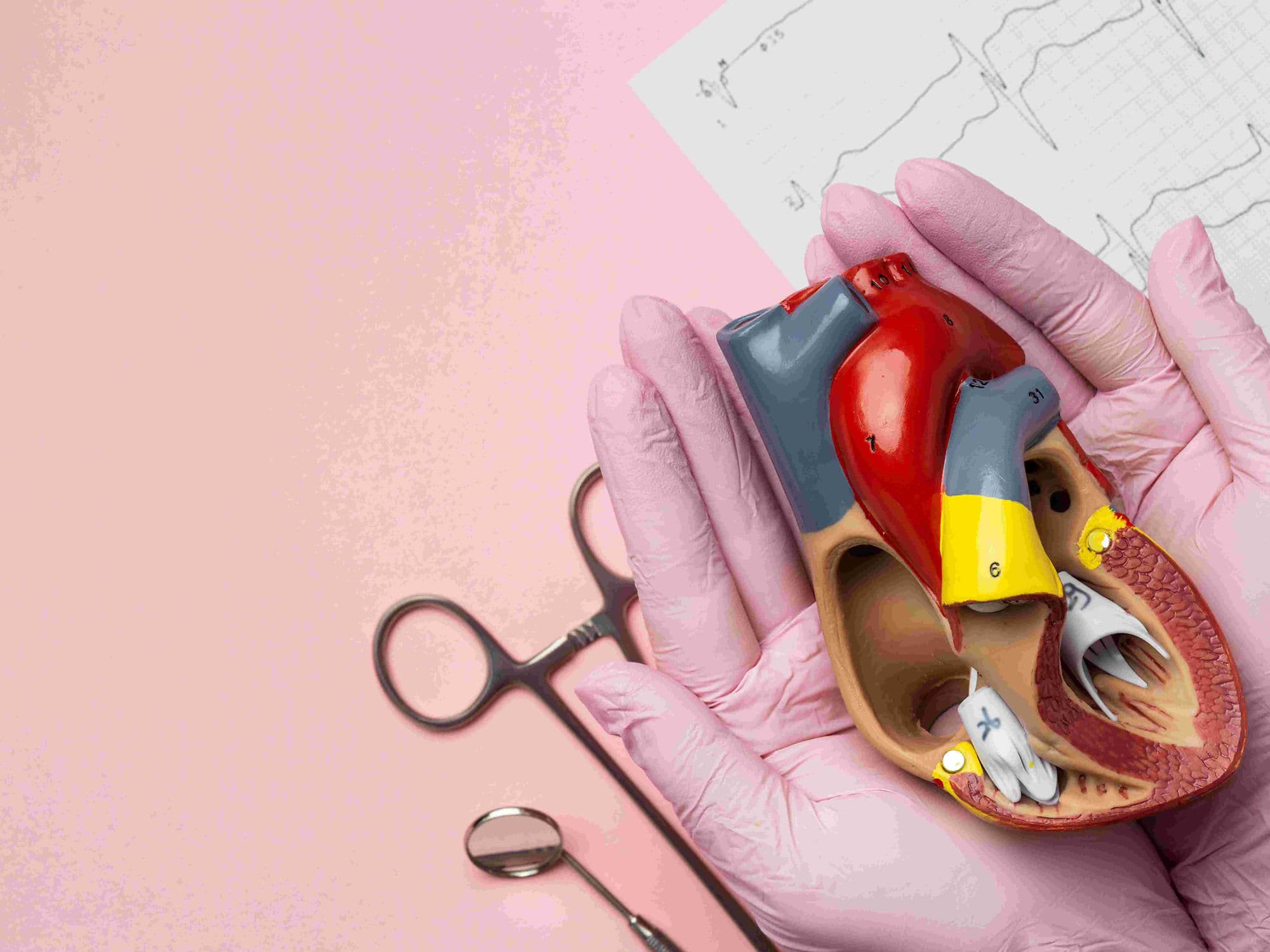
VSD Device Closure
VSD Device Closure
VSD (Ventricular Septal Defect) device closure is a minimally invasive, non-surgical procedure used to seal a "hole in the heart" between the two lower chambers (ventricles). Unlike traditional open-heart surgery, this procedure is performed entirely through a catheter, resulting in no chest scars and a significantly faster recovery. This advanced technique allows for the permanent repair of the heart's internal wall without the need for a heart-lung bypass machine.
When You Should Consider VSD Device Closure
Muscular VSDs: This is the primary treatment for holes located in the muscular portion of the ventricular septum.
Symptom Management: For children or adults experiencing poor weight gain, frequent lung infections, or persistent shortness of breath.
Heart Protection: To prevent the left side of the heart from overworking, which can lead to an enlarged heart (cardiomegaly).
Pulmonary Hypertension Prevention: To reduce the risk of developing dangerously high blood pressure in the lung arteries.
Heart Failure Prevention: Correcting the defect before it leads to more serious long-term cardiac complications.
How It Is Performed
Access: A small incision is made in the groin to access the femoral vein or artery. No large incisions are made on the chest.
Anesthesia: The procedure is performed in a specialized Cardiac Catheterization Lab (Cath Lab) under general anesthesia or heavy sedation, typically taking 1 to 2 hours.
Guidance: A thin, flexible tube (catheter) is threaded through the blood vessels into the heart, guided by real-time X-ray (Fluoroscopy) and detailed ultrasound (Transesophageal Echo).
Measurement: The specialist measures the exact size and location of the hole to select a custom-sized Nitinol mesh device.
Deployment: A folded, umbrella-like device is pushed through the catheter. Once it reaches the hole, it is carefully unfolded to "sandwich" the defect from both sides.
Verification: Once the device is securely in place and the hole is confirmed to be sealed, the catheter is removed and the small puncture in the groin is closed.
Pre-Procedure Preparation
Echocardiogram: A detailed ultrasound of the heart to map the VSD's size and its proximity to the heart's valves.
Transesophageal Echo (TEE): A specialized ultrasound performed through the esophagus for high-resolution images of the defect.
Dental Clearance: Ensuring there are no active dental infections, which could increase the risk of heart infection (endocarditis) after the device is placed.
Fasting: Following "nothing by mouth" instructions for 8 hours prior to the procedure.
Medication Audit: You may be asked to adjust or stop certain medications, particularly blood thinners, a few days before the procedure.
Tests Before VSD Device Closure
Chest X-ray: To evaluate the current size of the heart and check for any fluid in the lungs.
Electrocardiogram (ECG): A baseline check of the heart's electrical system to identify any pre-existing arrhythmias.
Blood Panels: A routine check of your blood count, electrolytes, and kidney function.
Cardiac MRI or CT: Occasionally used to provide a 3D model of the heart for complex or multiple VSDs.
Life After VSD Device Closure
Hospital Stay: Most patients stay for one night for observation and are discharged the next day.
Medication: You will typically take blood-thinning medication (usually Aspirin) for 6 months to prevent clots from forming on the device while the heart lining grows over it.
Activity Restrictions: Most patients can return to school or light work within 3 to 5 days. You should avoid strenuous exercise and heavy lifting for at least 2 weeks.
Dental Care Precautions: For the first 6 months post-procedure, you must take preventive antibiotics before any dental work to prevent heart infections.
Long-term Integration: Over 3–6 months, the heart's natural lining (endocardium) grows completely over the device, making it a permanent and seamless part of your heart.
Why Specialized Treatment Is Highly Effective
Scar-Free Recovery: By avoiding a sternotomy (opening the chest), patients experience much less pain and have no permanent surgical scars.
Rapid Return to Normalcy: Recovery is measured in days rather than the months required for open-heart surgery.
High Success Rates: Device closure is a highly reliable method for sealing muscular VSDs with a very low risk of the hole reopening.
Protects Electrical System: Advanced imaging ensures the device is positioned to minimize pressure on the heart's natural "wiring."
Permanent Solution: The Nitinol mesh is designed to last a lifetime, providing a durable repair that grows with the patient.