Mole Removal
Mole Removal
Mole removal is a common procedure in 2026 performed for cosmetic reasons, physical comfort (to stop snagging on clothes), or medical necessity (to check for skin cancer). While many methods exist, a professional evaluation is always the first step to ensure the mole is not malignant before any cosmetic or corrective removal occurs.
When You Should Consider Mole Removal
Suspicious Appearance: Moles following the ABCDE rule (Asymmetry, irregular Border, varied Color, Diameter over 6mm, or Evolving over time).
Physical Irritation: Moles located in areas where they are frequently rubbed by clothing, jewelry, or razor blades.
Aesthetic Preference: Seeking a smoother skin surface or the removal of prominent facial moles.
Early Intervention: Removing atypical (dysplastic) moles to lower the risk of future melanoma development.
Psychological Comfort: Addressing moles that cause self-consciousness or impact confidence.
Methods of Mole Removal in 2026
Surgical Shave: The surgeon uses a thin blade to shave the mole flush with the skin, typically used for raised, benign moles and requiring no stitches.
Surgical Excision: Cutting out the entire mole and a small healthy margin, then closing with sutures—essential for suspected skin cancer.
Laser Removal: A 2026 favorite for small, flat, non-cancerous moles; uses light energy to break down pigment with minimal scarring risk.
Radiofrequency (RF) Surgery: Utilizing high-frequency energy to "shave" the mole with high precision and significantly reduced bleeding.
Cryotherapy: Freezing small, shallow, non-cancerous spots with liquid nitrogen, causing them to blister and fall off naturally.
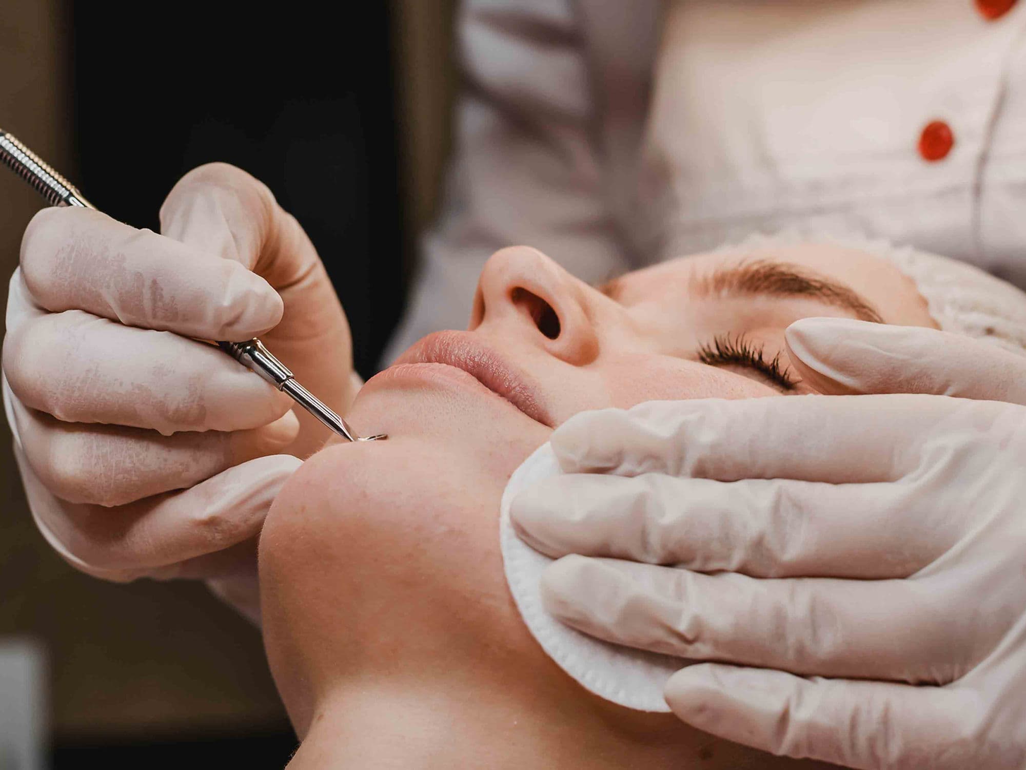
How Mole Removal Is Performed
Skin Cleansing: The area is thoroughly sanitized to prevent infection.
Local Anesthesia: A numbing agent is injected or applied topically; the patient remains awake but feels no pain during the 5–20 minute procedure.
Active Removal: The specialist uses the chosen tool (blade, laser, or RF wand) to remove the tissue.
Hemostasis: Any minor bleeding is controlled using pressure, chemical agents, or electrocautery.
Dressing: A sterile bandage and a thin layer of protective ointment are applied to the site.
Pre-Procedure Preparation
Professional Screening: A full-body skin check by a dermatologist to identify any other concerning lesions.
Medication Review: Stopping blood thinners or certain supplements (like Fish Oil) 3 days prior to reduce the risk of bruising.
Skin Hygiene: Arriving with clean, product-free skin at the treatment site.
Anxiety Management: Most patients can drive themselves home immediately; no significant fasting is required for local anesthesia.
Tests and Evaluations for Mole Removal
Pathological Biopsy: The standard in 2026; nearly all surgically removed moles are sent to a lab to be examined by a pathologist for cancerous cells.
Dermoscopy: Use of a specialized magnifying lens to view the mole's structure before removal.
Digital Mapping: In 2026, many clinics use AI-driven imaging to track the evolution of multiple moles before deciding which ones to remove.
Margin Check: For excisions, confirming "clear margins" ensures no abnormal cells were left behind.
Life After Mole Removal (Recovery)
Days 1–3: A small scab forms; the area must be kept clean and dry to allow initial "clotting" and repair.
Weeks 1–2: Stitches (if any) are removed; the scab naturally falls off to reveal fresh, pink skin underneath.
Months 1–6: The redness fades as the scar matures and blends with the surrounding skin tone.
The "One Year" Rule: While the surface heals in weeks, the underlying skin tissue continues to remodel for up to a full year for a final result.
Benefits of Professional Mole Removal
Peace of Mind: Immediate removal of potentially dangerous or cancerous lesions through lab-verified biopsy.
Cosmetic Improvement: Achieves a clearer complexion with techniques optimized for minimal scarring.
Enhanced Comfort: Eliminates physical snagging, itching, or bleeding caused by raised moles.
Fast and Convenient: Most procedures are completed in under 30 minutes with zero downtime.
Expert Monitoring: Professional removal ensures that if a mole returns, it is managed by a specialist who understands the risk of recurrence.