Scoliosis Correction Surgery
Scoliosis Correction Surgery
Scoliosis Correction Surgery is a major reconstructive procedure used to treat an abnormal sideways curvature of the spine. The latest 2026 surgical standards focus on maximizing the degree of correction while utilizing motion-preserving technologies and advanced safety monitoring to ensure optimal patient outcomes.
When You Should Consider Scoliosis Correction
Surgery is typically recommended when the spinal curve is progressive or has reached a severity that impacts physical health. Indications include:
Significant Curvature: Sideways curves generally exceeding 45 to 50 degrees.
Respiratory/Cardiac Impact: Severe curves that compress the lungs or heart, affecting breathing and stamina.
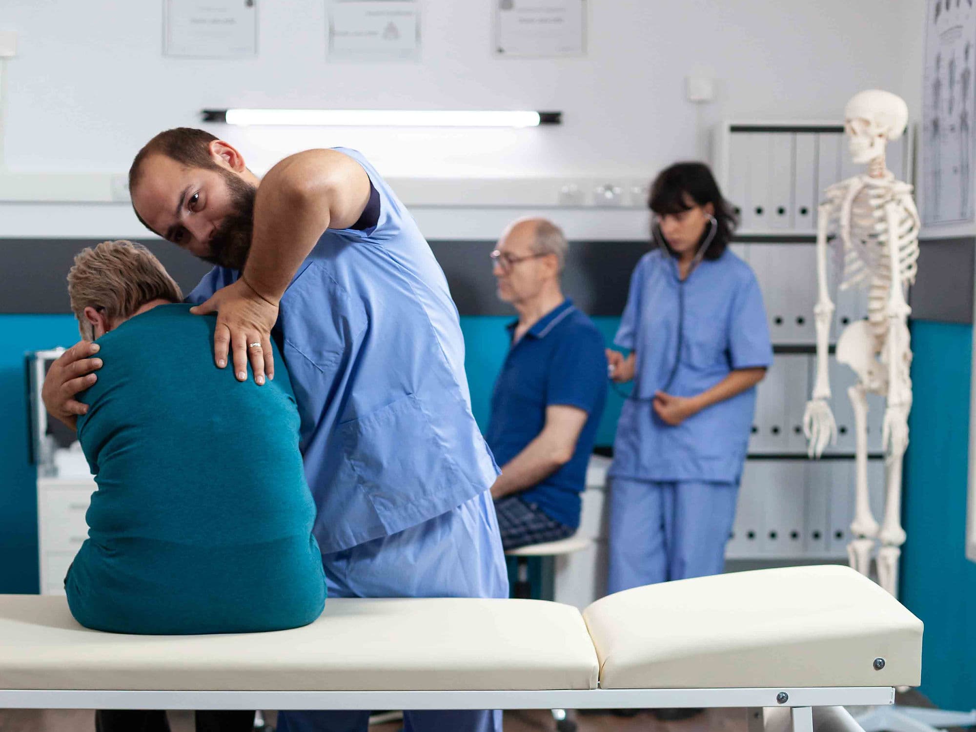
Visible Deformity: Significant rib humps, uneven shoulders, or a tilted pelvis that affects balance and gait.
Failed Conservative Treatment: When bracing or physical therapy has failed to stop the progression of the curve.
Methods of Scoliosis Correction
Spinal Fusion: The most established method, using bone grafts, metal rods, and pedicle screws to hold the spine straight while the vertebrae grow together.
Vertebral Body Tethering (VBT): A modern, motion-preserving option for growing patients that uses a flexible cord to guide the spine's growth into alignment.
Magnetically Controlled Growing Rods (MAGEC): Designed for young children, these rods are lengthened periodically using an external magnet, eliminating the need for repeat surgeries.
Posterior Spinal Instrumentation: A technique focused on the back of the spine to provide maximum stabilization and rotational correction.
How Scoliosis Correction Is Performed
Surgical Access: The surgeon makes an incision along the midline of the back (or side for VBT) to reach the affected vertebral segments.
Curve Realignment: Using rods and screws, the surgeon carefully maneuvers the vertebrae to reduce the sideways curve and correct any rotation.
Bone Grafting: For fusion cases, the surface of the vertebrae is prepared and bone graft material is added to facilitate the permanent joining of the bones.
Safety Monitoring: Throughout the procedure, real-time neuromonitoring tracks nerve signals to ensure the spinal cord is protected during the straightening process.
Pre-Procedure Preparation
Fasting: Patients are required to fast for 8–12 hours before the surgery.
Medical Clearances: Extensive blood work, ECG, and pulmonary function tests are performed to ensure the heart and lungs are healthy for anesthesia.
Blood Preparation: Use of "cell saver" technology is planned to collect and recycle the patient's own blood during the procedure.
Medication Review: Discussing all supplements and medications with the surgical team to minimize bleeding risks.
Tests Before Scoliosis Correction
Full-Spine X-rays: Taken while standing and bending to measure the "Cobb angle" and determine the flexibility of the curve.
MRI Scan: Used to rule out any underlying spinal cord abnormalities or syrinx before the correction.
CT Scan (3D): Provides a high-resolution map of the vertebrae for use with intraoperative "GPS" navigation systems.
Pulmonary Function Test (PFT): Measures lung capacity to assess the impact of the scoliosis on the respiratory system.
Life After Scoliosis Correction (Recovery)
Hospital Stay: Typically involves a stay of 3 to 5 days, with mobilization (walking) starting as early as the first day post-op.
Short-Term Recovery: Most patients return to school or sedentary work within 3 to 4 weeks.
Activity Timeline: Light exercise like walking is encouraged at 6 weeks; swimming and jogging are typically permitted between 3 to 6 months.
Long-Term Outlook: Full bone fusion is usually achieved at the 1-year mark, allowing a return to most non-contact sports and normal activities.
Benefits of Scoliosis Correction
Curvature Reduction: Significantly flattens the sideways curve and corrects the rotational "rib hump."
Prevention of Progression: Stops the spine from curving further, protecting long-term heart and lung function.
Restored Body Balance: Realigns the head and shoulders over the pelvis, improving posture and reducing muscle strain.
Improved Quality of Life: Provides a long-term solution that allows patients to lead active, healthy lives without the limitations of a severe spinal deformity.