Medivisor India Treatment Logo
  • Home
              • Become a Partner

              +91 83407 80250

              Medivisor Logo

              Medivisor India Treatment is a trusted New Delhi–based medical travel partner connecting global patients to advanced, affordable, high-quality healthcare in India.

              • Medivisor House 359, Sector 1, Vaishali, Ghaziabad, (Delhi/NCR) India
              • +91 8340 780 250
              • info@medivisorhealth.com

              About Us

              • About Us
              • Our Services
              • Our Team
              • Our Medical Advisors
              • Our Safety Measures
              • Join Our Network
              • Contact Us

              India Treatment

              • Hospitals
              • Treatments
              • Treatment Cost
              • Treatment Process
              • Visa Process
              • Travel Guide
              • FAQs
              • Why Medivisor

              Gallery

              • Patient Testimonials
              • Patient Activities
              • News Coverage
              • Blog

              Get in Touch

              © 2026 Medivisor India Treatment. All rights reserved.

              Dr. Dr. Subhash Gupta

              Dr. Subhash Gupta

              Group Chairman - Centre for Liver & Biliary Sciences

              38+ years experience

              All India Institute of Medical Sciences, New Delhi
              1. Home
              2. Doctors
              3. Dr. Subhash Gupta

              About Dr. Subhash Gupta

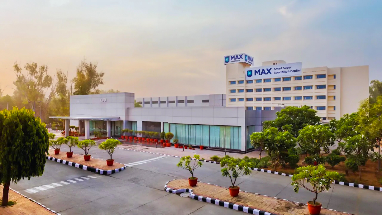
              Dr. Gupta is a globally recognized pioneer in liver transplantation and hepatopancreatobiliary (HPB) surgery, with over 30 years of extensive clinical experience. He currently serves as the Chairman of the Max Centre for Liver and Biliary Sciences, one of the most prolific and largest liver transplant centers in the world.

              Under his leadership, the center has achieved a landmark milestone of over 3,000 successful liver transplants. Currently, Dr. Gupta oversees a high-volume program that conducts more than 200 transplants annually. His expertise is foundational to the center’s reputation as a global destination for patients requiring life-saving hepatic interventions.

              Mastery in Complex HPB Oncology and Surgery

              Beyond transplantation, Dr. Gupta is a specialist in surgical gastroenterology and hepatopancreatobiliary oncology. He manages an additional 200 highly complex HPB cases each year, addressing intricate malignancies of the liver, gallbladder, and pancreas. His technical proficiency allows for the management of advanced tumors that require sophisticated surgical resection and reconstruction.

              Clinical Governance and Safety Protocols

              As Chairman, Dr. Gupta is responsible for maintaining rigorous safety and quality protocols across a multidisciplinary team. His oversight extends to specialized divisions including Hepatology, Transplant Anaesthesia, and Critical Care, ensuring a seamless and high-standard care pathway for patients undergoing major abdominal surgeries.

              Institutional Excellence and Experience

              Since joining Max Healthcare in 2017, Dr. Gupta has been instrumental in scaling the surgical gastroenterology services to international standards. His three decades of experience provide a seasoned perspective on the evolution of transplant medicine, making him a sought-after expert for both clinical excellence and institutional medical strategy.

              Prof. (Dr.) Subhash Gupta at a Glance

              • Chairman of the Max Centre for Liver and Biliary Sciences.

              • Over 30 years of experience in Liver Transplantation and Surgical Gastroenterology.

              • Leader of a program with a cumulative experience of 3,000+ transplants.

              • Expert in high-volume, difficult Hepatopancreaticobiliary (HPB) oncology cases.

              • Oversees comprehensive safety protocols in Anaesthesia, Hepatology, and Critical Care.

              • Recognized globally for pioneering work in living donor liver transplantation.

              All India Institute of Medical Sciences, New Delhi
              Specialization: Group Chairman - Centre for Liver & Biliary Sciences
              He has been honored with the position of Professorship in Surgery from Apollo Health Education
              Research Foundation The Institute of Postgraduate Education
              Medical Research, Kolkata, has also honored him with the position of Professor of Liver Transplantation Delhi Medical Association awarded him with a Gold Medal in
              The Rotary Association of India has honored him for excellence in clinical medicine in 2012 In 2012, the Delhi Medical Association honored him with the award of Vishist&nbsp
              Chikitsh Rattan (Distinguished Clinician) on Doctor’s Day In 2014, he &amp
              has team were one of the finalists for the category “Surgical team of the year” for BMJ India &nbsp
              awards Apollo Health foundation has made him an honorary Professor of Surgery In 2016, the Uttar Pradesh government has awarded him the prestigious “YASH BHARTI” award In 2016, the Medical Council of India awarded him the prestigious ‘Dr.B.C. Roy’ award In 2016, he was also awarded - The Honorary Professor of Kazakhstan

              Affiliated Hospitals

              Max Smart Super Specialty Hospital, Saket
              Max Smart Super Specialty Hospital, Saket

              Super Specialty

              New Delhi, Delhi NCR

              2013

              Estd.

              250+

              Beds

              300+

              Doctors

              Max Super Specialty Hospital, Saket
              Max Super Specialty Hospital, Saket

              Super Specialty

              New Delhi, Delhi NCR

              2006

              Estd.

              540+

              Beds

              450+

              Doctors

              Max Super Specialty Hospital, Vaishali
              Max Super Specialty Hospital, Vaishali

              Super Specialty

              Ghaziabad, Uttar Pradesh

              2008

              Estd.

              370+

              Beds

              260+

              Doctors

              May we help you?

              Enter digits only (6-15 digits). We'll add your country code.

              May we help you?

              Enter digits only (6-15 digits). We'll add your country code.

              Related Treatments

              Low Anterior Resection (Rectal Cancer)
              Low Anterior Resection (Rectal Cancer)

              Low Anterior Resection (LAR) Low Anterior Resection (LAR) is a major surgical procedure used to treat cancers located in the upper or middle parts of the rectum. The primary goal is to remove the cancerous section while preserving the anal sphincter, allowing for the maintenance of normal bowel continuity and avoiding a permanent stoma. The integration of Total Mesorectal Excision (TME) and robotic-assisted precision has made LAR the standard of care for sphincter-preserving rectal surgery. When You Should Consider LAR Mid-to-Upper Rectal Tumors: When the malignancy is located typically 5 cm or more from the anal verge. Sphincter Preservation: When the cancer has not invaded the anal sphincter muscles, allowing for a safe reconnection. Clear Distal Margins: When imaging confirms enough healthy tissue remains below the tumor to create a secure internal connection. Response to Chemoradiotherapy: For patients who have undergone neoadjuvant treatment to shrink a tumor into a resectable range. Non-Metastatic Localized Disease: When the primary goal is curative intent through the complete removal of the rectum and surrounding lymph nodes. Surgical Approaches Robotic-Assisted LAR: The 2026 preferred method for mid-to-low tumors. The robotic platform’s 3D high-definition vision and "wristed" instruments provide superior precision in the narrow male or female pelvis. Laparoscopic Surgery: A minimally invasive "keyhole" approach that offers faster recovery and less pain than open surgery. Open Surgery: Performed via a midline abdominal incision; reserved for very large tumors or complex cases involving multiple organ involvements. Total Mesorectal Excision (TME): A meticulous technique used during LAR to remove the rectum along with the intact fatty envelope (mesorectum) containing the lymph nodes, significantly reducing recurrence. Ultra-Low LAR: A specialized variation for tumors very close to the pelvic floor, where the connection is made almost at the level of the anal opening. How LAR Is Performed Anaesthesia: The procedure typically takes 3 to 6 hours under general anaesthesia. Mobilization: The surgeon frees the sigmoid colon and rectum from the surrounding pelvic structures and critical nerves. Vascular Control: The main artery supplying the rectum (Inferior Mesenteric Artery) is ligated to ensure a complete lymph node harvest. Stapled Anastomosis: Specialized circular staplers are used to connect the healthy colon to the remaining rectal stump. Air-Leak Testing: A routine safety check is performed during surgery to ensure the new connection is airtight and watertight. Defunctioning Ileostomy: A temporary stoma is often created to "divert" stool, allowing the internal connection (anastomosis) to heal without stress for 2–3 months. Pre-Surgery Preparation Neoadjuvant Therapy: Completing a 5-week course of chemoradiotherapy to shrink the tumor and "sterilize" the surgical field. Mechanical Bowel Prep: A thorough clearing of the bowels the day before surgery to minimize infection risks. Pelvic Floor Baseline: Consulting with a physiotherapist to establish pelvic muscle strength before surgery. Stoma Education: Meeting with a Wound, Ostomy, and Continence (WOC) nurse to mark a potential stoma site and learn about temporary bag management. Nutritional Loading: Adhering to a "pre-habilitation" diet to maintain protein levels, which is crucial for internal healing. Pre-Surgery Tests Pelvic MRI: The "gold standard" for staging rectal cancer and determining the exact distance of the tumor from the sphincter. Endorectal Ultrasound: To assess the depth of tumor invasion into the rectal wall layers. CEA Blood Test: To establish a baseline tumor marker level for post-operative monitoring. Rigid Proctoscopy: A physical measurement of the tumor's height to plan the exact level of the resection. CT Chest/Abdomen/Pelvis: To ensure the cancer has not spread to the liver or lungs before proceeding with major pelvic surgery. Life After LAR (Recovery & Risks) Hospital Stay: Usually 4 to 7 days, focusing on the return of bowel function and pain management. Anastomotic Leak: A serious complication (5–10% risk) where the internal connection fails; 2026 protocols use early CRP monitoring to detect this before symptoms appear. LARS (Low Anterior Resection Syndrome): A cluster of symptoms including urgency, frequency, and "clustering" of bowel movements that typically improves over 6–12 months. Pelvic Nerve Preservation: While robotic surgery reduces risk, some may experience temporary urinary or sexual dysfunction due to the proximity of the autonomic nerves. Low-Residue Diet: A temporary post-operative diet low in fiber to allow the bowel connections to heal without irritation. Why Specialized Treatment Is Highly Effective Lower Recurrence Rates: TME technique combined with neoadjuvant therapy has reduced local recurrence to below 5% in specialized centers. Permanent Stoma Avoidance: Modern LAR techniques allow over 90% of mid-rectal cancer patients to avoid a permanent colostomy. Robotic Precision: 2026 data shows that robotic LAR leads to better preservation of sexual and bladder function compared to traditional methods. Enhanced Recovery (ERAS): Specialized protocols allow patients to walk and eat sooner, reducing the risk of blood clots and pneumonia. Biologically Tailored Care: Integration of tumor genetic profiling helps determine if a patient needs further "mop-up" chemotherapy after a successful LAR.

              Gastrectomy (Stomach Cancer)
              Gastrectomy (Stomach Cancer)

              Stomach Cancer Treatment Stomach Cancer Treatment, also known as Gastric Cancer Treatment, involves a range of medical and surgical interventions to eliminate malignant growths within the stomach lining. Clinical protocols prioritize the removal of adenocarcinomas while preserving digestive function, utilizing precision therapies to target specific genetic markers and improve long-term outcomes for patients. When You Should Consider Screening Persistent Indigestion: A lasting burning sensation in the upper abdomen. Early Satiety: Feeling unusually full or bloated after consuming only small meals. Nausea and Vomiting: Chronic episodes, particularly if blood is present. Dysphagia: Difficulty swallowing or a sensation of food being "stuck" in the esophagus. Tarry Stools: Black, sticky stools which may indicate internal gastrointestinal bleeding. Unexplained Fatigue: Persistent weakness often associated with anemia. Conditions That Require Specialized Care Gastric Adenocarcinomas: Originating in the glandular cells of the stomach mucosa. HER2-Positive Tumors: Stomach cancers that require specific targeted medications. CLDN18.2-Positive Cancers: Identified through modern molecular profiling. H. pylori Infections: Chronic bacterial infections that have led to malignant cellular changes. Genetic Syndromes: Diffuse gastric cancer related to inherited conditions or Lynch syndrome. How Is Performed Endoscopy: An upper endoscopy is conducted to visualize the stomach lining and obtain tissue biopsies. Staging: Advanced imaging and endoscopic ultrasounds are used to determine the depth of the tumor. Surgical Resection: Intervention is performed to remove part or all of the affected stomach tissue. Perioperative Therapy: Chemotherapy is often administered to shrink the tumor before the primary procedure. Targeted Biology: Biological therapies are introduced to attack specific proteins on the cancer cells. Systematic Immunotherapy: Utilized to enhance the body's natural response to the malignancy. Types of Stomach Cancer Treatment Partial Gastrectomy: The surgical removal of the specific portion of the stomach containing the tumor and nearby lymph nodes. Total Gastrectomy: A procedure where the entire stomach is removed and the esophagus is connected directly to the small intestine. Targeted Therapy: The use of specialized drugs like Trastuzumab or Zolbetuximab to attack specific cancer cell markers. Immunotherapy: Checkpoint inhibitors that empower the immune system to recognize and destroy gastric cancer cells. Endoscopic Mucosal Resection: A minimally invasive technique used to remove very early-stage tumors during an endoscopy. HIPEC (Heated Chemotherapy): The delivery of heated chemotherapy directly into the abdominal cavity during surgery for advanced cases. Pre-Procedure Preparation Nutritional Counseling: Consult with a specialized dietitian to manage intake and prepare for digestive changes. Physical Assessment: A comprehensive evaluation to ensure readiness for general anesthesia. Medication Audit: Adhering to specific adjustments as advised by the surgical oncology team. Fasting Protocols: Strict adherence to fasting in the hours leading up to the scheduled procedure. Support Planning: Arranging for assistance during the transition to a modified eating schedule. Tests Before Stomach Cancer Treatment Upper GI Endoscopy: To provide a direct view of the gastric environment and tumor site. Endoscopic Ultrasound (EUS): To evaluate how far the cancer has invaded the stomach wall. CT and PET Scans: To check for the spread of cells to the liver, lungs, or lymph nodes. Biomarker Testing: HER2 and CLDN18.2 testing to determine the most effective drug therapies. Blood Work: Complete Blood Count (CBC) and iron studies to assess for internal blood loss. Life After Stomach Cancer Treatment Dietary Adaptation: Adopting a lifestyle of eating small, frequent, and nutrient-dense meals throughout the day. Supplementation: Regular vitamin and mineral support, particularly Vitamin B12, following a gastrectomy. Routine Surveillance: Ongoing monitoring through imaging and endoscopy to ensure continued remission. Weight Management: Consultation with nutritionists to maintain a healthy weight and energy levels. Survivorship Support: Participation in programs to manage the emotional and physical impact of the disease. Why Specialized Treatment Is Highly Effective Precision Guidelines: Matches therapies with the tumor's specific molecular profile. Perioperative Success: Significantly reduces the risk of cancer recurrence through timed therapy. Minimally Invasive Options: Results in faster recovery and less discomfort for eligible patients. Targeted Strategies: Provides life-prolonging options even for advanced or metastatic conditions. Integrated Care: Maintains high standards of digestive and nutritional health through multidisciplinary teams.

              Colectomy (Colon Cancer)
              Colectomy (Colon Cancer)

              Colon Cancer Treatment Colon Cancer Treatment involves a combination of specialized surgical, medical, and radiological interventions to remove malignant tumors from the large intestine. As of 2026, clinical standards emphasize a precision-based approach, utilizing robotic surgery for localized tumors and advanced immunotherapy for cases with specific genetic biomarkers (MSI-H/dMMR), aiming to achieve long-term remission while preserving bowel function. When You Should Consider Screening Persistent changes in bowel habits, such as diarrhea, constipation, or narrowing of the stool. Presence of bright red blood or dark, tarry stools during bowel movements. Ongoing abdominal discomfort, including persistent cramps, gas, or localized pain. A frequent feeling that the bowel does not empty completely after a movement. Unexplained weight loss accompanied by chronic fatigue or weakness. Signs of anemia, such as paleness and lack of energy, due to hidden internal bleeding. Conditions That Require Specialized Care Adenomatous Polyps that show signs of high-grade dysplasia or early transformation. Localized Colon Adenocarcinoma confined to the inner layers of the intestinal wall. Hereditary Syndromes including Lynch Syndrome or Familial Adenomatous Polyposis (FAP). Inflammatory Bowel Disease (IBD) such as long-term Ulcerative Colitis or Crohn’s disease. Advanced or metastatic colon cancer that has spread to the liver, lungs, or peritoneum. How Colon Cancer Treatment Is Performed Diagnostic colonoscopy is utilized to visualize the colon and remove precancerous polyps. High-resolution CT, MRI, or PET scans are performed to map the tumor’s size and spread. A partial or total colectomy is surgically performed to remove the malignant section of the colon. Adjuvant chemotherapy is administered after surgery to eliminate any remaining microscopic cells. Systematic immunotherapy is introduced for tumors with specific genetic MSI-H or dMMR markers. Targeted drug therapy is used to block the proteins that allow cancer cells to grow and spread. Types of Colon Cancer Treatment Laparoscopic or Robotic Colectomy A minimally invasive surgery to remove the cancerous part of the colon through small abdominal incisions. Polypectomy The removal of small, early-stage cancerous growths directly through a colonoscope without external incisions. Immunotherapy (Pembrolizumab/Nivolumab) Advanced biological treatments that help the immune system identify and attack cancer cells. Targeted Therapy (Bevacizumab/Cetuximab) Precision medications that target specific growth factors or blood vessel formation in the tumor. Cytoreductive Surgery with HIPEC A specialized procedure where the abdominal cavity is bathed in heated chemotherapy after tumor removal. Colostomy or Ileostomy A surgical procedure to create a stoma (opening) in the abdomen to allow waste to exit the body temporarily or permanently. Pre-Surgery Preparation Complete a "bowel prep" to thoroughly clear the colon of stool before the procedure. Consult with a dietitian to establish a low-residue diet in the days leading up to surgery. Adjust or stop blood-thinning medications as instructed by the surgical oncology team. Undergo a preoperative physical to ensure readiness for general anesthesia and recovery. Meet with an ostomy nurse if there is a possibility of needing a temporary or permanent stoma. Pre-Surgery Tests Colonoscopy with biopsy to confirm the specific pathology and grade of the cancer. CT Scan of the chest, abdomen, and pelvis to determine the clinical stage of the disease. CEA (Carcinoembryonic Antigen) Blood Test to establish a baseline for post-treatment monitoring. MSI/dMMR Biomarker Testing to identify if the tumor will respond to modern immunotherapies. Complete Blood Count (CBC) to check for anemia or other blood-related issues before surgery. Why Specialized Treatment Is Highly Effective Features a 91% five-year survival rate for localized cases through early detection and surgery. Utilizes 2026 precision medicine to match treatments to the tumor's unique genetic profile. Offers minimally invasive robotic options that significantly reduce recovery time and pain. Employs neoadjuvant therapies to shrink large tumors, making them easier to remove safely. Integrates multidisciplinary care to manage complex cases that have spread to other organs. Recovery After Treatment In-hospital monitoring for 3–5 days to ensure the return of normal bowel function. Personalized pain management and early mobilization to prevent blood clots and lung issues. Gradual transition from a liquid diet to soft foods as the digestive tract heals. Specialized wound care and training for patients who require a temporary or permanent stoma. Follow-up visits with the surgical and oncology teams to monitor healing and plan next steps. Life After Colon Cancer Treatment Regular surveillance through colonoscopies and blood tests every 6–12 months for the first few years. Adoption of a high-fiber diet rich in fruits, vegetables, and whole grains to support colon health. Commitment to regular physical activity and maintaining a healthy weight to reduce recurrence risk. Avoidance of tobacco products and limiting alcohol consumption as part of a cancer-prevention lifestyle. Participation in survivorship programs to manage the emotional and physical transition after treatment.

              Gastrointestinal Cancer Surgery
              Gastrointestinal Cancer Surgery

              Gastrointestinal (GI) Cancer Surgery Gastrointestinal (GI) Cancer Surgery refers to a broad category of operations used to remove tumors from the digestive tract, including the esophagus, stomach, liver, pancreas, and intestines. The primary goal is curative resection, where the surgeon removes the tumor along with a surrounding margin of healthy tissue and nearby lymph nodes. These procedures are increasingly performed using robotic platforms to enhance precision and protect delicate internal structure. When You Should Consider GI Cancer Surgery Esophageal Malignancies: When cancer is located in the tube connecting the throat to the stomach. Gastric Adenocarcinoma: When a tumor is identified in the lining of the stomach. Localized Pancreatic/Biliary Tumors: For cancers in the head of the pancreas or the bile ducts. Colorectal Cancer: When malignant growths are found in the large intestine or the rectum. Primary or Metastatic Liver Cancer: When tumors are confined to specific segments of the liver, allowing for safe removal. Gastrointestinal Stromal Tumors (GIST): For specialized mesenchymal tumors found anywhere along the GI tract. Types of GI Cancer Surgery Oesophagectomy: Removal of part or most of the esophagus. The stomach is typically shaped into a tube and pulled up into the chest to replace the missing section. Gastrectomy: The removal of either a portion (subtotal) or the entirety of the stomach. In a total gastrectomy, the esophagus is connected directly to the small intestine. Whipple Procedure: A complex "re-plumbing" of the digestive system used for pancreatic head tumors, involving the removal of the pancreas head, duodenum, and gallbladder. Hepatectomy: Surgical removal of a portion of the liver. This is highly effective because the liver can regenerate to nearly its full size within weeks. Colectomy: Removal of a diseased section of the colon (large intestine), followed by an anastomosis (reconnection) of the healthy ends. Low Anterior Resection (LAR): A sphincter-preserving surgery for rectal cancer that avoids the need for a permanent stoma. How GI Cancer Surgery Is Performed Anesthesia: All major GI surgeries are performed under general anesthesia, often supplemented with an epidural for post-operative pain control. Minimally Invasive Approaches: Most modern procedures utilize laparoscopic or robotic-assisted techniques through small "keyhole" incisions. Lymphadenectomy: A critical step where surgeons remove specific groups of lymph nodes (e.g., D2 dissection in stomach cancer) to check for microscopic spread. Anastomosis: The process of reconnecting the digestive tract using specialized surgical staples or hand-sewn sutures to ensure a watertight seal. Stoma Creation (Optional): In some colorectal cases, a temporary or permanent opening (stoma) is created on the abdomen to allow waste to exit the body while the internal connections heal. Pathologic Verification: All removed tissue is sent for immediate and long-term analysis to ensure "R0" margins (no cancer cells left behind). Pre-Surgery Preparation Nutritional Optimization: Many patients require a high-protein diet or specialized supplements to combat "cancer cachexia" before a major operation. Bowel Preparation: For colorectal surgery, a mechanical bowel prep (drinking a clearing solution) is necessary to reduce the risk of infection. Endoscopic Staging: Undergoing a final EUS (Endoscopic Ultrasound) to confirm the tumor depth and nodal involvement. Cardiopulmonary Clearance: Ensuring the heart and lungs are strong enough to tolerate the shifts in fluid and circulation during long GI procedures. Cessation Protocols: Strictly adhering to tobacco and alcohol cessation to improve the healing of new internal connections. Pre-Surgery Tests Multi-Phase CT or MRI: High-resolution imaging to map the tumor’s relationship with major abdominal blood vessels (like the mesenteric artery). PET-CT Scan: To rule out any spread of the cancer to the bones or lungs, ensuring surgery remains the correct curative path. Endoscopy / Colonoscopy: To physically visualize the tumor and mark its location for the surgeon using "clipping" or tattooing. Tumor Markers: Blood tests for markers like CEA, CA 19-9, or AFP to establish a baseline for post-operative monitoring. Biopsy Verification: Confirming the cellular grade of the tumor to determine if chemotherapy should be given before surgery (neoadjuvant). Life After GI Cancer Surgery (Recovery & Risks) Hospital Stay: Varies by procedure—3–5 days for a colectomy, but 7–14 days for more complex cases like a Whipple or Oesophagectomy. Anastomotic Leak: The most serious risk; modern protocols involve early monitoring of inflammatory markers to catch and treat leaks quickly. Dietary Transition: Starting with clear liquids and slowly progressing to soft, small, frequent meals as the bowel "wakes up." Nutritional Monitoring: Patients may need lifelong vitamin supplements (like B12) if large portions of the stomach or intestine were removed. Long-Term Activity: Most patients return to light activities in 6 weeks, but full core strength and bowel habit stabilization can take 6–12 months. Why Specialized Treatment Is Highly Effective Curative Foundation: Surgery remains the primary treatment for most localized GI cancers and provides the best chance for long-term survival. D2 Lymph Node Clearance: Specialized centers use advanced techniques to remove regional nodes, significantly lowering the risk of local recurrence. Organ Regeneration & Adaptation: The liver's ability to regrow and the intestine's ability to adapt allow for extensive resections with minimal long-term impact on lifestyle. Minimally Invasive Recovery: Robotic-assisted techniques lead to less internal scarring (adhesions) and a much faster return to normal eating. Multidisciplinary Care: When integrated with modern neoadjuvant and adjuvant protocols, GI surgery outcomes have reached historic highs.

              Whipple Surgery (Pancreatic Cancer)
              Whipple Surgery (Pancreatic Cancer)

              Whipple Surgery The Whipple procedure, or pancreaticoduodenectomy, is one of the most complex and demanding abdominal surgeries. It is primarily performed to treat tumors in the head of the pancreas, the bile duct, or the duodenum (the first part of the small intestine). Many of these procedures are now performed using robotic-assisted platforms to improve precision and shorten recovery times for this intricate "re-plumbing" of the digestive tract. When You Should Consider the Whipple Procedure Pancreatic Head Tumors: When a malignancy is localized in the widest part of the pancreas (the head). Bile Duct Cancer: For tumors located in the distal (lower) portion of the bile duct. Duodenal Cancer: When cancer is found in the first section of the small intestine. Ampullary Cancer: For tumors at the "Ampulla of Vater," where the bile and pancreatic ducts join. Localized Management: When imaging confirms the tumor has not yet encased major arteries, making it surgically resectable. Phase 1: Resection (Removal) Pancreas Head: The surgeon removes the right, widest part of the pancreas where the tumor is located. Duodenum: The first 25–30 cm of the small intestine is removed to ensure clear margins. Biliary System: The gallbladder and the common bile duct are removed as they are physically attached to the pancreatic head. Stomach (Variable): In a "classic" Whipple, the lower portion of the stomach is removed. In a "pylorus-preserving" Whipple, the entire stomach and its exit valve are kept intact. Lymph Nodes: Surrounding nodes are excised to check for cancer spread and ensure the most accurate staging. Phase 2: Reconstruction ("Re-plumbing") Pancreaticojejunostomy: The remaining tail and body of the pancreas are attached to the small intestine so digestive enzymes can reach food. Hepaticojejunostomy: The remaining bile duct is reconnected to the small intestine to allow bile from the liver to drain properly. Gastrojejunostomy: The stomach (or remaining duodenum) is attached to the small intestine so food can pass through the digestive system. Vascular Reconstruction: Specialized surgeons can often replace or repair nearby veins if the tumor has minimally invaded them. How Is Performed Anesthesia: The operation is performed under general anesthesia and typically lasts between 4 to 12 hours depending on complexity. Surgical Approach: Can be performed via a traditional "open" incision or through minimally invasive robotic-assisted surgery. Margin Assessment: Real-time pathology (frozen sections) is often used during the surgery to ensure all edges of the removed tissue are cancer-free. Drain Placement: Small tubes (drains) are placed near the new connections to monitor for fluid leaks during the first few days of recovery. Feeding Tube: In some cases, a temporary feeding tube is placed to ensure nutrition while the new stomach-to-intestine connection heals. Pre-Procedure Preparation Biliary Decompression: If you have severe jaundice, a stent may be placed in the bile duct a few weeks before surgery to allow the liver to recover. Pre-habilitation: Engaging in a guided walking and breathing exercise program to improve heart and lung stamina before the long surgery. Nutritional Loading: Following a high-protein diet to prevent muscle wasting and ensure the body has the resources to heal complex internal sutures. Medication Audit: Pausing blood thinners or certain herbal supplements that can increase bleeding risks during the extensive resection. Cardiac Clearance: A thorough heart evaluation to ensure you can safely tolerate several hours of general anesthesia. Tests Before Whipple Surgery Multi-Phase CT (Pancreas Protocol): A specialized scan to see exactly how the tumor sits against the mesenteric veins and arteries. Endoscopic Ultrasound (EUS): Used to take a biopsy and look at the tumor's proximity to the "re-plumbing" site. CA 19-9 Marker: A blood test to establish a baseline for monitoring the cancer's response to surgery and future treatments. Chest CT: To confirm the lungs are clear of any metastatic activity before proceeding with the abdominal resection. Complete Metabolic Panel: Assessing liver and kidney function to ensure the body can process the medications used during and after surgery. Life After the Whipple Procedure (Recovery & Risks) Hospital Stay: Usually 7 to 14 days in a specialized surgical unit to monitor the stability of the new connections. Pancreatic Fistula: The most serious common risk, where pancreatic juice leaks from a connection; this requires careful drain management. Delayed Gastric Emptying (DGE): The stomach may temporarily lose its ability to push food into the intestine, causing nausea for 7–10 days. Enzyme Replacement (PERT): Many patients require daily pancreatic enzyme supplements for life to help digest fats and proteins. New-Onset Diabetes: If a significant portion of the pancreas was removed, you may need insulin or oral meds to manage blood sugar. Benefits of the Whipple Procedure Significantly Higher Survival: For pancreatic cancer, the 5-year survival rate after a successful Whipple is 20–25%, much higher than without surgery. Potential for Cure: It remains the only treatment that offers a definitive chance to completely remove a localized pancreatic tumor. Pain Relief: Removing a tumor that is pressing on the celiac plexus (nerves) can significantly reduce chronic abdominal and back pain. Robotic Precision: Modern robotic techniques have reduced the incidence of post-operative infections and shortened hospital stays. Multidisciplinary Success: When paired with modern chemotherapy, the Whipple procedure provides the strongest foundation for long-term remission.

              Liver Resection (Cancer)
              Liver Resection (Cancer)

              Liver Resection (Cancer) Liver Resection (also known as a Hepatectomy) is a major surgical procedure to remove malignant tumours from the liver. Because the liver is the only internal organ capable of regeneration, surgeons can safely remove a large portion of it, and the remaining healthy tissue will grow back to nearly its original size within 6 to 10 weeks. This remains the "gold standard" for curative intent when cancer is confined to the liver. When You Should Consider Liver Resection Hepatocellular Carcinoma (HCC): The most common primary liver cancer, particularly in patients with a history of hepatitis or cirrhosis. Intrahepatic Cholangiocarcinoma: When cancer originates in the bile ducts located within the liver tissue. Metastatic Colorectal Cancer: If colorectal cancer has spread only to the liver, a resection can still be a curative pathway. Clear Margins: When imaging confirms the tumour can be removed while leaving a healthy "rim" of tissue behind. Solitary or Limited Tumours: When the malignancy is confined to specific segments that allow for a safe "Future Liver Remnant." Surgical Techniques Anatomic Resection: Removing a specific functional segment or lobe (the liver has 8 segments) along with its dedicated blood supply and bile duct. Non-Anatomic (Wedge) Resection: Removing the tumour plus a 1-cm "rim" of healthy tissue; typically used for small tumours near the surface. Open Surgery: Performed via a "Mercedes-Benz" or "J-shaped" incision; used for large or centrally located tumours. Laparoscopic/Robotic Surgery: Performed through several 1-cm punctures. This 2026 standard offers faster recovery and less pain for suitable tumour locations. Two-Stage Hepatectomy: In complex cases, surgeons may remove tumours from one side, allow it to regenerate, and then remove the rest in a second surgery. How Liver Resection Is Performed Anaesthesia: The procedure typically takes 3 to 7 hours under general anaesthesia. Intraoperative Ultrasound: Surgeons use a specialized probe directly on the liver during surgery to find hidden tumours and map blood vessels. Transection: The liver tissue is carefully divided using advanced tools (like ultrasonic aspirators) that seal blood vessels and bile ducts as they cut. Pringle Manoeuvre: A technique used to temporarily "clamp" blood flow to the liver to prevent heavy bleeding during the removal phase. Drain Placement: Small tubes may be left in the abdomen to monitor for any bile leaks or fluid buildup during the first few days of recovery. Pre-Surgery Preparation Future Liver Remnant (FLR) Assessment: Ensuring that the amount of healthy liver left after surgery (at least 25–30% for healthy livers) is sufficient for survival. Portal Vein Embolization (PVE): If the planned remnant is too small, a procedure is done weeks prior to "trick" the healthy side into growing larger before the operation. Nutritional Optimization: Following a specific diet to reduce liver fat (steatosis), which improves the organ's ability to regenerate. Cardiovascular Clearance: Undergoing a stress test to ensure the heart can handle the circulatory shifts that occur during liver surgery. Medication Audit: Strictly stopping all blood-thinners and certain herbal supplements at least one week before the procedure. Pre-Surgery Tests Tri-Phasic CT or MRI: To visualize the liver's blood supply and precisely locate tumours in relation to the eight segments. Indocyanine Green (ICG) Clearance: A specialized test to measure how well the liver filters dye, predicting its post-operative function. AFP (Alpha-fetoprotein) Test: A blood marker used to establish a baseline for monitoring primary liver cancer. FibroScan: To assess the degree of underlying scarring (cirrhosis), which dictates how much liver can safely be removed. PET-CT Scan: To ensure there is no hidden cancer outside the liver that would make surgery ineffective. Life After Liver Resection (Recovery & Risks) Regeneration Timeline: The most intense growth happens in the first 14 days, with the liver returning to nearly full size within 2 months. Haemorrhage Risk: Because the liver is highly vascular, significant bleeding is the primary risk during the surgery and the immediate recovery phase. Bile Leak: A 5–10% risk where bile leaks from the cut surface; most are managed with temporary plastic drains. Post-Hepatectomy Liver Failure (PHLF): A serious risk if the remaining liver is too small or weak to filter toxins and produce clotting factors. Pleural Effusion: Fluid buildup around the right lung is common after right-sided surgery and is monitored closely in the hospital. Why Specialized Treatment Is Highly Effective Unique Regenerative Power: The liver’s ability to grow back allows for the removal of up to 75% of the organ while maintaining life. Curative Intent: For colorectal metastases, the 5-year survival rate after a successful resection is approximately 40–60%. 2026 Robotic Precision: Minimally invasive techniques have significantly reduced the "Mercedes-Benz" scar and shortened hospital stays to 5 days. PVE Advancements: Portal Vein Embolization now allows patients who were previously "inoperable" to become candidates for surgery. Multidisciplinary Success: When paired with modern chemotherapy, resection offers the best long-term outlook for primary and metastatic liver cancers.

              Esophageal Cancer Treatment
              Esophageal Cancer Treatment

              Esophageal Cancer Treatment Esophageal Cancer Treatment involves a range of advanced medical and surgical procedures designed to eliminate malignant tumors within the esophagus—the muscular tube connecting the throat to the stomach. Clinical protocols emphasize a personalized, multimodal approach to restore swallowing function and prevent the systemic spread of cancer cells while prioritizing the patient’s overall quality of life. When You Should Consider Screening Persistent difficulty or pain while swallowing solid foods or liquids. Unexplained weight loss without changes in diet or physical activity. Chronic chest pain or a sensation of pressure behind the breastbone. Frequent or worsening symptoms of acid reflux and heartburn. Persistent hoarseness or a chronic cough that does not resolve. Frequent choking on food or a feeling of something being stuck in the throat. Conditions That Require Specialized Care Adenocarcinoma occurring primarily in the lower part of the esophagus. Squamous Cell Carcinoma affecting the thin, flat cells of the upper and middle esophagus. Barrett’s Esophagus with high-grade dysplasia that may lead to malignancy. Gastroesophageal Junction (GEJ) tumors affecting the area where the esophagus meets the stomach. Recurrent esophageal cancer following previous chemotherapy or surgical interventions. How Esophageal Cancer Treatment Is Performed Diagnostic upper endoscopy and biopsies are utilized to identify the cellular subtype. Imaging scans including PET and CT are conducted to map the extent of the tumor. Minimally invasive surgery is performed to remove the diseased segment of the esophagus. Immunotherapy infusions are administered to help the immune system identify malignant cells. Precision radiation is applied to the tumor site to reduce its size before surgical removal. Real-time monitoring of tumor markers in the blood is used to guide post-operative recovery decisions. Types of Esophageal Cancer Treatment Robotic-Assisted Esophagectomy A minimally invasive surgical procedure to remove the tumor and nearby lymph nodes with high precision. Radiofrequency Ablation (RFA) The use of thermal energy to destroy precancerous tissue in patients with Barrett’s esophagus. Immunotherapy Modern checkpoint inhibitors like pembrolizumab and tislelizumab that empower the immune system to fight advanced cancer. FLOT Chemotherapy Regimen A specialized drug protocol used to achieve superior systemic control in cases of adenocarcinoma. Endoscopic Mucosal Resection (EMR) A procedure used to remove small, early-stage tumors from the lining of the esophagus without major surgery. Targeted Therapy Medications designed to attack specific molecular markers found on esophageal cancer cells. Pre-Surgery Preparation Engage in nutritional counseling to ensure the body is strong enough for the recovery process. Adhere to breathing exercises and pulmonary rehabilitation to optimize lung function. Follow specific guidelines for fasting and hydration as directed by the anesthesia team. Consult with the surgical team regarding the use of feeding tubes for post-operative support. Complete all cardiovascular clearances to ensure safe management during the procedure. Pre-Surgery Tests Upper Endoscopy to visually inspect the esophagus and collect tissue samples. Endoscopic Ultrasound (EUS) to determine how deeply the tumor has invaded the esophageal wall. CT and PET Scans to evaluate whether the cancer has spread to distant organs. Circulating Tumor DNA (ctDNA) test to establish a baseline for molecular monitoring. Pulmonary Function Tests to assess the strength of the respiratory system before surgery. Why Specialized Treatment Is Highly Effective Incorporates 2026 breakthroughs in immunotherapy for first-line treatment success. Utilizes robotic technology to reduce surgical trauma and shorten hospital stays. Employs "guided" ablative therapies to treat early-stage cancer without major incisions. Targets systemic disease more effectively through modern chemotherapy combinations. Provides a more accurate prognosis using molecular monitoring and precision imaging. Recovery After Treatment Intensive monitoring of swallowing function and respiratory health in a clinical setting. Early mobilization and physical therapy to prevent complications and restore strength. Gradual transition from liquid diets to soft foods under the supervision of a dietitian. Specialized care for surgical incisions to ensure proper healing and prevent infection. Follow-up evaluations with the oncology team to manage any side effects of systemic therapy. Life After Esophageal Cancer Treatment Permanent adoption of smaller, more frequent meals to accommodate changes in digestive capacity. Maintaining an elevated sleeping position to prevent reflux and respiratory irritation. Regular follow-up appointments with periodic imaging to ensure continued remission. Ongoing monitoring of tumor markers through non-invasive blood-based liquid biopsies. Commitment to a lifestyle free from tobacco and excessive alcohol to reduce recurrence risk.

              Low Anterior Resection (Rectal Cancer)
              Low Anterior Resection (Rectal Cancer)

              Low Anterior Resection (LAR) Low Anterior Resection (LAR) is a major surgical procedure used to treat cancers located in the upper or middle parts of the rectum. The primary goal is to remove the cancerous section while preserving the anal sphincter, allowing for the maintenance of normal bowel continuity and avoiding a permanent stoma. The integration of Total Mesorectal Excision (TME) and robotic-assisted precision has made LAR the standard of care for sphincter-preserving rectal surgery. When You Should Consider LAR Mid-to-Upper Rectal Tumors: When the malignancy is located typically 5 cm or more from the anal verge. Sphincter Preservation: When the cancer has not invaded the anal sphincter muscles, allowing for a safe reconnection. Clear Distal Margins: When imaging confirms enough healthy tissue remains below the tumor to create a secure internal connection. Response to Chemoradiotherapy: For patients who have undergone neoadjuvant treatment to shrink a tumor into a resectable range. Non-Metastatic Localized Disease: When the primary goal is curative intent through the complete removal of the rectum and surrounding lymph nodes. Surgical Approaches Robotic-Assisted LAR: The 2026 preferred method for mid-to-low tumors. The robotic platform’s 3D high-definition vision and "wristed" instruments provide superior precision in the narrow male or female pelvis. Laparoscopic Surgery: A minimally invasive "keyhole" approach that offers faster recovery and less pain than open surgery. Open Surgery: Performed via a midline abdominal incision; reserved for very large tumors or complex cases involving multiple organ involvements. Total Mesorectal Excision (TME): A meticulous technique used during LAR to remove the rectum along with the intact fatty envelope (mesorectum) containing the lymph nodes, significantly reducing recurrence. Ultra-Low LAR: A specialized variation for tumors very close to the pelvic floor, where the connection is made almost at the level of the anal opening. How LAR Is Performed Anaesthesia: The procedure typically takes 3 to 6 hours under general anaesthesia. Mobilization: The surgeon frees the sigmoid colon and rectum from the surrounding pelvic structures and critical nerves. Vascular Control: The main artery supplying the rectum (Inferior Mesenteric Artery) is ligated to ensure a complete lymph node harvest. Stapled Anastomosis: Specialized circular staplers are used to connect the healthy colon to the remaining rectal stump. Air-Leak Testing: A routine safety check is performed during surgery to ensure the new connection is airtight and watertight. Defunctioning Ileostomy: A temporary stoma is often created to "divert" stool, allowing the internal connection (anastomosis) to heal without stress for 2–3 months. Pre-Surgery Preparation Neoadjuvant Therapy: Completing a 5-week course of chemoradiotherapy to shrink the tumor and "sterilize" the surgical field. Mechanical Bowel Prep: A thorough clearing of the bowels the day before surgery to minimize infection risks. Pelvic Floor Baseline: Consulting with a physiotherapist to establish pelvic muscle strength before surgery. Stoma Education: Meeting with a Wound, Ostomy, and Continence (WOC) nurse to mark a potential stoma site and learn about temporary bag management. Nutritional Loading: Adhering to a "pre-habilitation" diet to maintain protein levels, which is crucial for internal healing. Pre-Surgery Tests Pelvic MRI: The "gold standard" for staging rectal cancer and determining the exact distance of the tumor from the sphincter. Endorectal Ultrasound: To assess the depth of tumor invasion into the rectal wall layers. CEA Blood Test: To establish a baseline tumor marker level for post-operative monitoring. Rigid Proctoscopy: A physical measurement of the tumor's height to plan the exact level of the resection. CT Chest/Abdomen/Pelvis: To ensure the cancer has not spread to the liver or lungs before proceeding with major pelvic surgery. Life After LAR (Recovery & Risks) Hospital Stay: Usually 4 to 7 days, focusing on the return of bowel function and pain management. Anastomotic Leak: A serious complication (5–10% risk) where the internal connection fails; 2026 protocols use early CRP monitoring to detect this before symptoms appear. LARS (Low Anterior Resection Syndrome): A cluster of symptoms including urgency, frequency, and "clustering" of bowel movements that typically improves over 6–12 months. Pelvic Nerve Preservation: While robotic surgery reduces risk, some may experience temporary urinary or sexual dysfunction due to the proximity of the autonomic nerves. Low-Residue Diet: A temporary post-operative diet low in fiber to allow the bowel connections to heal without irritation. Why Specialized Treatment Is Highly Effective Lower Recurrence Rates: TME technique combined with neoadjuvant therapy has reduced local recurrence to below 5% in specialized centers. Permanent Stoma Avoidance: Modern LAR techniques allow over 90% of mid-rectal cancer patients to avoid a permanent colostomy. Robotic Precision: 2026 data shows that robotic LAR leads to better preservation of sexual and bladder function compared to traditional methods. Enhanced Recovery (ERAS): Specialized protocols allow patients to walk and eat sooner, reducing the risk of blood clots and pneumonia. Biologically Tailored Care: Integration of tumor genetic profiling helps determine if a patient needs further "mop-up" chemotherapy after a successful LAR.

              Gastrointestinal Cancer Surgery
              Gastrointestinal Cancer Surgery

              Gastrointestinal (GI) Cancer Surgery Gastrointestinal (GI) Cancer Surgery refers to a broad category of operations used to remove tumors from the digestive tract, including the esophagus, stomach, liver, pancreas, and intestines. The primary goal is curative resection, where the surgeon removes the tumor along with a surrounding margin of healthy tissue and nearby lymph nodes. These procedures are increasingly performed using robotic platforms to enhance precision and protect delicate internal structure. When You Should Consider GI Cancer Surgery Esophageal Malignancies: When cancer is located in the tube connecting the throat to the stomach. Gastric Adenocarcinoma: When a tumor is identified in the lining of the stomach. Localized Pancreatic/Biliary Tumors: For cancers in the head of the pancreas or the bile ducts. Colorectal Cancer: When malignant growths are found in the large intestine or the rectum. Primary or Metastatic Liver Cancer: When tumors are confined to specific segments of the liver, allowing for safe removal. Gastrointestinal Stromal Tumors (GIST): For specialized mesenchymal tumors found anywhere along the GI tract. Types of GI Cancer Surgery Oesophagectomy: Removal of part or most of the esophagus. The stomach is typically shaped into a tube and pulled up into the chest to replace the missing section. Gastrectomy: The removal of either a portion (subtotal) or the entirety of the stomach. In a total gastrectomy, the esophagus is connected directly to the small intestine. Whipple Procedure: A complex "re-plumbing" of the digestive system used for pancreatic head tumors, involving the removal of the pancreas head, duodenum, and gallbladder. Hepatectomy: Surgical removal of a portion of the liver. This is highly effective because the liver can regenerate to nearly its full size within weeks. Colectomy: Removal of a diseased section of the colon (large intestine), followed by an anastomosis (reconnection) of the healthy ends. Low Anterior Resection (LAR): A sphincter-preserving surgery for rectal cancer that avoids the need for a permanent stoma. How GI Cancer Surgery Is Performed Anesthesia: All major GI surgeries are performed under general anesthesia, often supplemented with an epidural for post-operative pain control. Minimally Invasive Approaches: Most modern procedures utilize laparoscopic or robotic-assisted techniques through small "keyhole" incisions. Lymphadenectomy: A critical step where surgeons remove specific groups of lymph nodes (e.g., D2 dissection in stomach cancer) to check for microscopic spread. Anastomosis: The process of reconnecting the digestive tract using specialized surgical staples or hand-sewn sutures to ensure a watertight seal. Stoma Creation (Optional): In some colorectal cases, a temporary or permanent opening (stoma) is created on the abdomen to allow waste to exit the body while the internal connections heal. Pathologic Verification: All removed tissue is sent for immediate and long-term analysis to ensure "R0" margins (no cancer cells left behind). Pre-Surgery Preparation Nutritional Optimization: Many patients require a high-protein diet or specialized supplements to combat "cancer cachexia" before a major operation. Bowel Preparation: For colorectal surgery, a mechanical bowel prep (drinking a clearing solution) is necessary to reduce the risk of infection. Endoscopic Staging: Undergoing a final EUS (Endoscopic Ultrasound) to confirm the tumor depth and nodal involvement. Cardiopulmonary Clearance: Ensuring the heart and lungs are strong enough to tolerate the shifts in fluid and circulation during long GI procedures. Cessation Protocols: Strictly adhering to tobacco and alcohol cessation to improve the healing of new internal connections. Pre-Surgery Tests Multi-Phase CT or MRI: High-resolution imaging to map the tumor’s relationship with major abdominal blood vessels (like the mesenteric artery). PET-CT Scan: To rule out any spread of the cancer to the bones or lungs, ensuring surgery remains the correct curative path. Endoscopy / Colonoscopy: To physically visualize the tumor and mark its location for the surgeon using "clipping" or tattooing. Tumor Markers: Blood tests for markers like CEA, CA 19-9, or AFP to establish a baseline for post-operative monitoring. Biopsy Verification: Confirming the cellular grade of the tumor to determine if chemotherapy should be given before surgery (neoadjuvant). Life After GI Cancer Surgery (Recovery & Risks) Hospital Stay: Varies by procedure—3–5 days for a colectomy, but 7–14 days for more complex cases like a Whipple or Oesophagectomy. Anastomotic Leak: The most serious risk; modern protocols involve early monitoring of inflammatory markers to catch and treat leaks quickly. Dietary Transition: Starting with clear liquids and slowly progressing to soft, small, frequent meals as the bowel "wakes up." Nutritional Monitoring: Patients may need lifelong vitamin supplements (like B12) if large portions of the stomach or intestine were removed. Long-Term Activity: Most patients return to light activities in 6 weeks, but full core strength and bowel habit stabilization can take 6–12 months. Why Specialized Treatment Is Highly Effective Curative Foundation: Surgery remains the primary treatment for most localized GI cancers and provides the best chance for long-term survival. D2 Lymph Node Clearance: Specialized centers use advanced techniques to remove regional nodes, significantly lowering the risk of local recurrence. Organ Regeneration & Adaptation: The liver's ability to regrow and the intestine's ability to adapt allow for extensive resections with minimal long-term impact on lifestyle. Minimally Invasive Recovery: Robotic-assisted techniques lead to less internal scarring (adhesions) and a much faster return to normal eating. Multidisciplinary Care: When integrated with modern neoadjuvant and adjuvant protocols, GI surgery outcomes have reached historic highs.

              Whipple Surgery (Pancreatic Cancer)
              Whipple Surgery (Pancreatic Cancer)

              Whipple Surgery The Whipple procedure, or pancreaticoduodenectomy, is one of the most complex and demanding abdominal surgeries. It is primarily performed to treat tumors in the head of the pancreas, the bile duct, or the duodenum (the first part of the small intestine). Many of these procedures are now performed using robotic-assisted platforms to improve precision and shorten recovery times for this intricate "re-plumbing" of the digestive tract. When You Should Consider the Whipple Procedure Pancreatic Head Tumors: When a malignancy is localized in the widest part of the pancreas (the head). Bile Duct Cancer: For tumors located in the distal (lower) portion of the bile duct. Duodenal Cancer: When cancer is found in the first section of the small intestine. Ampullary Cancer: For tumors at the "Ampulla of Vater," where the bile and pancreatic ducts join. Localized Management: When imaging confirms the tumor has not yet encased major arteries, making it surgically resectable. Phase 1: Resection (Removal) Pancreas Head: The surgeon removes the right, widest part of the pancreas where the tumor is located. Duodenum: The first 25–30 cm of the small intestine is removed to ensure clear margins. Biliary System: The gallbladder and the common bile duct are removed as they are physically attached to the pancreatic head. Stomach (Variable): In a "classic" Whipple, the lower portion of the stomach is removed. In a "pylorus-preserving" Whipple, the entire stomach and its exit valve are kept intact. Lymph Nodes: Surrounding nodes are excised to check for cancer spread and ensure the most accurate staging. Phase 2: Reconstruction ("Re-plumbing") Pancreaticojejunostomy: The remaining tail and body of the pancreas are attached to the small intestine so digestive enzymes can reach food. Hepaticojejunostomy: The remaining bile duct is reconnected to the small intestine to allow bile from the liver to drain properly. Gastrojejunostomy: The stomach (or remaining duodenum) is attached to the small intestine so food can pass through the digestive system. Vascular Reconstruction: Specialized surgeons can often replace or repair nearby veins if the tumor has minimally invaded them. How Is Performed Anesthesia: The operation is performed under general anesthesia and typically lasts between 4 to 12 hours depending on complexity. Surgical Approach: Can be performed via a traditional "open" incision or through minimally invasive robotic-assisted surgery. Margin Assessment: Real-time pathology (frozen sections) is often used during the surgery to ensure all edges of the removed tissue are cancer-free. Drain Placement: Small tubes (drains) are placed near the new connections to monitor for fluid leaks during the first few days of recovery. Feeding Tube: In some cases, a temporary feeding tube is placed to ensure nutrition while the new stomach-to-intestine connection heals. Pre-Procedure Preparation Biliary Decompression: If you have severe jaundice, a stent may be placed in the bile duct a few weeks before surgery to allow the liver to recover. Pre-habilitation: Engaging in a guided walking and breathing exercise program to improve heart and lung stamina before the long surgery. Nutritional Loading: Following a high-protein diet to prevent muscle wasting and ensure the body has the resources to heal complex internal sutures. Medication Audit: Pausing blood thinners or certain herbal supplements that can increase bleeding risks during the extensive resection. Cardiac Clearance: A thorough heart evaluation to ensure you can safely tolerate several hours of general anesthesia. Tests Before Whipple Surgery Multi-Phase CT (Pancreas Protocol): A specialized scan to see exactly how the tumor sits against the mesenteric veins and arteries. Endoscopic Ultrasound (EUS): Used to take a biopsy and look at the tumor's proximity to the "re-plumbing" site. CA 19-9 Marker: A blood test to establish a baseline for monitoring the cancer's response to surgery and future treatments. Chest CT: To confirm the lungs are clear of any metastatic activity before proceeding with the abdominal resection. Complete Metabolic Panel: Assessing liver and kidney function to ensure the body can process the medications used during and after surgery. Life After the Whipple Procedure (Recovery & Risks) Hospital Stay: Usually 7 to 14 days in a specialized surgical unit to monitor the stability of the new connections. Pancreatic Fistula: The most serious common risk, where pancreatic juice leaks from a connection; this requires careful drain management. Delayed Gastric Emptying (DGE): The stomach may temporarily lose its ability to push food into the intestine, causing nausea for 7–10 days. Enzyme Replacement (PERT): Many patients require daily pancreatic enzyme supplements for life to help digest fats and proteins. New-Onset Diabetes: If a significant portion of the pancreas was removed, you may need insulin or oral meds to manage blood sugar. Benefits of the Whipple Procedure Significantly Higher Survival: For pancreatic cancer, the 5-year survival rate after a successful Whipple is 20–25%, much higher than without surgery. Potential for Cure: It remains the only treatment that offers a definitive chance to completely remove a localized pancreatic tumor. Pain Relief: Removing a tumor that is pressing on the celiac plexus (nerves) can significantly reduce chronic abdominal and back pain. Robotic Precision: Modern robotic techniques have reduced the incidence of post-operative infections and shortened hospital stays. Multidisciplinary Success: When paired with modern chemotherapy, the Whipple procedure provides the strongest foundation for long-term remission.

              Liver Resection (Cancer)
              Liver Resection (Cancer)

              Liver Resection (Cancer) Liver Resection (also known as a Hepatectomy) is a major surgical procedure to remove malignant tumours from the liver. Because the liver is the only internal organ capable of regeneration, surgeons can safely remove a large portion of it, and the remaining healthy tissue will grow back to nearly its original size within 6 to 10 weeks. This remains the "gold standard" for curative intent when cancer is confined to the liver. When You Should Consider Liver Resection Hepatocellular Carcinoma (HCC): The most common primary liver cancer, particularly in patients with a history of hepatitis or cirrhosis. Intrahepatic Cholangiocarcinoma: When cancer originates in the bile ducts located within the liver tissue. Metastatic Colorectal Cancer: If colorectal cancer has spread only to the liver, a resection can still be a curative pathway. Clear Margins: When imaging confirms the tumour can be removed while leaving a healthy "rim" of tissue behind. Solitary or Limited Tumours: When the malignancy is confined to specific segments that allow for a safe "Future Liver Remnant." Surgical Techniques Anatomic Resection: Removing a specific functional segment or lobe (the liver has 8 segments) along with its dedicated blood supply and bile duct. Non-Anatomic (Wedge) Resection: Removing the tumour plus a 1-cm "rim" of healthy tissue; typically used for small tumours near the surface. Open Surgery: Performed via a "Mercedes-Benz" or "J-shaped" incision; used for large or centrally located tumours. Laparoscopic/Robotic Surgery: Performed through several 1-cm punctures. This 2026 standard offers faster recovery and less pain for suitable tumour locations. Two-Stage Hepatectomy: In complex cases, surgeons may remove tumours from one side, allow it to regenerate, and then remove the rest in a second surgery. How Liver Resection Is Performed Anaesthesia: The procedure typically takes 3 to 7 hours under general anaesthesia. Intraoperative Ultrasound: Surgeons use a specialized probe directly on the liver during surgery to find hidden tumours and map blood vessels. Transection: The liver tissue is carefully divided using advanced tools (like ultrasonic aspirators) that seal blood vessels and bile ducts as they cut. Pringle Manoeuvre: A technique used to temporarily "clamp" blood flow to the liver to prevent heavy bleeding during the removal phase. Drain Placement: Small tubes may be left in the abdomen to monitor for any bile leaks or fluid buildup during the first few days of recovery. Pre-Surgery Preparation Future Liver Remnant (FLR) Assessment: Ensuring that the amount of healthy liver left after surgery (at least 25–30% for healthy livers) is sufficient for survival. Portal Vein Embolization (PVE): If the planned remnant is too small, a procedure is done weeks prior to "trick" the healthy side into growing larger before the operation. Nutritional Optimization: Following a specific diet to reduce liver fat (steatosis), which improves the organ's ability to regenerate. Cardiovascular Clearance: Undergoing a stress test to ensure the heart can handle the circulatory shifts that occur during liver surgery. Medication Audit: Strictly stopping all blood-thinners and certain herbal supplements at least one week before the procedure. Pre-Surgery Tests Tri-Phasic CT or MRI: To visualize the liver's blood supply and precisely locate tumours in relation to the eight segments. Indocyanine Green (ICG) Clearance: A specialized test to measure how well the liver filters dye, predicting its post-operative function. AFP (Alpha-fetoprotein) Test: A blood marker used to establish a baseline for monitoring primary liver cancer. FibroScan: To assess the degree of underlying scarring (cirrhosis), which dictates how much liver can safely be removed. PET-CT Scan: To ensure there is no hidden cancer outside the liver that would make surgery ineffective. Life After Liver Resection (Recovery & Risks) Regeneration Timeline: The most intense growth happens in the first 14 days, with the liver returning to nearly full size within 2 months. Haemorrhage Risk: Because the liver is highly vascular, significant bleeding is the primary risk during the surgery and the immediate recovery phase. Bile Leak: A 5–10% risk where bile leaks from the cut surface; most are managed with temporary plastic drains. Post-Hepatectomy Liver Failure (PHLF): A serious risk if the remaining liver is too small or weak to filter toxins and produce clotting factors. Pleural Effusion: Fluid buildup around the right lung is common after right-sided surgery and is monitored closely in the hospital. Why Specialized Treatment Is Highly Effective Unique Regenerative Power: The liver’s ability to grow back allows for the removal of up to 75% of the organ while maintaining life. Curative Intent: For colorectal metastases, the 5-year survival rate after a successful resection is approximately 40–60%. 2026 Robotic Precision: Minimally invasive techniques have significantly reduced the "Mercedes-Benz" scar and shortened hospital stays to 5 days. PVE Advancements: Portal Vein Embolization now allows patients who were previously "inoperable" to become candidates for surgery. Multidisciplinary Success: When paired with modern chemotherapy, resection offers the best long-term outlook for primary and metastatic liver cancers.

              Esophageal Cancer Treatment
              Esophageal Cancer Treatment

              Esophageal Cancer Treatment Esophageal Cancer Treatment involves a range of advanced medical and surgical procedures designed to eliminate malignant tumors within the esophagus—the muscular tube connecting the throat to the stomach. Clinical protocols emphasize a personalized, multimodal approach to restore swallowing function and prevent the systemic spread of cancer cells while prioritizing the patient’s overall quality of life. When You Should Consider Screening Persistent difficulty or pain while swallowing solid foods or liquids. Unexplained weight loss without changes in diet or physical activity. Chronic chest pain or a sensation of pressure behind the breastbone. Frequent or worsening symptoms of acid reflux and heartburn. Persistent hoarseness or a chronic cough that does not resolve. Frequent choking on food or a feeling of something being stuck in the throat. Conditions That Require Specialized Care Adenocarcinoma occurring primarily in the lower part of the esophagus. Squamous Cell Carcinoma affecting the thin, flat cells of the upper and middle esophagus. Barrett’s Esophagus with high-grade dysplasia that may lead to malignancy. Gastroesophageal Junction (GEJ) tumors affecting the area where the esophagus meets the stomach. Recurrent esophageal cancer following previous chemotherapy or surgical interventions. How Esophageal Cancer Treatment Is Performed Diagnostic upper endoscopy and biopsies are utilized to identify the cellular subtype. Imaging scans including PET and CT are conducted to map the extent of the tumor. Minimally invasive surgery is performed to remove the diseased segment of the esophagus. Immunotherapy infusions are administered to help the immune system identify malignant cells. Precision radiation is applied to the tumor site to reduce its size before surgical removal. Real-time monitoring of tumor markers in the blood is used to guide post-operative recovery decisions. Types of Esophageal Cancer Treatment Robotic-Assisted Esophagectomy A minimally invasive surgical procedure to remove the tumor and nearby lymph nodes with high precision. Radiofrequency Ablation (RFA) The use of thermal energy to destroy precancerous tissue in patients with Barrett’s esophagus. Immunotherapy Modern checkpoint inhibitors like pembrolizumab and tislelizumab that empower the immune system to fight advanced cancer. FLOT Chemotherapy Regimen A specialized drug protocol used to achieve superior systemic control in cases of adenocarcinoma. Endoscopic Mucosal Resection (EMR) A procedure used to remove small, early-stage tumors from the lining of the esophagus without major surgery. Targeted Therapy Medications designed to attack specific molecular markers found on esophageal cancer cells. Pre-Surgery Preparation Engage in nutritional counseling to ensure the body is strong enough for the recovery process. Adhere to breathing exercises and pulmonary rehabilitation to optimize lung function. Follow specific guidelines for fasting and hydration as directed by the anesthesia team. Consult with the surgical team regarding the use of feeding tubes for post-operative support. Complete all cardiovascular clearances to ensure safe management during the procedure. Pre-Surgery Tests Upper Endoscopy to visually inspect the esophagus and collect tissue samples. Endoscopic Ultrasound (EUS) to determine how deeply the tumor has invaded the esophageal wall. CT and PET Scans to evaluate whether the cancer has spread to distant organs. Circulating Tumor DNA (ctDNA) test to establish a baseline for molecular monitoring. Pulmonary Function Tests to assess the strength of the respiratory system before surgery. Why Specialized Treatment Is Highly Effective Incorporates 2026 breakthroughs in immunotherapy for first-line treatment success. Utilizes robotic technology to reduce surgical trauma and shorten hospital stays. Employs "guided" ablative therapies to treat early-stage cancer without major incisions. Targets systemic disease more effectively through modern chemotherapy combinations. Provides a more accurate prognosis using molecular monitoring and precision imaging. Recovery After Treatment Intensive monitoring of swallowing function and respiratory health in a clinical setting. Early mobilization and physical therapy to prevent complications and restore strength. Gradual transition from liquid diets to soft foods under the supervision of a dietitian. Specialized care for surgical incisions to ensure proper healing and prevent infection. Follow-up evaluations with the oncology team to manage any side effects of systemic therapy. Life After Esophageal Cancer Treatment Permanent adoption of smaller, more frequent meals to accommodate changes in digestive capacity. Maintaining an elevated sleeping position to prevent reflux and respiratory irritation. Regular follow-up appointments with periodic imaging to ensure continued remission. Ongoing monitoring of tumor markers through non-invasive blood-based liquid biopsies. Commitment to a lifestyle free from tobacco and excessive alcohol to reduce recurrence risk.

              Low Anterior Resection (Rectal Cancer)
              Low Anterior Resection (Rectal Cancer)

              Low Anterior Resection (LAR) Low Anterior Resection (LAR) is a major surgical procedure used to treat cancers located in the upper or middle parts of the rectum. The primary goal is to remove the cancerous section while preserving the anal sphincter, allowing for the maintenance of normal bowel continuity and avoiding a permanent stoma. The integration of Total Mesorectal Excision (TME) and robotic-assisted precision has made LAR the standard of care for sphincter-preserving rectal surgery. When You Should Consider LAR Mid-to-Upper Rectal Tumors: When the malignancy is located typically 5 cm or more from the anal verge. Sphincter Preservation: When the cancer has not invaded the anal sphincter muscles, allowing for a safe reconnection. Clear Distal Margins: When imaging confirms enough healthy tissue remains below the tumor to create a secure internal connection. Response to Chemoradiotherapy: For patients who have undergone neoadjuvant treatment to shrink a tumor into a resectable range. Non-Metastatic Localized Disease: When the primary goal is curative intent through the complete removal of the rectum and surrounding lymph nodes. Surgical Approaches Robotic-Assisted LAR: The 2026 preferred method for mid-to-low tumors. The robotic platform’s 3D high-definition vision and "wristed" instruments provide superior precision in the narrow male or female pelvis. Laparoscopic Surgery: A minimally invasive "keyhole" approach that offers faster recovery and less pain than open surgery. Open Surgery: Performed via a midline abdominal incision; reserved for very large tumors or complex cases involving multiple organ involvements. Total Mesorectal Excision (TME): A meticulous technique used during LAR to remove the rectum along with the intact fatty envelope (mesorectum) containing the lymph nodes, significantly reducing recurrence. Ultra-Low LAR: A specialized variation for tumors very close to the pelvic floor, where the connection is made almost at the level of the anal opening. How LAR Is Performed Anaesthesia: The procedure typically takes 3 to 6 hours under general anaesthesia. Mobilization: The surgeon frees the sigmoid colon and rectum from the surrounding pelvic structures and critical nerves. Vascular Control: The main artery supplying the rectum (Inferior Mesenteric Artery) is ligated to ensure a complete lymph node harvest. Stapled Anastomosis: Specialized circular staplers are used to connect the healthy colon to the remaining rectal stump. Air-Leak Testing: A routine safety check is performed during surgery to ensure the new connection is airtight and watertight. Defunctioning Ileostomy: A temporary stoma is often created to "divert" stool, allowing the internal connection (anastomosis) to heal without stress for 2–3 months. Pre-Surgery Preparation Neoadjuvant Therapy: Completing a 5-week course of chemoradiotherapy to shrink the tumor and "sterilize" the surgical field. Mechanical Bowel Prep: A thorough clearing of the bowels the day before surgery to minimize infection risks. Pelvic Floor Baseline: Consulting with a physiotherapist to establish pelvic muscle strength before surgery. Stoma Education: Meeting with a Wound, Ostomy, and Continence (WOC) nurse to mark a potential stoma site and learn about temporary bag management. Nutritional Loading: Adhering to a "pre-habilitation" diet to maintain protein levels, which is crucial for internal healing. Pre-Surgery Tests Pelvic MRI: The "gold standard" for staging rectal cancer and determining the exact distance of the tumor from the sphincter. Endorectal Ultrasound: To assess the depth of tumor invasion into the rectal wall layers. CEA Blood Test: To establish a baseline tumor marker level for post-operative monitoring. Rigid Proctoscopy: A physical measurement of the tumor's height to plan the exact level of the resection. CT Chest/Abdomen/Pelvis: To ensure the cancer has not spread to the liver or lungs before proceeding with major pelvic surgery. Life After LAR (Recovery & Risks) Hospital Stay: Usually 4 to 7 days, focusing on the return of bowel function and pain management. Anastomotic Leak: A serious complication (5–10% risk) where the internal connection fails; 2026 protocols use early CRP monitoring to detect this before symptoms appear. LARS (Low Anterior Resection Syndrome): A cluster of symptoms including urgency, frequency, and "clustering" of bowel movements that typically improves over 6–12 months. Pelvic Nerve Preservation: While robotic surgery reduces risk, some may experience temporary urinary or sexual dysfunction due to the proximity of the autonomic nerves. Low-Residue Diet: A temporary post-operative diet low in fiber to allow the bowel connections to heal without irritation. Why Specialized Treatment Is Highly Effective Lower Recurrence Rates: TME technique combined with neoadjuvant therapy has reduced local recurrence to below 5% in specialized centers. Permanent Stoma Avoidance: Modern LAR techniques allow over 90% of mid-rectal cancer patients to avoid a permanent colostomy. Robotic Precision: 2026 data shows that robotic LAR leads to better preservation of sexual and bladder function compared to traditional methods. Enhanced Recovery (ERAS): Specialized protocols allow patients to walk and eat sooner, reducing the risk of blood clots and pneumonia. Biologically Tailored Care: Integration of tumor genetic profiling helps determine if a patient needs further "mop-up" chemotherapy after a successful LAR.

              Gastrectomy (Stomach Cancer)
              Gastrectomy (Stomach Cancer)

              Stomach Cancer Treatment Stomach Cancer Treatment, also known as Gastric Cancer Treatment, involves a range of medical and surgical interventions to eliminate malignant growths within the stomach lining. Clinical protocols prioritize the removal of adenocarcinomas while preserving digestive function, utilizing precision therapies to target specific genetic markers and improve long-term outcomes for patients. When You Should Consider Screening Persistent Indigestion: A lasting burning sensation in the upper abdomen. Early Satiety: Feeling unusually full or bloated after consuming only small meals. Nausea and Vomiting: Chronic episodes, particularly if blood is present. Dysphagia: Difficulty swallowing or a sensation of food being "stuck" in the esophagus. Tarry Stools: Black, sticky stools which may indicate internal gastrointestinal bleeding. Unexplained Fatigue: Persistent weakness often associated with anemia. Conditions That Require Specialized Care Gastric Adenocarcinomas: Originating in the glandular cells of the stomach mucosa. HER2-Positive Tumors: Stomach cancers that require specific targeted medications. CLDN18.2-Positive Cancers: Identified through modern molecular profiling. H. pylori Infections: Chronic bacterial infections that have led to malignant cellular changes. Genetic Syndromes: Diffuse gastric cancer related to inherited conditions or Lynch syndrome. How Is Performed Endoscopy: An upper endoscopy is conducted to visualize the stomach lining and obtain tissue biopsies. Staging: Advanced imaging and endoscopic ultrasounds are used to determine the depth of the tumor. Surgical Resection: Intervention is performed to remove part or all of the affected stomach tissue. Perioperative Therapy: Chemotherapy is often administered to shrink the tumor before the primary procedure. Targeted Biology: Biological therapies are introduced to attack specific proteins on the cancer cells. Systematic Immunotherapy: Utilized to enhance the body's natural response to the malignancy. Types of Stomach Cancer Treatment Partial Gastrectomy: The surgical removal of the specific portion of the stomach containing the tumor and nearby lymph nodes. Total Gastrectomy: A procedure where the entire stomach is removed and the esophagus is connected directly to the small intestine. Targeted Therapy: The use of specialized drugs like Trastuzumab or Zolbetuximab to attack specific cancer cell markers. Immunotherapy: Checkpoint inhibitors that empower the immune system to recognize and destroy gastric cancer cells. Endoscopic Mucosal Resection: A minimally invasive technique used to remove very early-stage tumors during an endoscopy. HIPEC (Heated Chemotherapy): The delivery of heated chemotherapy directly into the abdominal cavity during surgery for advanced cases. Pre-Procedure Preparation Nutritional Counseling: Consult with a specialized dietitian to manage intake and prepare for digestive changes. Physical Assessment: A comprehensive evaluation to ensure readiness for general anesthesia. Medication Audit: Adhering to specific adjustments as advised by the surgical oncology team. Fasting Protocols: Strict adherence to fasting in the hours leading up to the scheduled procedure. Support Planning: Arranging for assistance during the transition to a modified eating schedule. Tests Before Stomach Cancer Treatment Upper GI Endoscopy: To provide a direct view of the gastric environment and tumor site. Endoscopic Ultrasound (EUS): To evaluate how far the cancer has invaded the stomach wall. CT and PET Scans: To check for the spread of cells to the liver, lungs, or lymph nodes. Biomarker Testing: HER2 and CLDN18.2 testing to determine the most effective drug therapies. Blood Work: Complete Blood Count (CBC) and iron studies to assess for internal blood loss. Life After Stomach Cancer Treatment Dietary Adaptation: Adopting a lifestyle of eating small, frequent, and nutrient-dense meals throughout the day. Supplementation: Regular vitamin and mineral support, particularly Vitamin B12, following a gastrectomy. Routine Surveillance: Ongoing monitoring through imaging and endoscopy to ensure continued remission. Weight Management: Consultation with nutritionists to maintain a healthy weight and energy levels. Survivorship Support: Participation in programs to manage the emotional and physical impact of the disease. Why Specialized Treatment Is Highly Effective Precision Guidelines: Matches therapies with the tumor's specific molecular profile. Perioperative Success: Significantly reduces the risk of cancer recurrence through timed therapy. Minimally Invasive Options: Results in faster recovery and less discomfort for eligible patients. Targeted Strategies: Provides life-prolonging options even for advanced or metastatic conditions. Integrated Care: Maintains high standards of digestive and nutritional health through multidisciplinary teams.

              Colectomy (Colon Cancer)
              Colectomy (Colon Cancer)

              Colon Cancer Treatment Colon Cancer Treatment involves a combination of specialized surgical, medical, and radiological interventions to remove malignant tumors from the large intestine. As of 2026, clinical standards emphasize a precision-based approach, utilizing robotic surgery for localized tumors and advanced immunotherapy for cases with specific genetic biomarkers (MSI-H/dMMR), aiming to achieve long-term remission while preserving bowel function. When You Should Consider Screening Persistent changes in bowel habits, such as diarrhea, constipation, or narrowing of the stool. Presence of bright red blood or dark, tarry stools during bowel movements. Ongoing abdominal discomfort, including persistent cramps, gas, or localized pain. A frequent feeling that the bowel does not empty completely after a movement. Unexplained weight loss accompanied by chronic fatigue or weakness. Signs of anemia, such as paleness and lack of energy, due to hidden internal bleeding. Conditions That Require Specialized Care Adenomatous Polyps that show signs of high-grade dysplasia or early transformation. Localized Colon Adenocarcinoma confined to the inner layers of the intestinal wall. Hereditary Syndromes including Lynch Syndrome or Familial Adenomatous Polyposis (FAP). Inflammatory Bowel Disease (IBD) such as long-term Ulcerative Colitis or Crohn’s disease. Advanced or metastatic colon cancer that has spread to the liver, lungs, or peritoneum. How Colon Cancer Treatment Is Performed Diagnostic colonoscopy is utilized to visualize the colon and remove precancerous polyps. High-resolution CT, MRI, or PET scans are performed to map the tumor’s size and spread. A partial or total colectomy is surgically performed to remove the malignant section of the colon. Adjuvant chemotherapy is administered after surgery to eliminate any remaining microscopic cells. Systematic immunotherapy is introduced for tumors with specific genetic MSI-H or dMMR markers. Targeted drug therapy is used to block the proteins that allow cancer cells to grow and spread. Types of Colon Cancer Treatment Laparoscopic or Robotic Colectomy A minimally invasive surgery to remove the cancerous part of the colon through small abdominal incisions. Polypectomy The removal of small, early-stage cancerous growths directly through a colonoscope without external incisions. Immunotherapy (Pembrolizumab/Nivolumab) Advanced biological treatments that help the immune system identify and attack cancer cells. Targeted Therapy (Bevacizumab/Cetuximab) Precision medications that target specific growth factors or blood vessel formation in the tumor. Cytoreductive Surgery with HIPEC A specialized procedure where the abdominal cavity is bathed in heated chemotherapy after tumor removal. Colostomy or Ileostomy A surgical procedure to create a stoma (opening) in the abdomen to allow waste to exit the body temporarily or permanently. Pre-Surgery Preparation Complete a "bowel prep" to thoroughly clear the colon of stool before the procedure. Consult with a dietitian to establish a low-residue diet in the days leading up to surgery. Adjust or stop blood-thinning medications as instructed by the surgical oncology team. Undergo a preoperative physical to ensure readiness for general anesthesia and recovery. Meet with an ostomy nurse if there is a possibility of needing a temporary or permanent stoma. Pre-Surgery Tests Colonoscopy with biopsy to confirm the specific pathology and grade of the cancer. CT Scan of the chest, abdomen, and pelvis to determine the clinical stage of the disease. CEA (Carcinoembryonic Antigen) Blood Test to establish a baseline for post-treatment monitoring. MSI/dMMR Biomarker Testing to identify if the tumor will respond to modern immunotherapies. Complete Blood Count (CBC) to check for anemia or other blood-related issues before surgery. Why Specialized Treatment Is Highly Effective Features a 91% five-year survival rate for localized cases through early detection and surgery. Utilizes 2026 precision medicine to match treatments to the tumor's unique genetic profile. Offers minimally invasive robotic options that significantly reduce recovery time and pain. Employs neoadjuvant therapies to shrink large tumors, making them easier to remove safely. Integrates multidisciplinary care to manage complex cases that have spread to other organs. Recovery After Treatment In-hospital monitoring for 3–5 days to ensure the return of normal bowel function. Personalized pain management and early mobilization to prevent blood clots and lung issues. Gradual transition from a liquid diet to soft foods as the digestive tract heals. Specialized wound care and training for patients who require a temporary or permanent stoma. Follow-up visits with the surgical and oncology teams to monitor healing and plan next steps. Life After Colon Cancer Treatment Regular surveillance through colonoscopies and blood tests every 6–12 months for the first few years. Adoption of a high-fiber diet rich in fruits, vegetables, and whole grains to support colon health. Commitment to regular physical activity and maintaining a healthy weight to reduce recurrence risk. Avoidance of tobacco products and limiting alcohol consumption as part of a cancer-prevention lifestyle. Participation in survivorship programs to manage the emotional and physical transition after treatment.

              Gastrointestinal Cancer Surgery
              Gastrointestinal Cancer Surgery

              Gastrointestinal (GI) Cancer Surgery Gastrointestinal (GI) Cancer Surgery refers to a broad category of operations used to remove tumors from the digestive tract, including the esophagus, stomach, liver, pancreas, and intestines. The primary goal is curative resection, where the surgeon removes the tumor along with a surrounding margin of healthy tissue and nearby lymph nodes. These procedures are increasingly performed using robotic platforms to enhance precision and protect delicate internal structure. When You Should Consider GI Cancer Surgery Esophageal Malignancies: When cancer is located in the tube connecting the throat to the stomach. Gastric Adenocarcinoma: When a tumor is identified in the lining of the stomach. Localized Pancreatic/Biliary Tumors: For cancers in the head of the pancreas or the bile ducts. Colorectal Cancer: When malignant growths are found in the large intestine or the rectum. Primary or Metastatic Liver Cancer: When tumors are confined to specific segments of the liver, allowing for safe removal. Gastrointestinal Stromal Tumors (GIST): For specialized mesenchymal tumors found anywhere along the GI tract. Types of GI Cancer Surgery Oesophagectomy: Removal of part or most of the esophagus. The stomach is typically shaped into a tube and pulled up into the chest to replace the missing section. Gastrectomy: The removal of either a portion (subtotal) or the entirety of the stomach. In a total gastrectomy, the esophagus is connected directly to the small intestine. Whipple Procedure: A complex "re-plumbing" of the digestive system used for pancreatic head tumors, involving the removal of the pancreas head, duodenum, and gallbladder. Hepatectomy: Surgical removal of a portion of the liver. This is highly effective because the liver can regenerate to nearly its full size within weeks. Colectomy: Removal of a diseased section of the colon (large intestine), followed by an anastomosis (reconnection) of the healthy ends. Low Anterior Resection (LAR): A sphincter-preserving surgery for rectal cancer that avoids the need for a permanent stoma. How GI Cancer Surgery Is Performed Anesthesia: All major GI surgeries are performed under general anesthesia, often supplemented with an epidural for post-operative pain control. Minimally Invasive Approaches: Most modern procedures utilize laparoscopic or robotic-assisted techniques through small "keyhole" incisions. Lymphadenectomy: A critical step where surgeons remove specific groups of lymph nodes (e.g., D2 dissection in stomach cancer) to check for microscopic spread. Anastomosis: The process of reconnecting the digestive tract using specialized surgical staples or hand-sewn sutures to ensure a watertight seal. Stoma Creation (Optional): In some colorectal cases, a temporary or permanent opening (stoma) is created on the abdomen to allow waste to exit the body while the internal connections heal. Pathologic Verification: All removed tissue is sent for immediate and long-term analysis to ensure "R0" margins (no cancer cells left behind). Pre-Surgery Preparation Nutritional Optimization: Many patients require a high-protein diet or specialized supplements to combat "cancer cachexia" before a major operation. Bowel Preparation: For colorectal surgery, a mechanical bowel prep (drinking a clearing solution) is necessary to reduce the risk of infection. Endoscopic Staging: Undergoing a final EUS (Endoscopic Ultrasound) to confirm the tumor depth and nodal involvement. Cardiopulmonary Clearance: Ensuring the heart and lungs are strong enough to tolerate the shifts in fluid and circulation during long GI procedures. Cessation Protocols: Strictly adhering to tobacco and alcohol cessation to improve the healing of new internal connections. Pre-Surgery Tests Multi-Phase CT or MRI: High-resolution imaging to map the tumor’s relationship with major abdominal blood vessels (like the mesenteric artery). PET-CT Scan: To rule out any spread of the cancer to the bones or lungs, ensuring surgery remains the correct curative path. Endoscopy / Colonoscopy: To physically visualize the tumor and mark its location for the surgeon using "clipping" or tattooing. Tumor Markers: Blood tests for markers like CEA, CA 19-9, or AFP to establish a baseline for post-operative monitoring. Biopsy Verification: Confirming the cellular grade of the tumor to determine if chemotherapy should be given before surgery (neoadjuvant). Life After GI Cancer Surgery (Recovery & Risks) Hospital Stay: Varies by procedure—3–5 days for a colectomy, but 7–14 days for more complex cases like a Whipple or Oesophagectomy. Anastomotic Leak: The most serious risk; modern protocols involve early monitoring of inflammatory markers to catch and treat leaks quickly. Dietary Transition: Starting with clear liquids and slowly progressing to soft, small, frequent meals as the bowel "wakes up." Nutritional Monitoring: Patients may need lifelong vitamin supplements (like B12) if large portions of the stomach or intestine were removed. Long-Term Activity: Most patients return to light activities in 6 weeks, but full core strength and bowel habit stabilization can take 6–12 months. Why Specialized Treatment Is Highly Effective Curative Foundation: Surgery remains the primary treatment for most localized GI cancers and provides the best chance for long-term survival. D2 Lymph Node Clearance: Specialized centers use advanced techniques to remove regional nodes, significantly lowering the risk of local recurrence. Organ Regeneration & Adaptation: The liver's ability to regrow and the intestine's ability to adapt allow for extensive resections with minimal long-term impact on lifestyle. Minimally Invasive Recovery: Robotic-assisted techniques lead to less internal scarring (adhesions) and a much faster return to normal eating. Multidisciplinary Care: When integrated with modern neoadjuvant and adjuvant protocols, GI surgery outcomes have reached historic highs.

              Whipple Surgery (Pancreatic Cancer)
              Whipple Surgery (Pancreatic Cancer)

              Whipple Surgery The Whipple procedure, or pancreaticoduodenectomy, is one of the most complex and demanding abdominal surgeries. It is primarily performed to treat tumors in the head of the pancreas, the bile duct, or the duodenum (the first part of the small intestine). Many of these procedures are now performed using robotic-assisted platforms to improve precision and shorten recovery times for this intricate "re-plumbing" of the digestive tract. When You Should Consider the Whipple Procedure Pancreatic Head Tumors: When a malignancy is localized in the widest part of the pancreas (the head). Bile Duct Cancer: For tumors located in the distal (lower) portion of the bile duct. Duodenal Cancer: When cancer is found in the first section of the small intestine. Ampullary Cancer: For tumors at the "Ampulla of Vater," where the bile and pancreatic ducts join. Localized Management: When imaging confirms the tumor has not yet encased major arteries, making it surgically resectable. Phase 1: Resection (Removal) Pancreas Head: The surgeon removes the right, widest part of the pancreas where the tumor is located. Duodenum: The first 25–30 cm of the small intestine is removed to ensure clear margins. Biliary System: The gallbladder and the common bile duct are removed as they are physically attached to the pancreatic head. Stomach (Variable): In a "classic" Whipple, the lower portion of the stomach is removed. In a "pylorus-preserving" Whipple, the entire stomach and its exit valve are kept intact. Lymph Nodes: Surrounding nodes are excised to check for cancer spread and ensure the most accurate staging. Phase 2: Reconstruction ("Re-plumbing") Pancreaticojejunostomy: The remaining tail and body of the pancreas are attached to the small intestine so digestive enzymes can reach food. Hepaticojejunostomy: The remaining bile duct is reconnected to the small intestine to allow bile from the liver to drain properly. Gastrojejunostomy: The stomach (or remaining duodenum) is attached to the small intestine so food can pass through the digestive system. Vascular Reconstruction: Specialized surgeons can often replace or repair nearby veins if the tumor has minimally invaded them. How Is Performed Anesthesia: The operation is performed under general anesthesia and typically lasts between 4 to 12 hours depending on complexity. Surgical Approach: Can be performed via a traditional "open" incision or through minimally invasive robotic-assisted surgery. Margin Assessment: Real-time pathology (frozen sections) is often used during the surgery to ensure all edges of the removed tissue are cancer-free. Drain Placement: Small tubes (drains) are placed near the new connections to monitor for fluid leaks during the first few days of recovery. Feeding Tube: In some cases, a temporary feeding tube is placed to ensure nutrition while the new stomach-to-intestine connection heals. Pre-Procedure Preparation Biliary Decompression: If you have severe jaundice, a stent may be placed in the bile duct a few weeks before surgery to allow the liver to recover. Pre-habilitation: Engaging in a guided walking and breathing exercise program to improve heart and lung stamina before the long surgery. Nutritional Loading: Following a high-protein diet to prevent muscle wasting and ensure the body has the resources to heal complex internal sutures. Medication Audit: Pausing blood thinners or certain herbal supplements that can increase bleeding risks during the extensive resection. Cardiac Clearance: A thorough heart evaluation to ensure you can safely tolerate several hours of general anesthesia. Tests Before Whipple Surgery Multi-Phase CT (Pancreas Protocol): A specialized scan to see exactly how the tumor sits against the mesenteric veins and arteries. Endoscopic Ultrasound (EUS): Used to take a biopsy and look at the tumor's proximity to the "re-plumbing" site. CA 19-9 Marker: A blood test to establish a baseline for monitoring the cancer's response to surgery and future treatments. Chest CT: To confirm the lungs are clear of any metastatic activity before proceeding with the abdominal resection. Complete Metabolic Panel: Assessing liver and kidney function to ensure the body can process the medications used during and after surgery. Life After the Whipple Procedure (Recovery & Risks) Hospital Stay: Usually 7 to 14 days in a specialized surgical unit to monitor the stability of the new connections. Pancreatic Fistula: The most serious common risk, where pancreatic juice leaks from a connection; this requires careful drain management. Delayed Gastric Emptying (DGE): The stomach may temporarily lose its ability to push food into the intestine, causing nausea for 7–10 days. Enzyme Replacement (PERT): Many patients require daily pancreatic enzyme supplements for life to help digest fats and proteins. New-Onset Diabetes: If a significant portion of the pancreas was removed, you may need insulin or oral meds to manage blood sugar. Benefits of the Whipple Procedure Significantly Higher Survival: For pancreatic cancer, the 5-year survival rate after a successful Whipple is 20–25%, much higher than without surgery. Potential for Cure: It remains the only treatment that offers a definitive chance to completely remove a localized pancreatic tumor. Pain Relief: Removing a tumor that is pressing on the celiac plexus (nerves) can significantly reduce chronic abdominal and back pain. Robotic Precision: Modern robotic techniques have reduced the incidence of post-operative infections and shortened hospital stays. Multidisciplinary Success: When paired with modern chemotherapy, the Whipple procedure provides the strongest foundation for long-term remission.