+91 83682 47758
info@medivisorhealth.com
Medivisor India Treatment Logo
  • Home
    • About Us
    • Our Services
    • Our Team
    • Our Safety Measures
    • Loading treatments...
    • Loading hospitals...
    • Loading doctors...
    • Loading tests & diagnoses...
    • Treatment Cost
    • Treatment Process
    • Visa Process
    • Travel Guide
    • FAQs
    • Why Medivisor
    • Success Stories
    • Patient Activities
    • Social Activities
    • News Coverage
    • Blog
  • Become a Partner

Medivisor India Treatment is a trusted New Delhi-based medical travel company assisting international patients with advanced, affordable, and seamless treatment in India.

  • Medivisor House 359, Sector 1, Vaishali, Ghaziabad, (Delhi/NCR) India
  • +91 8368 247 758
  • info@medivisorhealth.com

About Us

  • About Us
  • Our Services
  • Our Team
  • Our Safety Measures
  • Join Our Network
  • Contact Us

India Treatment

  • Hospitals
  • Treatments
  • Treatment Cost
  • Treatment Process
  • Visa Process
  • Travel Guide
  • FAQs
  • Why Medivisor

Gallery

  • Patient Testimonials
  • Patient Activities
  • News Coverage
  • Blog

Get in Touch

© 2026 Medivisor India Treatment. All rights reserved.

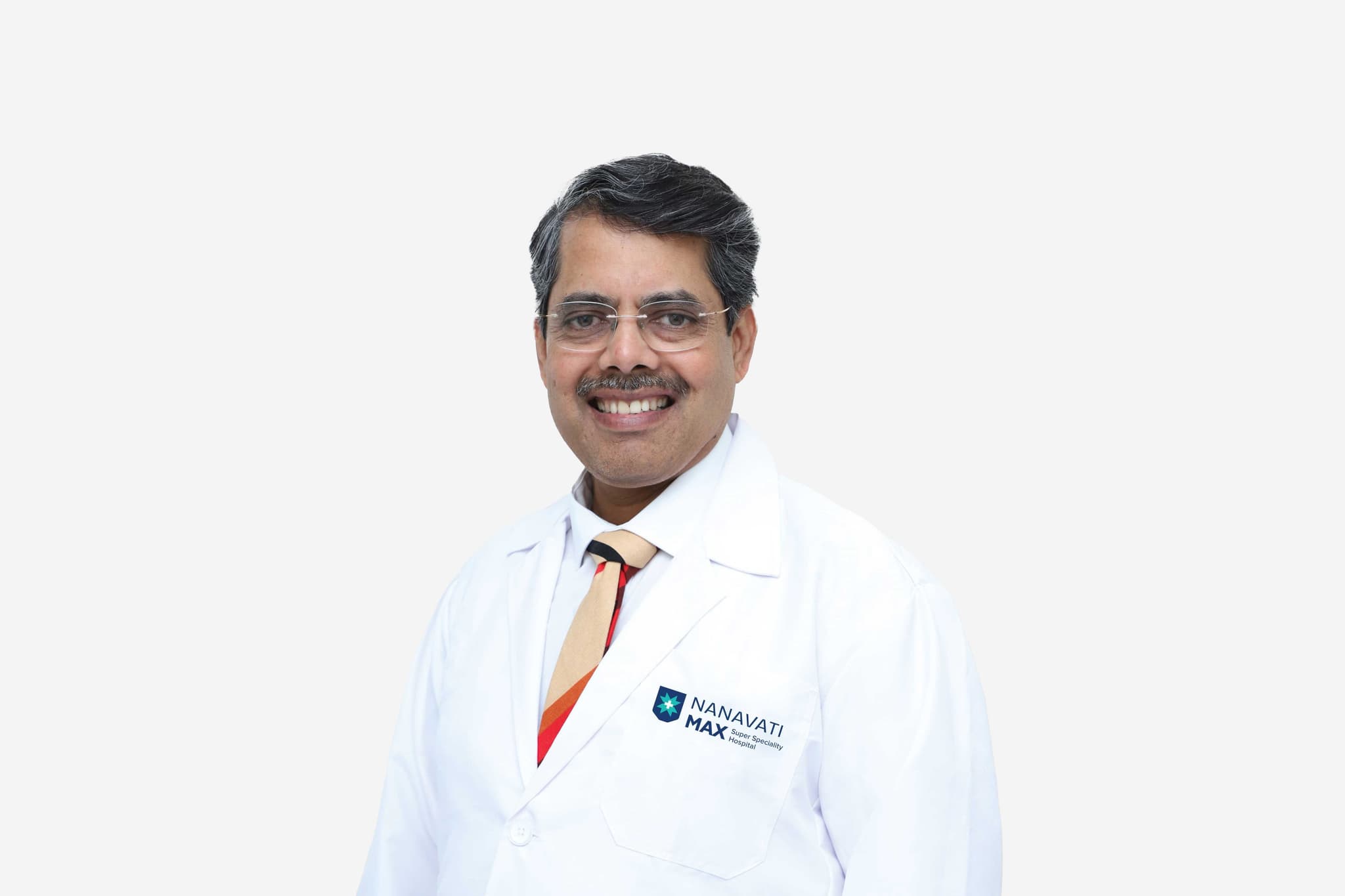
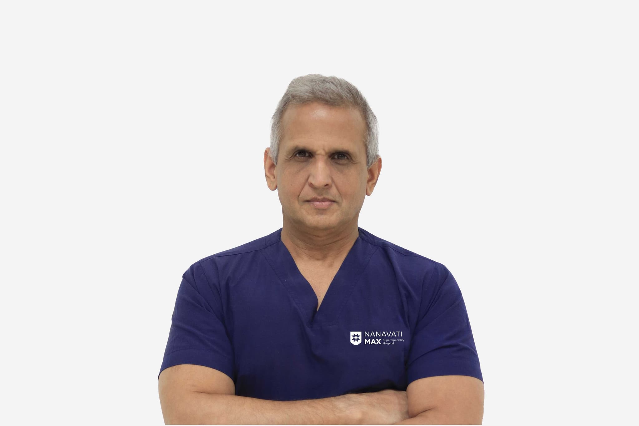
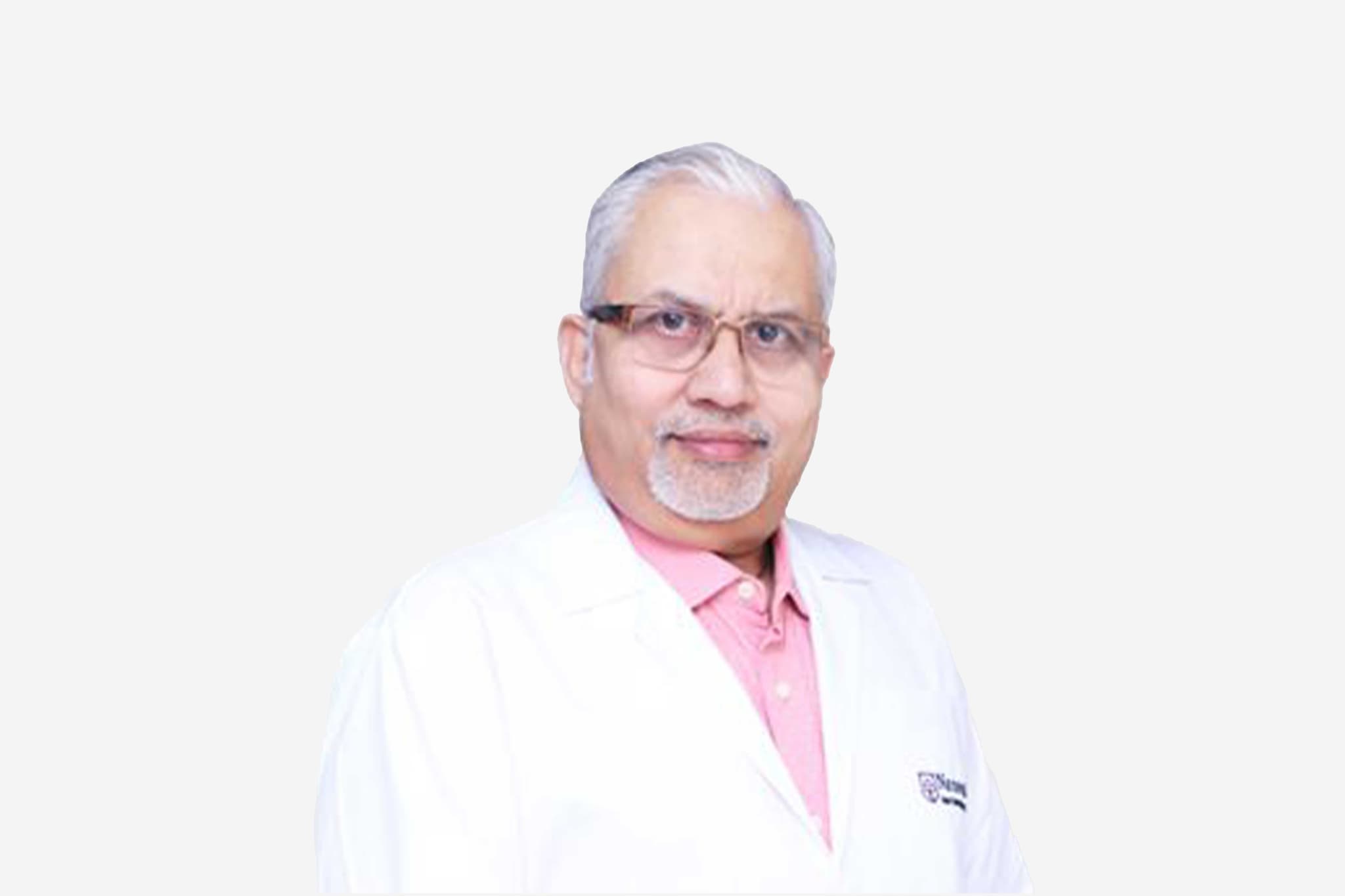
Dr Vishal Khullar

Director - CTVS and Heart and Lung Transplant

Nanavati-Max Super Specialty Hospital, Mumbai

32+ years experience
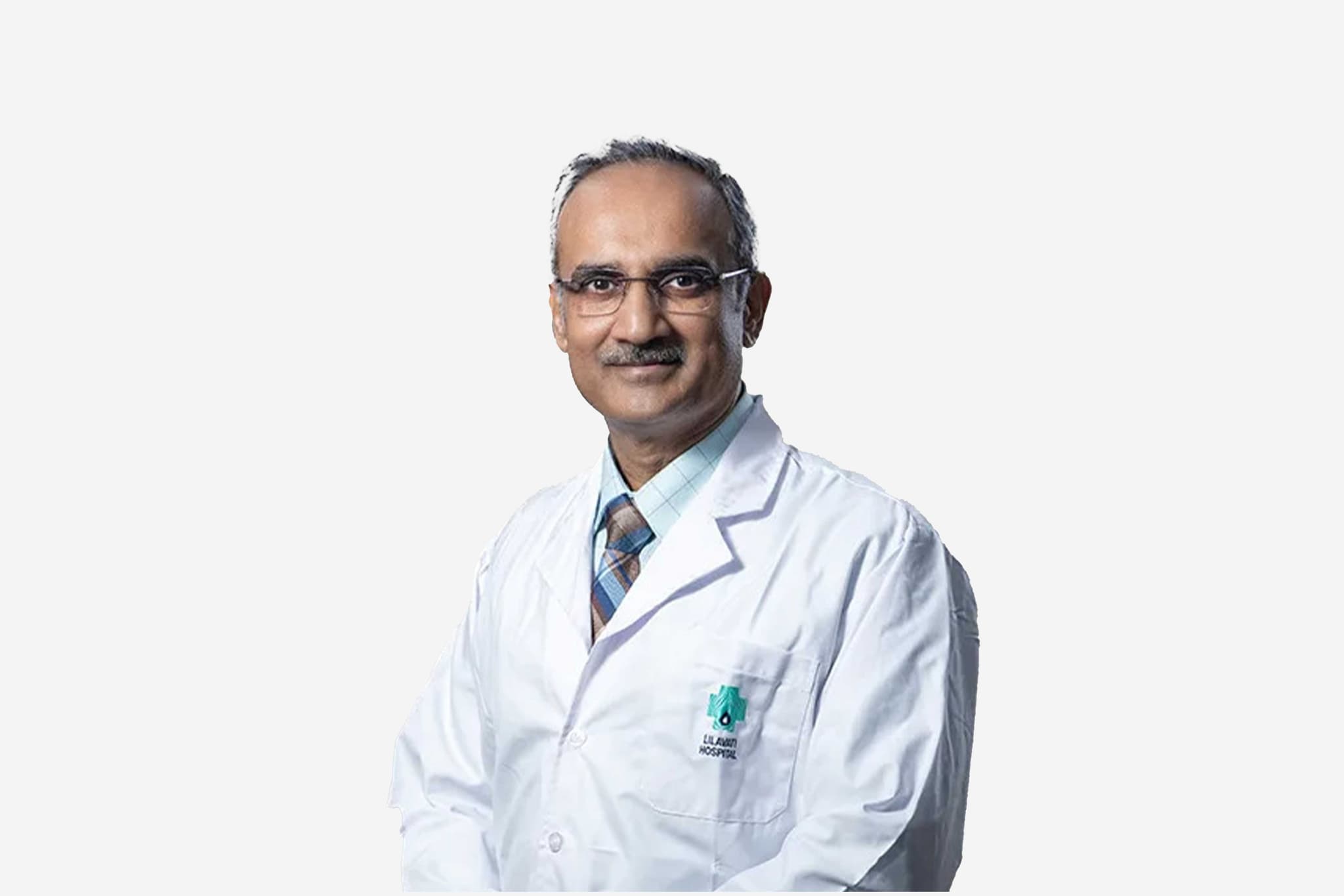
Dr Vishal Khullar, Director - CTVS and Heart and Lung Transplant at Nanavati-Max Super Specialty Hospital, Mumbai - Max Healthcare
  1. Home
  2. Doctors
  3. Dr Vishal Khullar

About Dr Vishal Khullar

Dr. Khullar is a pioneering figure in cardiovascular surgery, notably being the first surgeon in the Indian Subcontinent to hold the position of Senior Associate Consultant at the Mayo Clinic in Rochester, USA. With over 32 years of experience, he has served as an Assistant Professor of Surgery at the Mayo Clinic, bringing world-class surgical expertise to his clinical practice.

Mastery in Advanced Cardiovascular Procedures

Since 2017, Dr. Khullar has specialized in a broad range of high-complexity thoracic and cardiovascular interventions. His surgical repertoire includes aortic root and arch replacements, heart surgeries, and complex redo-sternotomies. He is particularly recognized for his technical proficiency in lung transplants and serves as an integral member of the Transplant Selection Committee.  

Areas of Clinical Interest

His clinical focus is dedicated to life-saving innovations and complex cardiac repairs. This includes beating heart bypass surgery, valve repairs, and redo heart surgeries. Additionally, he specializes in mechanical circulatory support and remains at the forefront of heart and lung transplantation.  

Academic Contributions and Excellence

Dr. Khullar's distinguished career is built upon a rigorous training foundation from premier medical institutions in Pune and Mumbai. His transition from a Senior Associate Consultant in the United States to a leading surgeon in India reflects a commitment to advancing the standards of thoracic surgery on a global scale.  

Dr. Vishal Khullar at a Glance

  • Over 32 years of total surgical experience.  

  • First surgeon from the Indian Subcontinent appointed as Senior Associate Consultant at Mayo Clinic, USA.  

  • Former Assistant Professor of Surgery at Mayo Clinic, Rochester.  

  • Expert in complex aortic surgeries, heart transplants, and lung transplants.  

  • Specialist in mechanical circulatory support and beating heart bypass surgery.  

  • Key member of the Transplant Selection Committee. 

MBBS: BJ Medical College, Pune (1992 Batch)
MS General Surgery: Seth GS Medical College and KEM Hospital, Mumbai
MCh Cardiothoracic Surgery: Bombay Hospital and Medical Research Centre, Mumbai

No awards & achievements available
May we help you?

Enter digits only (6-15 digits). We'll add your country code.

Related Treatments

Complex Congenital Heart Surgery

Complex Congenital Heart Surgery refers to a group of highly specialized operations performed to treat severe, often life-threatening structural heart defects present from birth. Unlike "simple" repairs, such as closing a small hole, complex surgeries often involve rearranging the entire circulatory system. These procedures are frequently performed in multiple stages over several years to allow the heart and lungs to adapt to new blood flow patterns.

When You Should Consider Complex Heart Surgery

  • Hypoplastic Left Heart Syndrome (HLHS): When the left side of the heart is severely underdeveloped and cannot pump enough blood to the body.

  • Transposition of the Great Arteries (TGA): A critical condition where the two main arteries leaving the heart are "switched," sending oxygen-poor blood to the body.

  • Tricuspid Atresia: When a missing heart valve prevents blood from flowing from the right atrium to the right ventricle, resulting in a "single ventricle" circulation.

  • Total Anomalous Pulmonary Venous Return (TAPVR): A defect where the veins bringing blood from the lungs attach to the wrong place in the heart.

  • Truncus Arteriosus: When a single large blood vessel stems from the heart instead of the separate aorta and pulmonary artery.

Common Complex Procedures

  • The Norwood Procedure (Stage 1 of 3): The first step in treating HLHS; the right ventricle is converted into the main pumping chamber, and the aorta is reconstructed to ensure the body receives blood.

  • Arterial Switch Operation (ASO): Performed for TGA; the aorta and pulmonary artery are disconnected and reattached to the correct ventricles, including the delicate transfer of coronary arteries.

  • The Fontan Procedure (Stage 3 of 3): The final stage for single-ventricle defects; oxygen-poor blood from the lower body is connected directly to the pulmonary artery, bypassing the heart.

  • The Glenn Procedure (Stage 2 of 3): Connects the large vein from the upper body (SVC) directly to the pulmonary artery to reduce the workload on a single working ventricle.

  • Ross Procedure: A sophisticated valve replacement where the patient’s own healthy pulmonary valve is moved to the aortic position, allowing it to grow as the child grows.

How Is Performed

  • Median Sternotomy: Under general anesthesia, a midline incision is made through the breastbone to allow the surgical team full access to the heart and great vessels.

  • Advanced Cardiopulmonary Bypass: The patient is connected to a heart-lung machine designed to manage the tiny blood volumes of newborns while maintaining oxygenation to the brain and organs.

  • Deep Hypothermic Circulatory Arrest (DHCA): For the most intricate repairs, the body temperature is lowered to approximately 18°C, and circulation is briefly stopped to provide a still, bloodless field for the surgeon.

  • Anatomical Reconstruction: Using the patient's own tissue or synthetic patches (Dacron/Gore-Tex), the surgeon "re-plumbs" the heart, enlarging vessels and closing internal defects.

  • Coronary Re-implantation: In "switch" procedures, the tiny coronary arteries—often the size of a needle—are meticulously moved to the new aortic root to ensure the heart muscle receives blood.

  • Delayed Chest Closure: In some newborn cases, the chest is left "open" for 2–3 days with a sterile covering to allow the heart to recover from swelling before the final closure.

Pre-Procedure Preparation

  • 3D Anatomical Modeling: Surgeons often use 3D-printed models of the patient's specific heart anatomy to "rehearse" and plan the complex reconstruction before surgery.

  • Prostaglandin Infusion: Many newborns are kept on a continuous IV medication (Alprostadil) to keep the ductus arteriosus open, ensuring survival until surgery can be performed.

  • Nutritional Optimization: Infants may require specialized high-calorie feeding or TPN (IV nutrition) to reach a stable weight and strength for the operation.

  • Cardiac Catheterization: A detailed study to measure internal heart pressures and resistance in the lung vessels, which is critical for planning "staged" procedures.

  • Fasting (NPO): Strict adherence to fasting guidelines is required to ensure safety during the induction of general anesthesia.

Tests Before Complex Heart Surgery

  • Fetal and Neonatal Echocardiogram: The primary diagnostic tool used to visualize the internal structures of the heart and the origin of the great vessels.

  • Cardiac MRI or CT: Provides high-resolution, three-dimensional images of the heart's relationship to the lungs and chest wall.

  • Genetic Screening: To check for associated syndromes (such as DiGeorge Syndrome) that may impact the child's overall surgical risk and recovery.

  • Cross-match Blood Work: Because these surgeries involve significant blood volumes, several units of specifically typed and screened blood are prepared in advance.

Life After Complex Heart Surgery

  • Cardiac ICU (CICU): Patients spend 7 to 21 days in a specialized ICU where heart function, rhythm, and oxygen levels are monitored second-by-second.

  • Inotropic Support: High doses of IV medications are often used for several days to help the "re-plumbed" heart pump effectively as it adapts to the new circulation.

  • Neurological Monitoring: Given the use of bypass and circulatory arrest, the medical team closely monitors for seizures or developmental milestones during recovery.

  • Wound and Bone Healing: For children, the breastbone typically heals within 6 to 8 weeks; parents are taught specific "lifting" techniques to protect the chest.

  • Lifelong CHD Specialist Care: These patients are considered "repaired" rather than "cured" and require lifelong surveillance to monitor for valve issues or rhythm changes.

Benefits Of Complex Heart Surgery

  • Life-Saving Intervention: Provides a definitive chance at survival for infants born with defects that would otherwise be fatal within days or weeks.

  • Improved Oxygenation: Corrects "cyanosis" (blueness), allowing the child’s brain and organs to receive the oxygen necessary for normal development.

  • Restores Physical Potential: Many children grow up to lead active lives, attend school, and participate in sports that would have been impossible without repair.

  • Growth and Development: Relieving the heart's workload allows the body to redirect energy toward physical growth and cognitive milestones.

  • Staged Success: The multi-stage approach (Norwood/Glenn/Fontan) allows the heart to grow and the lungs to mature, leading to better long-term outcomes in single-ventricle patients.

Starting Cost: $ 4000

Heart Bypass Surgery (CABG)

Coronary Artery Bypass Grafting (CABG), commonly called "heart bypass surgery," is a major surgical procedure used to treat severe coronary artery disease. It creates new pathways for blood to flow to the heart muscle by bypassing clogged or narrowed sections of the coronary arteries. By using healthy blood vessels from elsewhere in the body to "reroute" blood, CABG restores vital oxygen supply to the heart muscle and reduces the risk of a heart attack.

When You Should Consider CABG

  • Left Main Disease: A severe blockage in the main artery supplying the left side of the heart, which is considered high-risk.

  • Triple Vessel Disease: Significant blockages in all three major coronary arteries.

  • Diabetes: Patients with diabetes and multi-vessel disease often have better long-term outcomes with surgery than with stenting.

  • Complex Anatomy: Blockages that are too long, heavily calcified (hardened), or located in areas where a stent cannot be safely placed.

  • Failed Angioplasty: When previous attempts to open arteries with balloons or stents have not been successful or the artery has narrowed again.

Surgical Techniques

  • On-Pump CABG: The traditional method where a heart-lung bypass machine takes over the work of the heart and lungs, allowing the surgeon to operate on a still, non-beating heart.

  • Off-Pump (Beating Heart) CABG: The surgeon uses specialized stabilizers to operate while the heart continues to beat, avoiding the bypass machine. This is often preferred for patients at high risk for stroke.

  • Minimally Invasive (MIDCAB): Small incisions are made between the ribs rather than through the breastbone. This is typically used for bypassing one or two arteries on the front of the heart.

  • Endoscopic Vessel Harvesting (EVH): A 2026 standard where grafts from the leg or arm are removed through tiny incisions using a camera, reducing scarring and pain.

How CABG Is Performed

  • Incision: A midline incision is made, and the breastbone (sternum) is divided to access the heart.

  • Harvesting: Simultaneously, healthy vessels are harvested: the Internal Mammary Artery (chest), Saphenous Vein (leg), or Radial Artery (arm).

  • Bypass: One end of the graft is attached to the aorta (the main artery) and the other end below the blockage, creating a permanent "detour."

  • Restarting: Once the connections are tested for leaks, the heart is restarted (if it was stopped), and the bypass machine is disconnected.

  • Closing: The sternum is secured with permanent stainless steel wires, and the skin is closed with stitches or staples.

Pre-Procedure Preparation

  • Fasting for at least 8–12 hours before surgery, as it is performed under general anesthesia.

  • Extensive blood work, chest X-rays, and an ECG to ensure you are fit for major surgery.

  • Dental clearance is often required to ensure no hidden infections could travel to the heart.

  • Stopping or adjusting certain medications, especially blood thinners like Clopidogrel or Aspirin, as directed.

  • Shaving and surgical scrubbing of the chest, legs, and arms to prevent infection.

Tests Before CABG

  • Coronary Angiogram: The "roadmap" that shows exactly where the blockages are located.

  • Echocardiogram: To assess the heart's pumping strength (Ejection Fraction) and valve function.

  • Carotid Doppler: To check for blockages in the neck arteries that might increase the risk of stroke during surgery.

  • Pulmonary Function Test (PFT): To ensure the lungs are strong enough to handle anesthesia and recovery.

  • Vein Mapping: Ultrasound of the legs or arms to ensure the vessels are healthy enough to be used as grafts.

Life After CABG

  • ICU Stay: Expect to spend the first 24 hours in the Intensive Care Unit for close monitoring of heart rhythm and blood pressure.

  • Hospital Stay: Total recovery in the hospital usually lasts 5 to 7 days.

  • Sternal Precautions: For the first 6 weeks, you must avoid lifting anything heavier than 2–3 kg to allow the breastbone to heal properly.

  • Cardiac Rehabilitation: Starting around week 6, supervised exercise programs are highly recommended to rebuild strength.

  • Long-term Meds: You will likely remain on Aspirin and cholesterol-lowering medications (statins) indefinitely to keep the new grafts clear.

Benefits of CABG

  • Superior Longevity: Provides a long-term solution for complex multi-vessel disease, often outlasting stents.

  • Symptom Relief: Significant reduction or total elimination of chest pain (angina) and shortness of breath.

  • Reduced Heart Attack Risk: By restoring blood flow to large areas of the heart, the risk of a future major cardiac event is lowered.

  • Improved Quality of Life: Most patients return to an active lifestyle and can exercise more effectively than before surgery.

  • 2026 Success Rates: Elective CABG has a high survival rate (approx. 98–99%) due to advanced surgical and anesthesia protocols.

Starting Cost: $ 3000

Heart Transplant Surgery

A heart transplant is a major life-saving procedure in which a failing or diseased heart is replaced with a healthy donor heart. It restores normal heart function, improves quality of life, and is recommended when other treatments no longer work.

When You Should Consider a Heart Transplant

  • Severe heart failure not improving with medicines or procedures

  • Frequent hospital admissions due to worsening heart condition

  • Extreme fatigue, weakness, or breathlessness during simple tasks

  • Swelling in legs, ankles, or abdomen because of fluid buildup

  • Life-threatening arrhythmias that cannot be controlled

  • Poor heart pumping capacity despite advanced treatment

Conditions That May Require a Heart Transplant

  • Dilated or restrictive cardiomyopathy

  • Severe coronary artery disease with repeated heart attacks

  • Congenital heart defects not treatable with surgery

  • End-stage valvular heart disease

  • Severe myocarditis causing permanent heart damage

  • Heart failure after previous surgeries or device implants

How Heart Transplant Surgery Is Performed

  • General anesthesia is given for complete comfort

  • The weakened or failing heart is surgically removed

  • A healthy donor heart is connected to major blood vessels

  • The new heart is started carefully to ensure proper function

  • Monitoring lines and drains are placed for recovery

  • You are shifted to the ICU for close observation

Pre-Surgery Preparation

  • Heart function tests and blood/tissue matching

  • Follow all fasting and medication-related instructions

  • Maintain controlled blood pressure, blood sugar, and healthy weight

  • Avoid alcohol and stop smoking at least 3–4 weeks before surgery

  • Attend counseling to prepare mentally and physically

Pre-Surgery Tests

  • ECG to check heart rhythm

  • Echocardiography to assess pumping capacity

  • CT or MRI scans for detailed imaging

  • Pulmonary function tests for lung strength

  • Blood typing and tissue matching

  • Coronary angiography, if required

Life After a Heart Transplant

  • ICU stay: usually 3–5 days

  • Hospital recovery: around 2–3 weeks

  • Regular follow-ups to monitor organ acceptance

  • Anti-rejection medicines taken lifelong

  • Gradual return to daily activities in 8–12 weeks

  • Healthy diet, low-salt meals, and light exercise

  • Cardiac rehabilitation for long-term recovery

Benefits of a Heart Transplant

  • Better heart function and improved blood flow

  • Relief from breathlessness, fatigue, and swelling

  • Fewer hospital visits and emergency episodes

  • Better stamina and improved quality of life

  • Long-term survival with the right care and medicines

Starting Cost: $ 20000

Lung Transplant Surgery

A lung transplant is a major surgical procedure to replace one or both diseased lungs with healthy donor lungs. It is typically a treatment of last resort for end-stage lung diseases that no longer respond to other medical therapies. The goal is to improve the patient's quality of life and extend life expectancy when other options have been exhausted.

When You Should Consider a Lung Transplant

  • End-Stage COPD (Emphysema): When chronic obstructive pulmonary disease has caused such severe damage that breathing is difficult even with supplemental oxygen.

  • Idiopathic Pulmonary Fibrosis: Progressive scarring of the lung tissue that prevents the lungs from transferring oxygen into the bloodstream.

  • Cystic Fibrosis: A genetic condition causing thick, sticky mucus to build up in the lungs, leading to repeated, life-threatening infections.

  • Pulmonary Hypertension: High blood pressure in the arteries of the lungs that can eventually lead to right-sided heart failure.

  • Alpha-1 Antitrypsin Deficiency: A genetic disorder that can cause severe lung and liver disease.

Types of Lung Transplant

  • Single Lung Transplant: Replaces one damaged lung. This is often used for certain restrictive diseases like pulmonary fibrosis.

  • Double (Bilateral) Lung Transplant: Replaces both lungs simultaneously. This is the primary choice for infectious diseases like cystic fibrosis to ensure no infected tissue remains.

  • Heart-Lung Transplant: A rare procedure that replaces the heart and both lungs, usually for patients with severe pulmonary hypertension and concomitant heart failure.

How Is Performed

  • Anesthesia: The procedure is highly complex and performed under general anesthesia.

  • Duration: A single transplant usually takes 4 to 8 hours, while a double transplant can take 6 to 12 hours.

  • Incision: For a single lung, an incision (thoracotomy) is made on the side of the chest. For a double lung, a horizontal "clamshell" incision is often made across the chest.

  • Support: During the surgery, you may be connected to a heart-lung bypass machine or ECMO (Extracorporeal Membrane Oxygenation) to circulate blood and oxygen.

  • Connection: Surgeons meticulously sew the new lung's main airway (bronchus) and major blood vessels (pulmonary artery and veins) to your own.

Pre-Procedure Preparation

  • Transplant Evaluation: An extensive series of tests to ensure you are healthy enough for surgery and committed to a lifelong post-transplant regimen.

  • Psychological Assessment: To evaluate your support system and ability to manage complex medication schedules.

  • Pulmonary Rehab: Engaging in specialized exercise to keep your body as strong as possible while waiting for a donor match.

  • Vaccinations: Ensuring all immunizations are up to date, as your immune system will be suppressed after surgery.

  • The Waiting List: Once approved, you are placed on a national registry where donor lungs are matched based on blood type, organ size, and geographic distance.

Tests Before Lung Transplant

  • Pulmonary Function Tests (PFTs): To measure exactly how much air your lungs can hold and how well they move gases.

  • Cardiac Catheterization: To check the pressure in your lung arteries and the overall strength of your heart.

  • Chest CT Scan: Providing high-resolution 3D images of your lung structure and chest cavity.

  • Tissue Typing: Matching your tissue markers with potential donors to reduce the risk of immediate organ rejection.

  • ECG and Stress Test: Ensuring your heart can withstand the rigors of a multi-hour major surgery.

Life After Lung Transplant

  • Hospital Stay: Typically 1 to 3 weeks, with the first several days spent in the Intensive Care Unit (ICU) on a mechanical ventilator.

  • Lifelong Medication: You must take immunosuppressant (anti-rejection) drugs for the rest of your life to prevent your body from attacking the new lung.

  • Monitoring: Frequent follow-up visits, blood tests, and bronchoscopies (using a camera to look inside the lungs) are required, especially in the first year.

  • Physical Rehab: You will start walking within days of surgery and gradually progress to light exercise over 6 to 8 weeks.

  • Lifestyle Adjustments: Avoiding crowds during flu season, wearing masks in certain environments, and strict food safety are necessary to prevent infections.

Why Specialized Treatment Is Highly Effective

  • Significant Survival Benefit: For many patients with end-stage disease, a transplant can extend life expectancy by many years.

  • Restored Breathing: Patients often transition from being housebound on oxygen to walking, traveling, and engaging in physical hobbies.

  • Advanced Surgical Support: The use of ECMO technology allows surgeons to perform transplants on the most critically ill patients with greater safety.

  • Comprehensive Care Teams: Post-transplant care involves a dedicated team of pulmonologists, surgeons, pharmacists, and coordinators to manage every aspect of recovery.

  • Improved Quality of Life: Beyond just survival, a successful transplant provides the "gift of breath," allowing for a return to a much more normal and active lifestyle.

Starting Cost: $ 20000

Off-Pump Bypass (Beating Heart Surgery)

Off-Pump Coronary Artery Bypass (OPCAB), also known as "Beating Heart Surgery," is a specialized technique where the surgeon performs the bypass while the heart continues to beat. Unlike traditional CABG, it does not use a heart-lung bypass machine to stop the heart and take over its function. This approach is highly valued for reducing systemic inflammation and protecting vital organs, particularly in high-risk patients.

When You Should Consider OPCAB

  • Elderly Patients (70+ years): Those who may be more vulnerable to the systemic physiological stress of a heart-lung machine.

  • History of Stroke: Patients with a "porcelain" (heavily calcified) aorta where clamping the vessel during traditional surgery increases the risk of a stroke.

  • Chronic Kidney Disease: Maintaining natural blood pressure and pulsatile flow during surgery is generally safer for renal function.

  • Liver Disease or Blood Disorders: Patients who may face higher complications from the intense blood-thinning required for "on-pump" machines.

  • Lung Issues: Those with respiratory compromise who benefit from being taken off a ventilator as quickly as possible following the procedure.

The Core Technology: How It Works

  • Suction Stabilizers: Small, mechanical arms that "grip" a tiny area (1–2 cm) of the heart surface, keeping that specific spot perfectly still while the rest of the heart continues to pump.

  • Intracoronary Shunts: Tiny plastic tubes inserted into the artery during the stitching process to ensure blood continues to flow to the heart muscle while the surgeon sews the graft.

  • Heart Positioners: Suction devices used to gently lift and rotate the beating heart, allowing the surgeon to reach blockages on the side or back walls.

  • Transit Time Flow Measurement (TTFM): A clinical standard used during surgery to verify that blood flow through the new graft is perfect before closing the chest.

  • Deep Pericardial Stay Sutures: Specialized internal stitches that allow the surgeon to maneuver the heart safely into the necessary positions without stopping it.

How Is Performed

  • Surgical Access: Under general anesthesia, a standard midline incision is made through the breastbone (sternotomy) to reach the heart.

  • Graft Harvesting: Healthy vessels are prepared from the chest (internal mammary artery), leg (saphenous vein), or arm (radial artery) to be used as the new bypass routes.

  • Heart Positioning: The surgeon carefully maneuvers the beating heart using positioners to expose the specific blocked coronary arteries.

  • The Bypass: The stabilizer is applied to the target site, and the surgeon meticulously sews the graft onto the artery using ultra-fine sutures.

  • Verification & Closing: After confirming flow with TTFM, the stabilizer is removed, and the breastbone is secured with permanent stainless steel wires.

Pre-Procedure Preparation

  • Fasting (NPO): No food or drink for at least 8–12 hours before surgery to ensure safety during general anesthesia.

  • Baseline Diagnostics: Extensive blood tests, chest X-rays, and an ECG to assess overall surgical readiness and organ function.

  • Dental Clearance: A check to rule out any active oral infections that could travel through the bloodstream and compromise the heart surgery.

  • Medication Adjustment: Reviewing all prescriptions; anti-platelet drugs or blood thinners may need to be paused or adjusted several days prior.

  • Surgical Scrub: Shaving and antiseptic scrubbing of the chest and any potential graft harvest sites on the legs or arms.

Tests Before OPCAB

  • Coronary Angiogram: The essential "roadmap" that identifies the exact location and severity of blockages for the surgical team.

  • Echocardiogram: An ultrasound to evaluate the heart's pumping strength and identify any underlying valve issues.

  • Carotid Ultrasound: To assess stroke risk by checking the health of the arteries supplying blood to the brain.

  • CT Scan of the Aorta: Specifically used to check for heavy calcification (porcelain aorta) that would favor an off-pump approach.

  • Vein/Artery Mapping: Ultrasound imaging to ensure the quality and size of the blood vessels intended for use as bypass grafts.

Life After OPCAB

  • ICU Recovery: Patients typically spend the first 12 to 24 hours in the Intensive Care Unit for close hemodynamic monitoring.

  • Hospital Discharge: The total stay is usually 4 to 5 days, which is often 1–2 days shorter than traditional "on-pump" bypass surgery.

  • Sternal Precautions: To allow the breastbone to heal, patients must avoid lifting anything heavier than 2–3 kg (about 5 lbs) for 6 to 8 weeks.

  • Gradual Recovery: Most patients return to light daily activity quickly but require 2 to 3 months for a full return to strenuous levels.

  • Cardiac Rehab: Participating in a supervised exercise and education program starting around week 6 is vital for long-term cardiovascular health.

Benefits Of OPCAB

  • Reduced Stroke Risk: Avoiding the clamping of a calcified aorta minimizes the chance of dislodging plaque that could travel to the brain.

  • Organ Protection: Shorter ventilator times and more natural, pulsatile blood flow help protect the sensitive kidney and lung systems.

  • Less Bleeding: Beating heart surgery generally requires fewer blood transfusions than procedures involving a bypass machine.

  • Lower Inflammatory Response: Avoiding the heart-lung machine reduces the "whole-body" inflammation often seen after major cardiac surgery.

  • Faster Return to Normalcy: Many patients experience shorter hospital stays and a quicker initial recovery phase compared to traditional methods.

Starting Cost: $ 2500

Related Specialists

Dr Suresh Advani
Dr Suresh Advani
Consultant - Medical Oncology
Nanavati-Max Super Specialty Hospital, Mumbai
46+years experience
View Profile
Dr Pradeep B Bhosale
Chairman - Robotic Joint Replacement, Hip and Knee Surgery
Nanavati-Max Super Specialty Hospital, Mumbai
40+years experience
View Profile
Dr Niraj Vora
Dr Niraj Vora
Senior Director - Orthopedics Joint Replacement and Robotic Joint Replacement
Nanavati-Max Super Specialty Hospital, Mumbai
35+years experience
View Profile
Dr Manoj Jain
Dr Manoj Jain
Director - Gastrointestinal, Bariatric, Laparoscopic & Robotic Surgery
Nanavati-Max Super Specialty Hospital, Mumbai
25+years experience
View Profile
Dr Rajan Shah
Dr Rajan Shah
Senior Director- Neuro Surgery
Nanavati-Max Super Specialty Hospital, Mumbai
32+years experience
View Profile
Dr Neeta Shah
Dr Neeta Shah
Consultant
Nanavati-Max Super Specialty Hospital, Mumbai
43+years experience
View Profile
Dr Pradyumna J Oak
Dr Pradyumna J Oak
Principal Director - Neurology
Nanavati-Max Super Specialty Hospital, Mumbai
22+years experience
View Profile
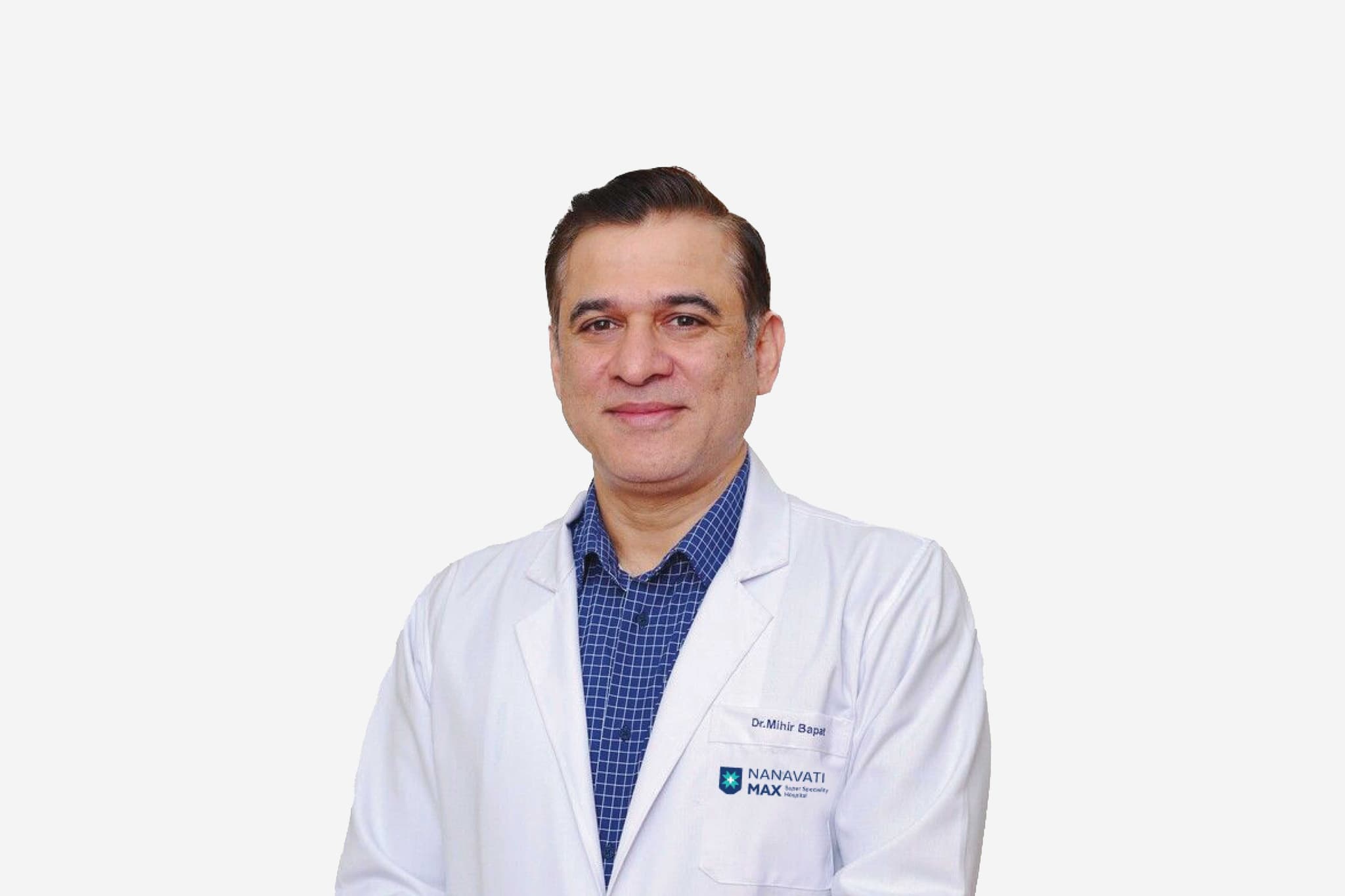
Dr Mihir Bapat
Dr Mihir Bapat
Vice Chairman - Orthopedics & Spinal Surgery
Nanavati-Max Super Specialty Hospital, Mumbai
25+years experience
View Profile
Dr Sanjiv Badhwar
Dr Sanjiv Badhwar
Senior Director – ENT, Program Head - Cochlear Implants & ENT Robotic Surgery
Nanavati-Max Super Specialty Hospital, Mumbai
34+years experience
View Profile
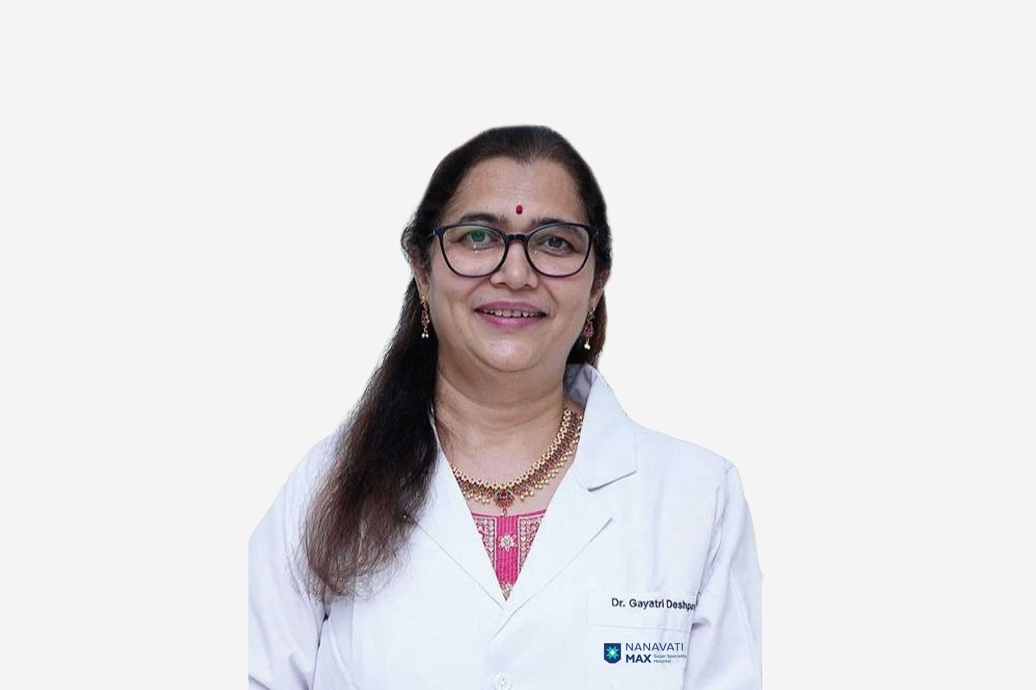
Dr Gayatri Deshpande
Director - Obstetrics & Gynecology
Nanavati-Max Super Specialty Hospital, Mumbai
32+years experience
View Profile
Dr Sanjay S Nabar
Dr Sanjay S Nabar
Consultant
Nanavati-Max Super Specialty Hospital, Mumbai
36+years experience
View Profile
Dr Avanish Arora
Dr Avanish Arora
Principal Director - Urology & Uro-Oncology
Nanavati-Max Super Specialty Hospital, Mumbai
26+years experience
View Profile
Dr Chandrashekhar Kulkarni
Visiting Consultant - Cardiothoracic & Vascular Surgery, Heart & Lung Transplant
Nanavati-Max Super Specialty Hospital, Mumbai
24+years experience
View Profile
Dr Jatin Kothari
Dr Jatin Kothari
Principal Director, Nephrology & Chief Consultant, Renal Transplant Medicine
Nanavati-Max Super Specialty Hospital, Mumbai
30+years experience
View Profile

May we help you?

Enter digits only (6-15 digits). We'll add your country code.