Medivisor India Treatment Logo
  • Home
              • Become a Partner

              +91 83407 80250

              Medivisor Logo

              Medivisor India Treatment is a trusted New Delhi–based medical travel partner connecting global patients to advanced, affordable, high-quality healthcare in India.

              • Medivisor House 359, Sector 1, Vaishali, Ghaziabad, (Delhi/NCR) India
              • +91 8340 780 250
              • info@medivisorhealth.com

              About Us

              • About Us
              • Our Services
              • Our Team
              • Our Medical Advisors
              • Our Safety Measures
              • Join Our Network
              • Contact Us

              India Treatment

              • Hospitals
              • Treatments
              • Treatment Cost
              • Treatment Process
              • Visa Process
              • Travel Guide
              • FAQs
              • Why Medivisor

              Gallery

              • Patient Testimonials
              • Patient Activities
              • News Coverage
              • Blog

              Get in Touch

              © 2026 Medivisor India Treatment. All rights reserved.

              Dr. Dr. Chandrashekhar Kulkarni

              Dr. Chandrashekhar Kulkarni

              Visiting Consultant - Cardiothoracic & Vascular Surgery, Heart & Lung Transplant

              24+ years experience

              Fellow (Cardio-Thoracic Surgery), Greenlane Hospital, New Zealand (2006-2008) M.Ch. (Cardio-Thoracic Surgery), KEM Hospital and Medical College, Mumbai (2001-2004) M.S. (General Surgery), Tata Memorial Hospital, Mumbai (1998-2001) DNB (General Surgery), Royal College of Surgeons of Edinburgh, UK (2001) M.B.B.S, LTM Medical College & Hospital, Mumbai (1992-1998)
              1. Home
              2. Doctors
              3. Dr. Chandrashekhar Kulkarni

              About Dr. Chandrashekhar Kulkarni

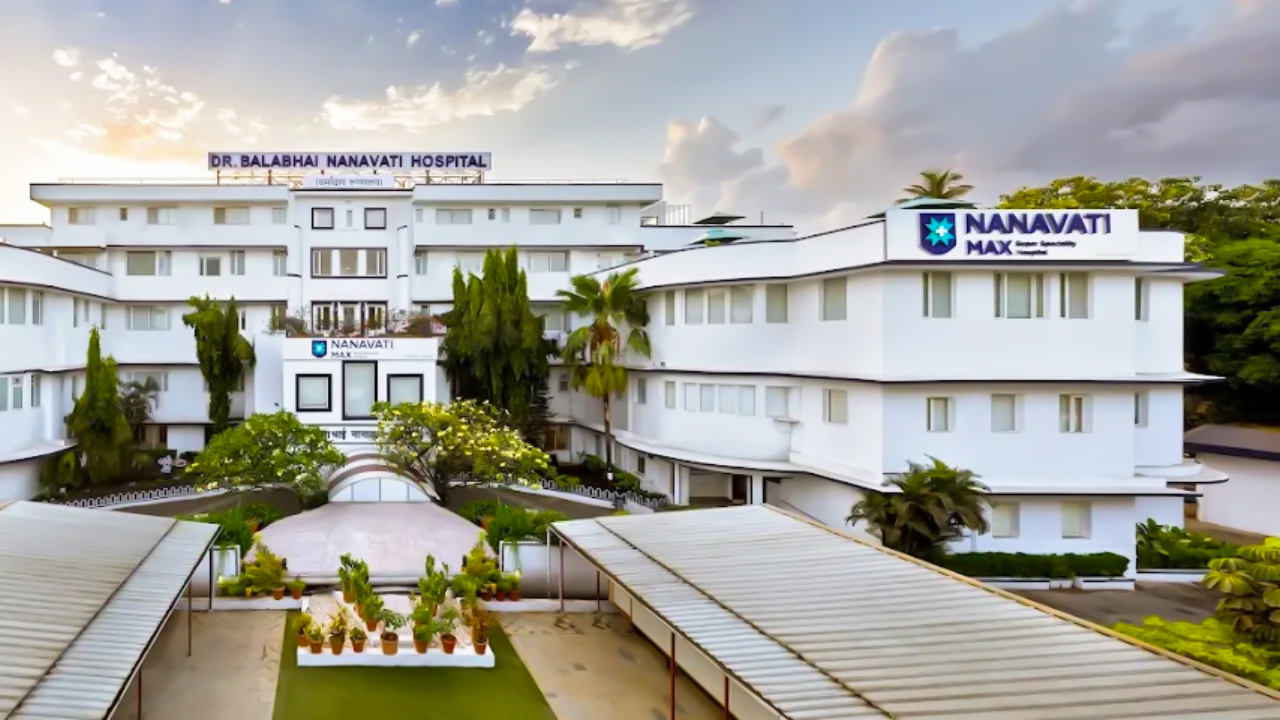
              Dr. Kulkarni is a highly distinguished specialist in cardiovascular and thoracic surgery based in Mumbai, bringing over 28 years of extensive clinical experience to his practice. He currently holds a major leadership role as the Director and Head of Cardiovascular & Thoracic Surgery and oversees the Heart & Lung Transplant Program at Nanavati Max Super Speciality Hospital.

              As the head of a premier transplant program, Dr. Kulkarni specializes in the complex management of end-stage heart and lung diseases. His expertise includes overseeing the clinical protocols for organ transplantation and implementing advanced surgical techniques within the thoracic and cardiovascular domain.

              Clinical Leadership and Specialized Procedures

              In his capacity as Director, he leads a multidisciplinary team to deliver high-standard surgical care for a wide range of cardiovascular conditions. His long-standing career is marked by a commitment to clinical excellence, focusing on both routine and highly specialized interventions for the heart and lungs.

              Academic Contributions and Excellence

              Throughout nearly three decades of practice, Dr. Kulkarni has been at the forefront of surgical innovation in India. His leadership at Nanavati Max Super Speciality Hospital ensures the integration of the latest medical advancements and safety protocols, making him a respected authority in the field of cardiovascular and thoracic surgery.

              Dr. Chandrashekhar Kulkarni at a Glance

              • Over 28 years of distinguished clinical experience in cardiovascular and thoracic surgery.

              • Director and Head of Cardiovascular & Thoracic Surgery at Nanavati Max Super Speciality Hospital.

              • Leads the specialized Heart & Lung Transplant Program.

              • Expert in managing complex cardiothoracic cases and advanced surgical interventions.

              • Prominent figure in the Mumbai medical community with a focus on transplant innovation.

              • Responsible for maintaining clinical excellence and research standards within the Max Healthcare network.

              Fellow (Cardio-Thoracic Surgery), Greenlane Hospital, New Zealand (2006-2008) M.Ch. (Cardio-Thoracic Surgery), KEM Hospital and Medical College, Mumbai (2001-2004) M.S. (General Surgery), Tata Memorial Hospital, Mumbai (1998-2001) DNB (General Surgery), Royal College of Surgeons of Edinburgh, UK (2001) M.B.B.S, LTM Medical College & Hospital, Mumbai (1992-1998)
              Specialization: Visiting Consultant - Cardiothoracic & Vascular Surgery, Heart & Lung Transplant

              No awards & achievements available

              Affiliated Hospitals

              Nanavati-Max Super Specialty Hospital, Mumbai
              Nanavati-Max Super Specialty Hospital, Mumbai

              Super Specialty

              Mumbai, Maharashtra

              1950

              Estd.

              595+

              Beds

              450+

              Doctors

              May we help you?

              Enter digits only (6-15 digits). We'll add your country code.

              May we help you?

              Enter digits only (6-15 digits). We'll add your country code.

              Related Treatments

              TMVI/TMVR (Transcatheter Mitral Valve Replacement)
              TMVI/TMVR (Transcatheter Mitral Valve Replacement)

              TMVI/TMVR (Transcatheter Mitral Valve Replacement) TMVI (Transcatheter Mitral Valve Implantation) and TMVR (Transcatheter Mitral Valve Replacement) are minimally invasive procedures used to replace a diseased mitral valve without the need for traditional open-heart surgery. These procedures are typically reserved for high-risk patients with severe Mitral Regurgitation (a leaking valve) or Mitral Stenosis (a narrowed valve) who may not tolerate a standard sternotomy. When You Should Consider TMVI / TMVR Severe Mitral Regurgitation: When the mitral valve does not close tightly, causing blood to flow backward into the lungs. Mitral Stenosis: When the valve leaflets become thick or stiff, restricting blood flow from the left atrium to the left ventricle. High Surgical Risk: For patients whose age or underlying health conditions (like lung or kidney disease) make traditional surgery too dangerous. Failed Previous Valve: A "Valve-in-Valve" procedure for patients whose previously implanted surgical biological valve has begun to wear out. Functional Mitral Disease: When heart failure has caused the heart to enlarge, pulling the mitral valve leaflets apart and causing a massive leak. How TMVI / TMVR Is Performed 3D Guidance: The surgical team uses a combination of real-time X-ray (fluoroscopy) and Transesophageal Echocardiography (TEE) to see the heart in three dimensions. Access Routes: * Transseptal: The most common approach; a catheter is guided from the groin vein, through the wall of the heart (septum), and into the mitral position. Transapical: A small incision is made between the ribs to access the valve directly through the tip (apex) of the heart. Valve Positioning: A collapsed artificial valve—constructed from biological tissue on a metal frame—is steered precisely into the center of the diseased native valve. Deployment: The new valve is expanded, either by a balloon or a self-expanding mechanism. This pushes the old valve leaflets aside and anchors the new valve firmly in place. Leak Check: Before finalizing the placement, the team checks for "paravalvular leaks" to ensure blood cannot escape around the edges of the new device. Pre-Procedure Preparation Cardiac CT Scan: A specialized high-resolution scan is mandatory to measure the "neo-LVOT"—ensuring the new valve frame won't block the heart's main exit path. Transesophageal Echocardiogram (TEE): An ultrasound probe passed down the esophagus to provide the clearest possible images of the valve structure. Heart Team Evaluation: A collaborative review by interventional cardiologists and cardiac surgeons to confirm this is the safest treatment path. Dental Clearance: To minimize the risk of bacteria entering the bloodstream and infecting the new heart valve (endocarditis). Fasting (NPO): No food or drink for at least 8 hours prior to the procedure, as it is performed under general anesthesia. Tests Before TMVI / TMVR 3D Cardiac CT: Essential for sizing the valve and mapping the internal dimensions of the left ventricle. Diagnostic Catheterization: To check for blockages in the coronary arteries that might need treatment at the same time. Blood Panels: To assess kidney function and ensure the blood's clotting ability is within a safe range for the procedure. Lung Function Tests: To evaluate the patient's overall respiratory health for anesthesia planning. Life After TMVI / TMVR Hospital Stay: Usually 2 to 5 days, which is significantly shorter than the recovery for open-heart surgery. Medication Adherence: Patients must take anticoagulants (blood thinners) for at least 3 to 6 months—and often indefinitely—to prevent clots from forming on the metal frame. Immediate Improvement: Most patients notice a dramatic reduction in shortness of breath and fatigue almost immediately after the procedure. Activity Restrictions: Heavy lifting and strenuous exercise are restricted for 2 to 4 weeks while the access site in the groin or chest heals. Long-Term Follow-up: Regular echocardiograms are required (at 30 days, 6 months, and annually) to ensure the valve remains functional and secure. Benefits of TMVI / TMVR No Sternotomy: Avoids the need to open the chest bone, resulting in significantly less pain and a lower risk of wound infection. Faster Mobilization: Patients are usually up and walking within a day of the procedure. Effective Symptom Relief: Successfully stops the "back-pressure" on the lungs, allowing for better breathing and increased energy levels. High Success Rate: Modern devices are highly effective at reducing or eliminating mitral leaks, even in the most complex heart geometries.

              Lung Biopsy (Surgical)
              Lung Biopsy (Surgical)

              Surgical Lung Biopsy A Surgical Lung Biopsy is an invasive procedure used to remove a sample of lung tissue for laboratory analysis, typically when less invasive methods—such as needle biopsies—cannot provide a definitive diagnosis. It is a critical diagnostic tool used to identify lung cancer, interstitial lung diseases, or chronic infections. By obtaining a larger tissue sample, pathologists can more accurately determine the exact nature of a lung abnormality and guide a specific treatment plan. When You Should Consider A Surgical Lung Biopsy Inconclusive Needle Biopsy: When previous, less invasive tests have failed to provide a clear diagnosis of a lung mass or nodule. Interstitial Lung Disease (ILD): To identify the specific pattern of scarring or inflammation in the lung tissue to determine the best course of medication. Complex Lung Infections: When a patient has a persistent infection that has not responded to standard treatments and the specific pathogen remains unknown. Staging Lung Cancer: To confirm if a known cancer has spread to different areas of the lung or to evaluate the characteristics of a secondary nodule. Unexplained Lung Nodules: For a suspicious spot on an X-ray or CT scan that is located in an area difficult to reach with a traditional biopsy needle. Methods Of A Surgical Lung Biopsy VATS (Video-Assisted Thoracoscopic Surgery): The preferred, minimally invasive method where a surgeon makes 1–3 small "keyhole" incisions to insert a camera (thoracoscope) and surgical tools. Open Lung Biopsy (Limited Thoracotomy): A traditional approach involving a larger incision between the ribs to access the lung directly; this is typically reserved for complex cases where VATS is not feasible. Robotic-Assisted Thoracoscopic Biopsy: A modern variation of VATS that uses robotic precision to navigate tight spaces within the chest cavity. Frozen Section Analysis: A technique where the removed tissue is immediately frozen and examined by a pathologist while the patient is still in surgery to guide the next surgical steps. Transbronchial Cryobiopsy: A specialized method using a bronchoscope and freezing probe; while less invasive than surgery, it is sometimes used in conjunction with surgical planning. How Is Performed Accessing the Chest: Under general anesthesia, the surgeon creates the necessary incisions (either keyhole for VATS or a single larger opening for an open biopsy). Lung Deflation: A specialized breathing tube is used to temporarily deflate the lung being biopsied, allowing the surgeon a clear view of the tissue. Tissue Resection: Using specialized surgical staplers or instruments, the surgeon removes a small, wedge-shaped piece of lung tissue containing the abnormality. Site Inspection: The surgeon checks the remaining lung tissue for bleeding or air leaks before the procedure is finalized. Chest Tube Placement: A plastic drainage tube is almost always inserted through the chest wall to drain air, blood, or fluid and help the lung re-expand. Incision Closure: The surgical incisions are closed with sutures or surgical staples, and a protective dressing is applied to the site. [Image showing the placement of a chest tube following lung surgery] Pre-Procedure Preparation Medication Adjustment: Blood thinners (such as Warfarin, Plavix, or Eliquis) must be stopped several days prior as instructed to minimize the risk of bleeding. Pulmonary Evaluation: Reviewing previous CT scans and X-rays to map the exact location of the tissue sample needed. Physical Assessment: A thorough exam and blood tests to ensure the patient is a safe candidate for general anesthesia. Smoking Cessation: Patients are strongly encouraged to stop smoking at least 4 weeks prior to surgery to reduce the risk of postoperative pneumonia. Fasting (NPO): Patients must typically fast for at least 8 hours before the procedure to ensure safety during anesthesia. Tests Before A Surgical Lung Biopsy High-Resolution CT Scan: To provide the surgeon with a detailed 3D map of the lung nodules or areas of interstitial disease. Pulmonary Function Tests (PFTs): To measure baseline lung capacity and ensure the patient can tolerate the temporary lung deflation during surgery. Electrocardiogram (EKG): To check heart health and ensure there are no underlying cardiac issues before undergoing a major procedure. Basic Metabolic Panel (BMP): Routine blood work to check kidney function and electrolyte levels. Life After A Surgical Lung Biopsy Hospital Stay: Patients usually remain hospitalized for 1 to 3 days to monitor lung expansion and manage the chest tube. Chest Tube Management: The drainage tube is typically removed once the surgeon confirms there are no air leaks and the lung remains fully inflated. Respiratory Care: Deep breathing exercises and the use of an incentive spirometer are essential to keep the lungs clear and prevent infection. Pain Management: Discomfort at the incision site and referred shoulder pain are common; these are managed with oral medications or IV drips. Activity Resumption: Patients are encouraged to walk within 24 hours of surgery, but strenuous activity and heavy lifting must be avoided for several weeks. Benefits Of A Surgical Lung Biopsy Definitive Diagnosis: Provides a much larger and more representative tissue sample than a needle biopsy, significantly increasing diagnostic accuracy. Guides Targeted Treatment: Allows doctors to identify the specific type of lung disease, ensuring the most effective medications or therapies are used. Immediate Surgical Decisions: If a "frozen section" confirms cancer, the surgeon can sometimes proceed immediately with a curative procedure like a lobectomy. Identifies Rare Conditions: Is often the only way to accurately diagnose complex interstitial lung diseases or rare fungal infections. Long-Term Peace of Mind: Resolves the uncertainty of suspicious lung findings that could not be identified through other means.

              Mediastinal Lymph Node Dissection (Cancer)
              Mediastinal Lymph Node Dissection (Cancer)

              Mediastinal Lymph Node Dissection Mediastinal Lymph Node Dissection (MLND) is a surgical procedure to remove the lymph nodes located in the mediastinum—the central area of the chest between the lungs. It is a critical component of lung cancer surgery. Rather than just taking a sample, the surgeon removes all the lymph nodes and surrounding fat within specific "stations" to ensure any microscopic cancer spread is captured. This procedure is the gold standard for accurate pathologic staging, which dictates whether a patient needs further treatment like immunotherapy or chemotherapy. When You Should Consider MLND Lung Cancer Surgery: Performed as a mandatory part of a lobectomy or pneumonectomy for Non-Small Cell Lung Cancer (NSCLC). Staging Accuracy: When imaging (PET-CT) suggests nodes might be involved, or even if they look normal but the primary tumor is large. Thymic Tumors: For patients with thymoma or thymic carcinoma to check for regional spread. Esophageal Cancer: Often included in an esophagectomy to clear the lymphatic drainage path of the esophagus. Diagnostic Uncertainty: When non-surgical biopsies (like EBUS) are inconclusive but suspicion of nodal involvement remains high. Methods Of MLND Robotic-Assisted (RATS) Dissection: The preferred modern tool for MLND. Its 3D magnification allows surgeons to see tiny nerves and vessels clearly, making it safer to remove nodes deep in the chest. Video-Assisted Thoracoscopic (VATS) Dissection: A minimally invasive approach using a camera and specialized instruments through small "keyhole" incisions. Open Thoracotomy Dissection: Usually performed through the same large incision used for an open lung resection, allowing for direct manual access to the mediastinum. Mediastinoscopy: A separate, smaller surgical procedure where a scope is inserted through a small notch at the base of the neck to reach the upper nodal stations. Systematic Nodal Sampling: A less extensive version where only representative nodes are taken, though full dissection (MLND) is preferred for more accurate staging. How Is Performed Surgical Access: The surgeon enters the chest cavity using the same approach selected for the primary lung or esophageal resection. Anatomical Exposure: The surgeon opens the thin lining (pleura) over the mediastinum to expose the fat pads containing the lymph nodes near the trachea, esophagus, and heart. Systematic Clearance: All lymphoid tissue and surrounding fat within the targeted "stations" are meticulously removed. Nerve Preservation: Great care is taken to identify and protect the Phrenic nerve (for breathing) and the Recurrent Laryngeal nerve (for the voice) that run through the mediastinum. Hemostasis: Using advanced energy devices like ultrasonic scalpels, the surgeon seals small lymphatic channels and blood vessels to prevent fluid buildup or "oozing." Pathology Review: The removed nodes are labeled by their specific station number and sent to a lab where a pathologist examines them under a microscope for cancer cells. Pre-Procedure Preparation PET-CT Scan: To identify which nodal stations show "metabolic activity," helping the surgeon prioritize specific areas for thorough dissection. EBUS-TBNA: Many patients undergo an Endobronchial Ultrasound biopsy before surgery to "pre-stage" the nodes and plan the extent of the dissection. Cardiovascular Review: Since the surgery occurs near the heart and great vessels, ensuring stable heart function is vital for a safe procedure. Anticoagulation Management: Stopping blood thinners is critical, as MLND involves working around highly vascular structures where bleeding must be strictly controlled. Incentive Spirometry: Strengthening the lungs before the procedure to ensure you can cough effectively and clear your airway post-operatively. Tests Before MLND High-Resolution Chest CT: To map the anatomy of the lymph nodes in relation to the laryngeal nerve and the superior vena cava. Endobronchial Ultrasound (EBUS): To provide a preliminary assessment of the nodes through the airway before the definitive surgical removal. Chest MRI: Sometimes used if nodes are near the spine or major nerves to evaluate if the tumor has invaded those structures. Blood Coagulation Profile: To ensure the body can effectively stop minor oozing from the lymphatic channels after the nodes are removed. Baseline Vocal Assessment: Since nerves controlling the voice box are located in the mediastinum, a baseline check of the voice is often performed for comparison after surgery. Life After MLND Chest Tube Management: You will have a chest tube for a few days to drain any fluid or air; it is removed once the drainage levels from the dissection site are safe. Vocal Cord Monitoring: A temporary hoarse voice can occur if the laryngeal nerve is irritated during the dissection; most cases recover with time and specialized therapy. Dietary Adjustments: In rare cases of "Chylothorax" (lymphatic fluid leak), a specific low-fat diet may be required for a short period to allow the duct to heal. Pain Management: Dissection near the ribs and spine can cause localized "aching" or soreness; this is managed with nerve blocks and oral medications. Follow-up Treatment: The final "nodal status" (Pathology Report) typically takes 5–7 days and is the most important factor in determining if you need follow-up chemotherapy or immunotherapy. Benefits Of MLND Definitive Staging: MLND provides the most accurate "N" (Nodal) stage, which is far more precise than a PET-CT or EBUS biopsy alone. Reduced Recurrence: Removing all nodes in a station (rather than just sampling) significantly lowers the chance of the cancer returning in the center of the chest. Adjuvant Guidance: Knowing exactly which nodes are involved allows oncologists to prescribe targeted therapies or immunotherapies that can significantly improve survival rates. Minimal Impact on Recovery: When performed robotically or thoracoscopically, adding MLND to a lung resection adds very little time to the hospital stay but provides invaluable data. Comprehensive Clearance: Ensures that any microscopic clusters of cancer cells in the regional lymph system are physically removed from the body.

              Pleurectomy / Decortication
              Pleurectomy / Decortication

              Pleurectomy and Decortication Pleurectomy and Decortication are major thoracic surgeries often performed together to treat diseases of the pleura (the lining of the lungs). While a pleurectomy involves the surgical removal of the diseased lining, decortication focuses on "peeling" off a thick layer of inflammatory or scar tissue—often called a "rind"—that is trapping the lung and preventing it from expanding. Together, these procedures aim to restore lung function and alleviate the chronic "heaviness" or shortness of breath caused by pleural disease. When You Should Consider Pleurectomy and Decortication Malignant Pleural Mesothelioma: Used as a lung-sparing surgical option to remove as much cancer as possible from the chest lining. Chronic Empyema: When a long-term infection or pus buildup has created a thick, restrictive layer of scar tissue around the lung. Persistent Pleural Effusions: For patients with recurring fluid buildup that has led to a "trapped lung" that can no longer expand on its own. Fibrothorax: When the lung is encased in a rigid layer of fibrous tissue following a previous injury, infection, or inflammatory condition. Chronic Hemothorax: To remove old, clotted blood and the resulting scar tissue that has formed after a traumatic chest injury. Methods Of Pleurectomy and Decortication Open Thoracotomy: The traditional and most common approach, involving a 6–10 inch incision on the side of the chest to provide the surgeon with maximum access for the meticulous "peeling" process. Video-Assisted Thoracoscopic Surgery (VATS): A minimally invasive method used in earlier stages of infection or cancer, utilizing small "keyhole" incisions and a camera. HIPE (Hyperthermic Intrathoracic Chemotherapy): An advanced technique where heated chemotherapy is circulated within the chest cavity during surgery to target remaining cancer cells. Extended Pleurectomy/Decortication: A more radical version that may include removing the diaphragm or the sac around the heart (pericardium) if the disease has spread to those areas. Robotic-Assisted Decortication: A modern variation of the minimally invasive approach that offers enhanced precision for separating delicate scar tissue from the lung surface. How Is Performed Surgical Access: Under general anesthesia, the surgeon enters the chest cavity—usually through a thoracotomy—and deflates the lung on the affected side. Pleurectomy: The surgeon meticulously strips away the parietal pleura (the lining attached to the ribs and chest wall), systematically removing the source of disease. Decortication: In this highly delicate stage, the surgeon "peels" the thick, restrictive fibrous rind off the surface of the lung (the visceral pleura). Lung Re-expansion: The surgeon gently inflates the lung to ensure it can now fill the chest cavity and that the fibrous "trap" has been successfully removed. Hemostasis and Air Leak Check: The lung surface is carefully inspected for tiny holes or bleeding points, which are sealed using surgical glues, staples, or sutures. Chest Tube Placement: Two or three large drainage tubes are placed in the chest to remove air, blood, and fluid, ensuring the lung remains expanded during the healing process. [Image showing a thoracotomy incision and the removal of the pleural lining] Pre-Procedure Preparation Imaging and Mapping: High-resolution CT scans or MRIs are mandatory to assess the thickness of the rind, while a PET scan may be used to evaluate cancer activity. Pulmonary Function Tests (PFTs): Essential tests to measure baseline lung capacity and ensure the patient can tolerate the temporary deflation of the lung during surgery. Smoking Cessation: Patients must stop smoking at least 4 weeks prior to the procedure to significantly reduce the risk of postoperative pneumonia. Nutritional Support: Because this is an extensive surgery, optimizing protein and calorie intake is vital to support complex tissue healing. Fasting (NPO): No food or drink for 8–12 hours before the surgery to ensure safety under general anesthesia. Tests Before Pleurectomy and Decortication Chest CT with Contrast: The primary tool used to visualize the "pleural peel" and plan the surgical approach. Quantitative V/Q Scan: Occasionally performed to predict exactly how much each lung is contributing to the patient’s overall breathing. Electrocardiogram (EKG): To ensure heart health, as the procedure involves working near the heart and major blood vessels. Complete Blood Count (CBC): To check for underlying infection (high white blood cell count) or anemia before a procedure where blood loss can be significant. Life After Pleurectomy and Decortication Hospital Stay: Typically 7 to 14 days; the stay depends heavily on how long it takes for the "air leaks" on the lung surface to seal and for the chest tubes to be removed. Pain Management: This is considered one of the most painful surgical recoveries; patients often receive an epidural or specialized nerve blocks for the first few days. Intensive Respiratory Therapy: Frequent use of an incentive spirometer and deep coughing exercises are mandatory to keep the lung expanded and prevent infection. Early Mobilization: Patients are encouraged to sit up and walk within 24 hours of surgery to improve circulation and prevent blood clots (DVT). Long-Term Recovery: It typically takes 8 to 12 weeks to return to normal energy levels, with dramatic improvements in breathing often felt once the chest wall has healed. Benefits Of Pleurectomy and Decortication Restores Lung Capacity: By removing the restrictive rind, the lung can once again expand and provide oxygen, significantly improving quality of life. Cytoreduction in Cancer: Effectively removes the vast majority of visible tumor in mesothelioma cases, allowing follow-up treatments to work more effectively. Clears Chronic Infection: Provides a definitive cure for trapped infections (empyema) that cannot be drained by simple needles or tubes. Reduces Chest Heaviness: Alleviates the chronic, "tight" sensation and pain associated with a thickened and scarred pleural lining. Lung-Sparing Approach: Unlike a pneumonectomy, this procedure preserves the lung tissue itself, maintaining a higher level of long-term respiratory function.

              Aortic Aneurysm Repair (Open)
              Aortic Aneurysm Repair (Open)

              Open Aortic Aneurysm Repair Open Aortic Aneurysm Repair is a major surgical procedure used to treat a life-threatening bulge in the aorta, the body's main artery. Unlike minimally invasive endovascular repair (EVAR), this traditional "open" approach involves a large incision to directly access the aorta, remove the diseased section, and replace it with a synthetic graft. It remains the "gold standard" for its durability and for treating complex aneurysms that are not suitable for stenting. When You Should Consider Open Aortic Aneurysm Repair Abdominal Aortic Aneurysm (AAA): When a bulge in the abdominal portion of the aorta reaches a critical size (typically 5.0–5.5 cm) or shows rapid growth. Thoracic Aortic Aneurysm (TAA): For aneurysms located in the chest cavity that carry a high risk of rupture or dissection. Complex Anatomy: When the shape or location of the aneurysm is too close to vital branching vessels, such as the renal (kidney) arteries, making a stent unfeasible. Younger, Fit Patients: Due to the graft's long-term durability, younger patients with a longer life expectancy often benefit from a one-time permanent repair. Ruptured Aneurysm: Open surgery remains a primary life-saving intervention for patients experiencing active internal bleeding from a burst aorta. Methods Of Open Aortic Aneurysm Repair Transperitoneal Approach: A long vertical incision made from the breastbone to below the belly button to access the abdominal aorta. Retroperitoneal Approach: A side incision often used for patients with previous abdominal surgeries or specific anatomical needs to reach the aorta from behind. Dacron Graft Interposition: The standard method of sewing a durable, woven polyester tube into the healthy parts of the aorta to replace the weakened section. Bifurcated Grafting: A specialized "Y-shaped" graft used when the aneurysm extends down into the iliac arteries that lead to the legs. Thoracoabdominal Repair: An extensive procedure involving both the chest and abdomen for aneurysms that span across the diaphragm. How Is Performed Surgical Access: Under general anesthesia, the surgeon makes a large incision (chest or abdomen) to provide direct visualization of the diseased aorta. Aortic Clamping: To stop blood flow during the repair, the surgeon places specialized clamps on the aorta above and below the aneurysm site. Organ Protection: During the clamping phase, techniques like mild hypothermia or selective perfusion are used to protect the kidneys and intestines from a lack of oxygen. Graft Insertion: The surgeon cuts open the weakened aortic wall and sews a synthetic tube (the graft) into the healthy tissue above and below the bulge. Aortic Wrap: The original, weakened aortic wall is often wrapped around the new synthetic graft to provide an extra layer of protection and support. Restoring Flow: The clamps are carefully removed to allow blood to flow through the new synthetic lining, and the surgeon checks all suture lines for leaks. Pre-Procedure Preparation Cardiac Clearance: Extensive heart testing, such as a stress test or echocardiogram, is mandatory to ensure the heart can handle the stress of aortic clamping. Advanced Imaging: High-resolution CT Angiography (CTA) is used to create a precise 3D map of the aneurysm and the branching arteries. Kidney Function Check: Blood tests to evaluate renal health, as the kidneys are temporarily affected by the change in blood flow during surgery. Smoking Cessation: Stopping smoking at least 4 weeks prior is critical to reduce the risk of postoperative lung complications and promote graft healing. Fasting (NPO): No food or drink for 8–12 hours prior to the procedure to ensure safety under general anesthesia. Tests Before Open Aortic Aneurysm Repair CT Angiogram (CTA): The primary tool for measuring the aneurysm's diameter and identifying its relationship to the renal and mesenteric arteries. Electrocardiogram (EKG): To check baseline heart rhythm and rule out underlying conditions before the major operation. Complete Blood Count (CBC): To ensure adequate hemoglobin levels and check for any signs of infection. Coagulation Profile: To confirm the blood's ability to clot normally, as this procedure carries a risk of significant blood loss. Life After Open Aortic Aneurysm Repair Hospital Stay: Expect to spend 5 to 10 days in the hospital, typically starting with the first 24–48 hours in the Intensive Care Unit (ICU). Pain Management: Significant abdominal or chest wall soreness is expected; patients are managed with IV pain relief initially, transitioning to oral medications. Incentive Spirometry: Deep breathing exercises are essential to prevent pneumonia, especially after a large abdominal or chest incision. Activity Restrictions: Walking is encouraged within 24 hours to prevent blood clots, but heavy lifting (over 10 lbs) is restricted for 6 to 12 weeks. Long-term Monitoring: Unlike EVAR, which requires annual scans, open repair usually requires less frequent follow-up imaging (often every 5 years) once the graft is secure. Benefits Of Open Aortic Aneurysm Repair Proven Durability: The synthetic graft is permanently sewn into place and is designed to last for the remainder of the patient's life. Lower Re-intervention Rate: Patients who undergo open repair are much less likely to need follow-up "fix-it" procedures compared to those with stents. Total Removal of Risk: By replacing the diseased section, the threat of a future rupture at that specific site is virtually eliminated. Versatility: Can treat complex aneurysms that are too tortuous or involve too many branching vessels for minimally invasive technology. Peace of Mind: Provides long-term security with a significantly lower requirement for frequent, life-long radiation-heavy CT surveillance.

              Aortic Valve Replacement AVR
              Aortic Valve Replacement AVR

              Aortic Valve Replacement (AVR) Aortic Valve Replacement (AVR) is an advanced cardiac procedure that replaces a damaged, stiff, or leaking aortic valve with a new mechanical or tissue valve. This restores healthy blood flow, improves heart pumping capacity, reduces symptoms, and prevents long-term heart failure or life-threatening complications. When You Should Consider AVR Severe or persistent shortness of breath that limits walking, climbing stairs, or daily activity. Chest pain, pressure, or heaviness due to the heart struggling to push blood through a narrowed valve. Extreme tiredness or low energy even during simple tasks. Dizziness or fainting episodes, especially during exertion. Irregular heartbeat or noticeable palpitations, indicating the heart is under stress. Swelling in the feet, legs, or ankles, a sign of poor blood circulation or early heart failure. Conditions That Require AVR Severe Aortic Stenosis – the valve becomes narrowed and heavily calcified, restricting blood flow. Severe Aortic Regurgitation – the valve leaks and allows blood to flow backward into the heart. Congenital valve abnormalities, including bicuspid valves. Infection-related valve damage (endocarditis) that weakens or destroys the valve. Aged, stiff, or heavily calcified aortic valve due to long-term wear and tear. How Aortic Valve Replacement Is Performed General anesthesia is given to ensure a pain-free and comfortable procedure. The surgeon makes either a full chest incision or a minimally invasive cut depending on your case. The damaged aortic valve is carefully removed. A new mechanical or biological valve is implanted to restore proper blood flow. The heart is restarted, and valve function is tested to ensure smooth operation. You are shifted to the ICU for continuous monitoring and early recovery. Types of Aortic Valve Replacement Mechanical Valve Replacement Long-lasting artificial valve; ideal for younger patients. Requires lifelong blood thinners to prevent clots. Biological (Tissue) Valve Replacement Made from natural tissue. Offers natural blood flow and usually requires minimal blood thinner use. Minimally Invasive AVR Smaller incisions, less pain, reduced blood loss, and faster healing. Robotic AVR Performed using robotic precision tools for high accuracy, minimal scars, and quicker recovery. TAVR (Transcatheter Aortic Valve Replacement) A non-surgical, catheter-based procedure performed through the groin. Ideal for elderly or high-risk patients. Pre-Surgery Preparation Quit smoking at least 2–3 weeks before surgery for better lung function. Keep blood pressure, diabetes, and heart rate well controlled. Follow fasting instructions before the procedure. Stop blood thinners only if your cardiologist advises. Complete all required heart and blood tests before the surgery date. Pre-Surgery Tests ECG to check heart rhythm. 2D Echocardiography to evaluate valve structure and pumping strength. CT scan or MRI for detailed imaging when needed. Coronary Angiography to detect any artery blockages. Chest X-ray to assess lung health. Routine blood tests including CBC, kidney/liver function, and clotting profile. Why AVR Is Highly Effective Restores normal forward blood flow from the heart. Reduces breathlessness and chest discomfort. Prevents the heart from becoming enlarged or weak. Improves daily stamina, energy levels, and activity tolerance. Provides long-lasting results with modern valve technology. Recovery After AVR ICU stay: Usually 1–2 days for close monitoring. Early walking begins within 24 hours. Tubes and drains are removed in 48–72 hours. Home recovery: Typically 4–8 weeks depending on the surgery type. Return to work: Usually within 6–10 weeks. Life After AVR Avoid smoking permanently to protect the new valve. Follow a heart-healthy, low-salt diet for lifelong cardiac wellness. Exercise daily with light walking, avoid heavy lifting initially. Take medications regularly, especially blood thinners if you have a mechanical valve. Join a cardiac rehabilitation program for guided recovery and long-term heart strength.

              Mitral Valve Replacement (MVR)
              Mitral Valve Replacement (MVR)

              Mitral Valve Replacement (MVR) (Cardiology) Mitral Valve Replacement (MVR) is a specialized heart procedure that restores healthy blood flow by replacing a diseased mitral valve with a mechanical or biological valve. This improves heart efficiency, reduces symptoms like breathlessness and fatigue, and prevents long-term complications such as heart failure. When You Should Consider MVR Shortness of breath during daily activities or while lying down. Chest discomfort or pressure caused by poor blood flow through the heart. Fatigue or low energy during simple tasks. Irregular heartbeat or palpitations due to valve dysfunction. Swelling in feet, legs, or ankles from fluid retention. Fainting or dizziness, especially during physical activity. Conditions That Require MVR Severe Mitral Stenosis – narrowing of the mitral valve restricting blood flow. Severe Mitral Regurgitation – leaking mitral valve causing backward blood flow. Congenital mitral valve defects present from birth. Valve damage from infection (endocarditis). Calcified or thickened mitral valve leading to poor heart function. How Mitral Valve Replacement Is Performed General anesthesia is administered for a safe, painless procedure. A chest or minimally invasive incision is made based on patient suitability. The damaged mitral valve is carefully removed. A mechanical or biological replacement valve is implanted. Heart function is tested before closing the incision. Patient is moved to the ICU for monitored recovery. Types of Mitral Valve Replacement Mechanical Valve Replacement Long-lasting artificial valve; requires lifelong blood thinners. Biological (Tissue) Valve Replacement Natural tissue valve; usually requires minimal blood thinner use. Minimally Invasive MVR Smaller incisions, less pain, quicker healing, and reduced scarring. Robotic MVR Uses robotic precision for high accuracy, minimal scarring, and faster recovery. Transcatheter Mitral Valve Replacement (TMVR) Non-surgical, catheter-based procedure for high-risk or elderly patients. Pre-Surgery Preparation Stop smoking 2–3 weeks before surgery. Maintain blood pressure, diabetes, and heart rate within target range. Follow fasting instructions as advised. Pause blood thinners only if instructed by your cardiologist. Complete all cardiac and routine blood tests prior to surgery. Pre-Surgery Tests ECG to check heart rhythm. Echocardiography (2D/3D) to evaluate mitral valve function. CT or MRI scans for detailed imaging if required. Coronary angiography to detect any blocked arteries. Chest X-ray to assess lung and heart health. Routine blood tests including CBC, kidney/liver function, and clotting profile. Why MVR Is Highly Effective Restores normal blood flow through the heart. Reduces shortness of breath, fatigue, and chest discomfort. Prevents heart enlargement and failure. Improves daily activity tolerance and quality of life. Provides long-lasting results with modern valve options. Recovery After MVR ICU stay: 1–2 days for close monitoring. Walking usually begins within 24 hours. Tubes and drains are removed in 48–72 hours. Home recovery: 4–8 weeks depending on the procedure type. Return to work: Typically 6–10 weeks, gradually increasing activity. Life After MVR Avoid smoking permanently. Follow a heart-healthy, low-salt diet. Engage in daily light exercise; avoid heavy lifting initially. Take prescribed medications regularly, especially blood thinners for mechanical valves. Join a cardiac rehabilitation program for optimal long-term recovery.

              TAVI/TAVR (Transcatheter Aortic Valve Replacement)
              TAVI/TAVR (Transcatheter Aortic Valve Replacement)

              TAVI/TAVR (Transcatheter Aortic Valve Replacement) (Cardiology) Transcatheter Aortic Valve Implantation (TAVI), also known as TAVR, is a minimally invasive procedure used to treat severe aortic stenosis. As of 2026, it has become a standard of care for patients across all surgical risk categories—from high-risk to low-risk—offering an alternative to traditional open-heart surgery. When You Should Consider TAVI/TAVR Diagnosis of severe aortic stenosis causing restricted blood flow Chest pain (angina) or tightness during physical activity Frequent shortness of breath or feeling easily winded Dizziness, lightheadedness, or fainting spells Symptoms of heart failure, such as swelling in the ankles or feet Key Benefits of TAVI/TAVR Minimally invasive approach with no need for a large chest incision Avoids the use of a heart-lung bypass machine in most cases Significantly shorter recovery time compared to open-heart surgery Faster improvement in breathing and energy levels Lower risk of certain complications like major bleeding or infection How the Procedure Is Performed Access: Usually performed through a tiny incision in the groin (transfemoral approach). Catheterization: A thin tube carries the collapsed replacement valve to the heart. Deployment: The new valve is expanded, pushing the old valve leaflets aside. Immediate Function: The new valve starts working instantly to restore blood flow. Anesthesia: Most procedures use conscious sedation rather than general anesthesia. 2026 Innovations in TAVI Care Universal Risk Application: Now available for low-risk patients as well as high-risk. Advanced Valve Materials: 2026 bioprosthetic valves are designed for greater durability. Conscious Sedation: Improved protocols allow for faster wake-up and recovery times. Cerebral Protection: Specialized filters are used during deployment to reduce stroke risk. Precision Imaging: 3D mapping ensures perfect valve placement and fit. Recovery and Expectations Hospital Stay: Most patients are ready to go home within 1 to 2 days. Post-Op Activity: Walking is encouraged almost immediately after the procedure. Incision Care: The small groin incision heals quickly with minimal scarring. Follow-up: Regular check-ups include an echocardiogram to monitor valve function. Return to Life: Most patients return to normal daily activities within a week. Living with Your New Valve Heart-Healthy Lifestyle: Balanced diet and light exercise support long-term success. Medication Management: Patients typically take blood-thinning medications for a short period. Infection Prevention: Always inform dentists and doctors about your valve before procedures. Regular Monitoring: Periodic imaging ensures the valve remains seated and functional. Immediate Relief: Most patients report a dramatic reduction in symptoms right away.

              TMVI/TMVR (Transcatheter Mitral Valve Replacement)
              TMVI/TMVR (Transcatheter Mitral Valve Replacement)

              TMVI/TMVR (Transcatheter Mitral Valve Replacement) TMVI (Transcatheter Mitral Valve Implantation) and TMVR (Transcatheter Mitral Valve Replacement) are minimally invasive procedures used to replace a diseased mitral valve without the need for traditional open-heart surgery. These procedures are typically reserved for high-risk patients with severe Mitral Regurgitation (a leaking valve) or Mitral Stenosis (a narrowed valve) who may not tolerate a standard sternotomy. When You Should Consider TMVI / TMVR Severe Mitral Regurgitation: When the mitral valve does not close tightly, causing blood to flow backward into the lungs. Mitral Stenosis: When the valve leaflets become thick or stiff, restricting blood flow from the left atrium to the left ventricle. High Surgical Risk: For patients whose age or underlying health conditions (like lung or kidney disease) make traditional surgery too dangerous. Failed Previous Valve: A "Valve-in-Valve" procedure for patients whose previously implanted surgical biological valve has begun to wear out. Functional Mitral Disease: When heart failure has caused the heart to enlarge, pulling the mitral valve leaflets apart and causing a massive leak. How TMVI / TMVR Is Performed 3D Guidance: The surgical team uses a combination of real-time X-ray (fluoroscopy) and Transesophageal Echocardiography (TEE) to see the heart in three dimensions. Access Routes: * Transseptal: The most common approach; a catheter is guided from the groin vein, through the wall of the heart (septum), and into the mitral position. Transapical: A small incision is made between the ribs to access the valve directly through the tip (apex) of the heart. Valve Positioning: A collapsed artificial valve—constructed from biological tissue on a metal frame—is steered precisely into the center of the diseased native valve. Deployment: The new valve is expanded, either by a balloon or a self-expanding mechanism. This pushes the old valve leaflets aside and anchors the new valve firmly in place. Leak Check: Before finalizing the placement, the team checks for "paravalvular leaks" to ensure blood cannot escape around the edges of the new device. Pre-Procedure Preparation Cardiac CT Scan: A specialized high-resolution scan is mandatory to measure the "neo-LVOT"—ensuring the new valve frame won't block the heart's main exit path. Transesophageal Echocardiogram (TEE): An ultrasound probe passed down the esophagus to provide the clearest possible images of the valve structure. Heart Team Evaluation: A collaborative review by interventional cardiologists and cardiac surgeons to confirm this is the safest treatment path. Dental Clearance: To minimize the risk of bacteria entering the bloodstream and infecting the new heart valve (endocarditis). Fasting (NPO): No food or drink for at least 8 hours prior to the procedure, as it is performed under general anesthesia. Tests Before TMVI / TMVR 3D Cardiac CT: Essential for sizing the valve and mapping the internal dimensions of the left ventricle. Diagnostic Catheterization: To check for blockages in the coronary arteries that might need treatment at the same time. Blood Panels: To assess kidney function and ensure the blood's clotting ability is within a safe range for the procedure. Lung Function Tests: To evaluate the patient's overall respiratory health for anesthesia planning. Life After TMVI / TMVR Hospital Stay: Usually 2 to 5 days, which is significantly shorter than the recovery for open-heart surgery. Medication Adherence: Patients must take anticoagulants (blood thinners) for at least 3 to 6 months—and often indefinitely—to prevent clots from forming on the metal frame. Immediate Improvement: Most patients notice a dramatic reduction in shortness of breath and fatigue almost immediately after the procedure. Activity Restrictions: Heavy lifting and strenuous exercise are restricted for 2 to 4 weeks while the access site in the groin or chest heals. Long-Term Follow-up: Regular echocardiograms are required (at 30 days, 6 months, and annually) to ensure the valve remains functional and secure. Benefits of TMVI / TMVR No Sternotomy: Avoids the need to open the chest bone, resulting in significantly less pain and a lower risk of wound infection. Faster Mobilization: Patients are usually up and walking within a day of the procedure. Effective Symptom Relief: Successfully stops the "back-pressure" on the lungs, allowing for better breathing and increased energy levels. High Success Rate: Modern devices are highly effective at reducing or eliminating mitral leaks, even in the most complex heart geometries.

              Lung Biopsy (Surgical)
              Lung Biopsy (Surgical)

              Surgical Lung Biopsy A Surgical Lung Biopsy is an invasive procedure used to remove a sample of lung tissue for laboratory analysis, typically when less invasive methods—such as needle biopsies—cannot provide a definitive diagnosis. It is a critical diagnostic tool used to identify lung cancer, interstitial lung diseases, or chronic infections. By obtaining a larger tissue sample, pathologists can more accurately determine the exact nature of a lung abnormality and guide a specific treatment plan. When You Should Consider A Surgical Lung Biopsy Inconclusive Needle Biopsy: When previous, less invasive tests have failed to provide a clear diagnosis of a lung mass or nodule. Interstitial Lung Disease (ILD): To identify the specific pattern of scarring or inflammation in the lung tissue to determine the best course of medication. Complex Lung Infections: When a patient has a persistent infection that has not responded to standard treatments and the specific pathogen remains unknown. Staging Lung Cancer: To confirm if a known cancer has spread to different areas of the lung or to evaluate the characteristics of a secondary nodule. Unexplained Lung Nodules: For a suspicious spot on an X-ray or CT scan that is located in an area difficult to reach with a traditional biopsy needle. Methods Of A Surgical Lung Biopsy VATS (Video-Assisted Thoracoscopic Surgery): The preferred, minimally invasive method where a surgeon makes 1–3 small "keyhole" incisions to insert a camera (thoracoscope) and surgical tools. Open Lung Biopsy (Limited Thoracotomy): A traditional approach involving a larger incision between the ribs to access the lung directly; this is typically reserved for complex cases where VATS is not feasible. Robotic-Assisted Thoracoscopic Biopsy: A modern variation of VATS that uses robotic precision to navigate tight spaces within the chest cavity. Frozen Section Analysis: A technique where the removed tissue is immediately frozen and examined by a pathologist while the patient is still in surgery to guide the next surgical steps. Transbronchial Cryobiopsy: A specialized method using a bronchoscope and freezing probe; while less invasive than surgery, it is sometimes used in conjunction with surgical planning. How Is Performed Accessing the Chest: Under general anesthesia, the surgeon creates the necessary incisions (either keyhole for VATS or a single larger opening for an open biopsy). Lung Deflation: A specialized breathing tube is used to temporarily deflate the lung being biopsied, allowing the surgeon a clear view of the tissue. Tissue Resection: Using specialized surgical staplers or instruments, the surgeon removes a small, wedge-shaped piece of lung tissue containing the abnormality. Site Inspection: The surgeon checks the remaining lung tissue for bleeding or air leaks before the procedure is finalized. Chest Tube Placement: A plastic drainage tube is almost always inserted through the chest wall to drain air, blood, or fluid and help the lung re-expand. Incision Closure: The surgical incisions are closed with sutures or surgical staples, and a protective dressing is applied to the site. [Image showing the placement of a chest tube following lung surgery] Pre-Procedure Preparation Medication Adjustment: Blood thinners (such as Warfarin, Plavix, or Eliquis) must be stopped several days prior as instructed to minimize the risk of bleeding. Pulmonary Evaluation: Reviewing previous CT scans and X-rays to map the exact location of the tissue sample needed. Physical Assessment: A thorough exam and blood tests to ensure the patient is a safe candidate for general anesthesia. Smoking Cessation: Patients are strongly encouraged to stop smoking at least 4 weeks prior to surgery to reduce the risk of postoperative pneumonia. Fasting (NPO): Patients must typically fast for at least 8 hours before the procedure to ensure safety during anesthesia. Tests Before A Surgical Lung Biopsy High-Resolution CT Scan: To provide the surgeon with a detailed 3D map of the lung nodules or areas of interstitial disease. Pulmonary Function Tests (PFTs): To measure baseline lung capacity and ensure the patient can tolerate the temporary lung deflation during surgery. Electrocardiogram (EKG): To check heart health and ensure there are no underlying cardiac issues before undergoing a major procedure. Basic Metabolic Panel (BMP): Routine blood work to check kidney function and electrolyte levels. Life After A Surgical Lung Biopsy Hospital Stay: Patients usually remain hospitalized for 1 to 3 days to monitor lung expansion and manage the chest tube. Chest Tube Management: The drainage tube is typically removed once the surgeon confirms there are no air leaks and the lung remains fully inflated. Respiratory Care: Deep breathing exercises and the use of an incentive spirometer are essential to keep the lungs clear and prevent infection. Pain Management: Discomfort at the incision site and referred shoulder pain are common; these are managed with oral medications or IV drips. Activity Resumption: Patients are encouraged to walk within 24 hours of surgery, but strenuous activity and heavy lifting must be avoided for several weeks. Benefits Of A Surgical Lung Biopsy Definitive Diagnosis: Provides a much larger and more representative tissue sample than a needle biopsy, significantly increasing diagnostic accuracy. Guides Targeted Treatment: Allows doctors to identify the specific type of lung disease, ensuring the most effective medications or therapies are used. Immediate Surgical Decisions: If a "frozen section" confirms cancer, the surgeon can sometimes proceed immediately with a curative procedure like a lobectomy. Identifies Rare Conditions: Is often the only way to accurately diagnose complex interstitial lung diseases or rare fungal infections. Long-Term Peace of Mind: Resolves the uncertainty of suspicious lung findings that could not be identified through other means.

              Mediastinal Lymph Node Dissection (Cancer)
              Mediastinal Lymph Node Dissection (Cancer)

              Mediastinal Lymph Node Dissection Mediastinal Lymph Node Dissection (MLND) is a surgical procedure to remove the lymph nodes located in the mediastinum—the central area of the chest between the lungs. It is a critical component of lung cancer surgery. Rather than just taking a sample, the surgeon removes all the lymph nodes and surrounding fat within specific "stations" to ensure any microscopic cancer spread is captured. This procedure is the gold standard for accurate pathologic staging, which dictates whether a patient needs further treatment like immunotherapy or chemotherapy. When You Should Consider MLND Lung Cancer Surgery: Performed as a mandatory part of a lobectomy or pneumonectomy for Non-Small Cell Lung Cancer (NSCLC). Staging Accuracy: When imaging (PET-CT) suggests nodes might be involved, or even if they look normal but the primary tumor is large. Thymic Tumors: For patients with thymoma or thymic carcinoma to check for regional spread. Esophageal Cancer: Often included in an esophagectomy to clear the lymphatic drainage path of the esophagus. Diagnostic Uncertainty: When non-surgical biopsies (like EBUS) are inconclusive but suspicion of nodal involvement remains high. Methods Of MLND Robotic-Assisted (RATS) Dissection: The preferred modern tool for MLND. Its 3D magnification allows surgeons to see tiny nerves and vessels clearly, making it safer to remove nodes deep in the chest. Video-Assisted Thoracoscopic (VATS) Dissection: A minimally invasive approach using a camera and specialized instruments through small "keyhole" incisions. Open Thoracotomy Dissection: Usually performed through the same large incision used for an open lung resection, allowing for direct manual access to the mediastinum. Mediastinoscopy: A separate, smaller surgical procedure where a scope is inserted through a small notch at the base of the neck to reach the upper nodal stations. Systematic Nodal Sampling: A less extensive version where only representative nodes are taken, though full dissection (MLND) is preferred for more accurate staging. How Is Performed Surgical Access: The surgeon enters the chest cavity using the same approach selected for the primary lung or esophageal resection. Anatomical Exposure: The surgeon opens the thin lining (pleura) over the mediastinum to expose the fat pads containing the lymph nodes near the trachea, esophagus, and heart. Systematic Clearance: All lymphoid tissue and surrounding fat within the targeted "stations" are meticulously removed. Nerve Preservation: Great care is taken to identify and protect the Phrenic nerve (for breathing) and the Recurrent Laryngeal nerve (for the voice) that run through the mediastinum. Hemostasis: Using advanced energy devices like ultrasonic scalpels, the surgeon seals small lymphatic channels and blood vessels to prevent fluid buildup or "oozing." Pathology Review: The removed nodes are labeled by their specific station number and sent to a lab where a pathologist examines them under a microscope for cancer cells. Pre-Procedure Preparation PET-CT Scan: To identify which nodal stations show "metabolic activity," helping the surgeon prioritize specific areas for thorough dissection. EBUS-TBNA: Many patients undergo an Endobronchial Ultrasound biopsy before surgery to "pre-stage" the nodes and plan the extent of the dissection. Cardiovascular Review: Since the surgery occurs near the heart and great vessels, ensuring stable heart function is vital for a safe procedure. Anticoagulation Management: Stopping blood thinners is critical, as MLND involves working around highly vascular structures where bleeding must be strictly controlled. Incentive Spirometry: Strengthening the lungs before the procedure to ensure you can cough effectively and clear your airway post-operatively. Tests Before MLND High-Resolution Chest CT: To map the anatomy of the lymph nodes in relation to the laryngeal nerve and the superior vena cava. Endobronchial Ultrasound (EBUS): To provide a preliminary assessment of the nodes through the airway before the definitive surgical removal. Chest MRI: Sometimes used if nodes are near the spine or major nerves to evaluate if the tumor has invaded those structures. Blood Coagulation Profile: To ensure the body can effectively stop minor oozing from the lymphatic channels after the nodes are removed. Baseline Vocal Assessment: Since nerves controlling the voice box are located in the mediastinum, a baseline check of the voice is often performed for comparison after surgery. Life After MLND Chest Tube Management: You will have a chest tube for a few days to drain any fluid or air; it is removed once the drainage levels from the dissection site are safe. Vocal Cord Monitoring: A temporary hoarse voice can occur if the laryngeal nerve is irritated during the dissection; most cases recover with time and specialized therapy. Dietary Adjustments: In rare cases of "Chylothorax" (lymphatic fluid leak), a specific low-fat diet may be required for a short period to allow the duct to heal. Pain Management: Dissection near the ribs and spine can cause localized "aching" or soreness; this is managed with nerve blocks and oral medications. Follow-up Treatment: The final "nodal status" (Pathology Report) typically takes 5–7 days and is the most important factor in determining if you need follow-up chemotherapy or immunotherapy. Benefits Of MLND Definitive Staging: MLND provides the most accurate "N" (Nodal) stage, which is far more precise than a PET-CT or EBUS biopsy alone. Reduced Recurrence: Removing all nodes in a station (rather than just sampling) significantly lowers the chance of the cancer returning in the center of the chest. Adjuvant Guidance: Knowing exactly which nodes are involved allows oncologists to prescribe targeted therapies or immunotherapies that can significantly improve survival rates. Minimal Impact on Recovery: When performed robotically or thoracoscopically, adding MLND to a lung resection adds very little time to the hospital stay but provides invaluable data. Comprehensive Clearance: Ensures that any microscopic clusters of cancer cells in the regional lymph system are physically removed from the body.

              Pleurectomy / Decortication
              Pleurectomy / Decortication

              Pleurectomy and Decortication Pleurectomy and Decortication are major thoracic surgeries often performed together to treat diseases of the pleura (the lining of the lungs). While a pleurectomy involves the surgical removal of the diseased lining, decortication focuses on "peeling" off a thick layer of inflammatory or scar tissue—often called a "rind"—that is trapping the lung and preventing it from expanding. Together, these procedures aim to restore lung function and alleviate the chronic "heaviness" or shortness of breath caused by pleural disease. When You Should Consider Pleurectomy and Decortication Malignant Pleural Mesothelioma: Used as a lung-sparing surgical option to remove as much cancer as possible from the chest lining. Chronic Empyema: When a long-term infection or pus buildup has created a thick, restrictive layer of scar tissue around the lung. Persistent Pleural Effusions: For patients with recurring fluid buildup that has led to a "trapped lung" that can no longer expand on its own. Fibrothorax: When the lung is encased in a rigid layer of fibrous tissue following a previous injury, infection, or inflammatory condition. Chronic Hemothorax: To remove old, clotted blood and the resulting scar tissue that has formed after a traumatic chest injury. Methods Of Pleurectomy and Decortication Open Thoracotomy: The traditional and most common approach, involving a 6–10 inch incision on the side of the chest to provide the surgeon with maximum access for the meticulous "peeling" process. Video-Assisted Thoracoscopic Surgery (VATS): A minimally invasive method used in earlier stages of infection or cancer, utilizing small "keyhole" incisions and a camera. HIPE (Hyperthermic Intrathoracic Chemotherapy): An advanced technique where heated chemotherapy is circulated within the chest cavity during surgery to target remaining cancer cells. Extended Pleurectomy/Decortication: A more radical version that may include removing the diaphragm or the sac around the heart (pericardium) if the disease has spread to those areas. Robotic-Assisted Decortication: A modern variation of the minimally invasive approach that offers enhanced precision for separating delicate scar tissue from the lung surface. How Is Performed Surgical Access: Under general anesthesia, the surgeon enters the chest cavity—usually through a thoracotomy—and deflates the lung on the affected side. Pleurectomy: The surgeon meticulously strips away the parietal pleura (the lining attached to the ribs and chest wall), systematically removing the source of disease. Decortication: In this highly delicate stage, the surgeon "peels" the thick, restrictive fibrous rind off the surface of the lung (the visceral pleura). Lung Re-expansion: The surgeon gently inflates the lung to ensure it can now fill the chest cavity and that the fibrous "trap" has been successfully removed. Hemostasis and Air Leak Check: The lung surface is carefully inspected for tiny holes or bleeding points, which are sealed using surgical glues, staples, or sutures. Chest Tube Placement: Two or three large drainage tubes are placed in the chest to remove air, blood, and fluid, ensuring the lung remains expanded during the healing process. [Image showing a thoracotomy incision and the removal of the pleural lining] Pre-Procedure Preparation Imaging and Mapping: High-resolution CT scans or MRIs are mandatory to assess the thickness of the rind, while a PET scan may be used to evaluate cancer activity. Pulmonary Function Tests (PFTs): Essential tests to measure baseline lung capacity and ensure the patient can tolerate the temporary deflation of the lung during surgery. Smoking Cessation: Patients must stop smoking at least 4 weeks prior to the procedure to significantly reduce the risk of postoperative pneumonia. Nutritional Support: Because this is an extensive surgery, optimizing protein and calorie intake is vital to support complex tissue healing. Fasting (NPO): No food or drink for 8–12 hours before the surgery to ensure safety under general anesthesia. Tests Before Pleurectomy and Decortication Chest CT with Contrast: The primary tool used to visualize the "pleural peel" and plan the surgical approach. Quantitative V/Q Scan: Occasionally performed to predict exactly how much each lung is contributing to the patient’s overall breathing. Electrocardiogram (EKG): To ensure heart health, as the procedure involves working near the heart and major blood vessels. Complete Blood Count (CBC): To check for underlying infection (high white blood cell count) or anemia before a procedure where blood loss can be significant. Life After Pleurectomy and Decortication Hospital Stay: Typically 7 to 14 days; the stay depends heavily on how long it takes for the "air leaks" on the lung surface to seal and for the chest tubes to be removed. Pain Management: This is considered one of the most painful surgical recoveries; patients often receive an epidural or specialized nerve blocks for the first few days. Intensive Respiratory Therapy: Frequent use of an incentive spirometer and deep coughing exercises are mandatory to keep the lung expanded and prevent infection. Early Mobilization: Patients are encouraged to sit up and walk within 24 hours of surgery to improve circulation and prevent blood clots (DVT). Long-Term Recovery: It typically takes 8 to 12 weeks to return to normal energy levels, with dramatic improvements in breathing often felt once the chest wall has healed. Benefits Of Pleurectomy and Decortication Restores Lung Capacity: By removing the restrictive rind, the lung can once again expand and provide oxygen, significantly improving quality of life. Cytoreduction in Cancer: Effectively removes the vast majority of visible tumor in mesothelioma cases, allowing follow-up treatments to work more effectively. Clears Chronic Infection: Provides a definitive cure for trapped infections (empyema) that cannot be drained by simple needles or tubes. Reduces Chest Heaviness: Alleviates the chronic, "tight" sensation and pain associated with a thickened and scarred pleural lining. Lung-Sparing Approach: Unlike a pneumonectomy, this procedure preserves the lung tissue itself, maintaining a higher level of long-term respiratory function.

              Aortic Aneurysm Repair (Open)
              Aortic Aneurysm Repair (Open)

              Open Aortic Aneurysm Repair Open Aortic Aneurysm Repair is a major surgical procedure used to treat a life-threatening bulge in the aorta, the body's main artery. Unlike minimally invasive endovascular repair (EVAR), this traditional "open" approach involves a large incision to directly access the aorta, remove the diseased section, and replace it with a synthetic graft. It remains the "gold standard" for its durability and for treating complex aneurysms that are not suitable for stenting. When You Should Consider Open Aortic Aneurysm Repair Abdominal Aortic Aneurysm (AAA): When a bulge in the abdominal portion of the aorta reaches a critical size (typically 5.0–5.5 cm) or shows rapid growth. Thoracic Aortic Aneurysm (TAA): For aneurysms located in the chest cavity that carry a high risk of rupture or dissection. Complex Anatomy: When the shape or location of the aneurysm is too close to vital branching vessels, such as the renal (kidney) arteries, making a stent unfeasible. Younger, Fit Patients: Due to the graft's long-term durability, younger patients with a longer life expectancy often benefit from a one-time permanent repair. Ruptured Aneurysm: Open surgery remains a primary life-saving intervention for patients experiencing active internal bleeding from a burst aorta. Methods Of Open Aortic Aneurysm Repair Transperitoneal Approach: A long vertical incision made from the breastbone to below the belly button to access the abdominal aorta. Retroperitoneal Approach: A side incision often used for patients with previous abdominal surgeries or specific anatomical needs to reach the aorta from behind. Dacron Graft Interposition: The standard method of sewing a durable, woven polyester tube into the healthy parts of the aorta to replace the weakened section. Bifurcated Grafting: A specialized "Y-shaped" graft used when the aneurysm extends down into the iliac arteries that lead to the legs. Thoracoabdominal Repair: An extensive procedure involving both the chest and abdomen for aneurysms that span across the diaphragm. How Is Performed Surgical Access: Under general anesthesia, the surgeon makes a large incision (chest or abdomen) to provide direct visualization of the diseased aorta. Aortic Clamping: To stop blood flow during the repair, the surgeon places specialized clamps on the aorta above and below the aneurysm site. Organ Protection: During the clamping phase, techniques like mild hypothermia or selective perfusion are used to protect the kidneys and intestines from a lack of oxygen. Graft Insertion: The surgeon cuts open the weakened aortic wall and sews a synthetic tube (the graft) into the healthy tissue above and below the bulge. Aortic Wrap: The original, weakened aortic wall is often wrapped around the new synthetic graft to provide an extra layer of protection and support. Restoring Flow: The clamps are carefully removed to allow blood to flow through the new synthetic lining, and the surgeon checks all suture lines for leaks. Pre-Procedure Preparation Cardiac Clearance: Extensive heart testing, such as a stress test or echocardiogram, is mandatory to ensure the heart can handle the stress of aortic clamping. Advanced Imaging: High-resolution CT Angiography (CTA) is used to create a precise 3D map of the aneurysm and the branching arteries. Kidney Function Check: Blood tests to evaluate renal health, as the kidneys are temporarily affected by the change in blood flow during surgery. Smoking Cessation: Stopping smoking at least 4 weeks prior is critical to reduce the risk of postoperative lung complications and promote graft healing. Fasting (NPO): No food or drink for 8–12 hours prior to the procedure to ensure safety under general anesthesia. Tests Before Open Aortic Aneurysm Repair CT Angiogram (CTA): The primary tool for measuring the aneurysm's diameter and identifying its relationship to the renal and mesenteric arteries. Electrocardiogram (EKG): To check baseline heart rhythm and rule out underlying conditions before the major operation. Complete Blood Count (CBC): To ensure adequate hemoglobin levels and check for any signs of infection. Coagulation Profile: To confirm the blood's ability to clot normally, as this procedure carries a risk of significant blood loss. Life After Open Aortic Aneurysm Repair Hospital Stay: Expect to spend 5 to 10 days in the hospital, typically starting with the first 24–48 hours in the Intensive Care Unit (ICU). Pain Management: Significant abdominal or chest wall soreness is expected; patients are managed with IV pain relief initially, transitioning to oral medications. Incentive Spirometry: Deep breathing exercises are essential to prevent pneumonia, especially after a large abdominal or chest incision. Activity Restrictions: Walking is encouraged within 24 hours to prevent blood clots, but heavy lifting (over 10 lbs) is restricted for 6 to 12 weeks. Long-term Monitoring: Unlike EVAR, which requires annual scans, open repair usually requires less frequent follow-up imaging (often every 5 years) once the graft is secure. Benefits Of Open Aortic Aneurysm Repair Proven Durability: The synthetic graft is permanently sewn into place and is designed to last for the remainder of the patient's life. Lower Re-intervention Rate: Patients who undergo open repair are much less likely to need follow-up "fix-it" procedures compared to those with stents. Total Removal of Risk: By replacing the diseased section, the threat of a future rupture at that specific site is virtually eliminated. Versatility: Can treat complex aneurysms that are too tortuous or involve too many branching vessels for minimally invasive technology. Peace of Mind: Provides long-term security with a significantly lower requirement for frequent, life-long radiation-heavy CT surveillance.

              Aortic Valve Replacement AVR
              Aortic Valve Replacement AVR

              Aortic Valve Replacement (AVR) Aortic Valve Replacement (AVR) is an advanced cardiac procedure that replaces a damaged, stiff, or leaking aortic valve with a new mechanical or tissue valve. This restores healthy blood flow, improves heart pumping capacity, reduces symptoms, and prevents long-term heart failure or life-threatening complications. When You Should Consider AVR Severe or persistent shortness of breath that limits walking, climbing stairs, or daily activity. Chest pain, pressure, or heaviness due to the heart struggling to push blood through a narrowed valve. Extreme tiredness or low energy even during simple tasks. Dizziness or fainting episodes, especially during exertion. Irregular heartbeat or noticeable palpitations, indicating the heart is under stress. Swelling in the feet, legs, or ankles, a sign of poor blood circulation or early heart failure. Conditions That Require AVR Severe Aortic Stenosis – the valve becomes narrowed and heavily calcified, restricting blood flow. Severe Aortic Regurgitation – the valve leaks and allows blood to flow backward into the heart. Congenital valve abnormalities, including bicuspid valves. Infection-related valve damage (endocarditis) that weakens or destroys the valve. Aged, stiff, or heavily calcified aortic valve due to long-term wear and tear. How Aortic Valve Replacement Is Performed General anesthesia is given to ensure a pain-free and comfortable procedure. The surgeon makes either a full chest incision or a minimally invasive cut depending on your case. The damaged aortic valve is carefully removed. A new mechanical or biological valve is implanted to restore proper blood flow. The heart is restarted, and valve function is tested to ensure smooth operation. You are shifted to the ICU for continuous monitoring and early recovery. Types of Aortic Valve Replacement Mechanical Valve Replacement Long-lasting artificial valve; ideal for younger patients. Requires lifelong blood thinners to prevent clots. Biological (Tissue) Valve Replacement Made from natural tissue. Offers natural blood flow and usually requires minimal blood thinner use. Minimally Invasive AVR Smaller incisions, less pain, reduced blood loss, and faster healing. Robotic AVR Performed using robotic precision tools for high accuracy, minimal scars, and quicker recovery. TAVR (Transcatheter Aortic Valve Replacement) A non-surgical, catheter-based procedure performed through the groin. Ideal for elderly or high-risk patients. Pre-Surgery Preparation Quit smoking at least 2–3 weeks before surgery for better lung function. Keep blood pressure, diabetes, and heart rate well controlled. Follow fasting instructions before the procedure. Stop blood thinners only if your cardiologist advises. Complete all required heart and blood tests before the surgery date. Pre-Surgery Tests ECG to check heart rhythm. 2D Echocardiography to evaluate valve structure and pumping strength. CT scan or MRI for detailed imaging when needed. Coronary Angiography to detect any artery blockages. Chest X-ray to assess lung health. Routine blood tests including CBC, kidney/liver function, and clotting profile. Why AVR Is Highly Effective Restores normal forward blood flow from the heart. Reduces breathlessness and chest discomfort. Prevents the heart from becoming enlarged or weak. Improves daily stamina, energy levels, and activity tolerance. Provides long-lasting results with modern valve technology. Recovery After AVR ICU stay: Usually 1–2 days for close monitoring. Early walking begins within 24 hours. Tubes and drains are removed in 48–72 hours. Home recovery: Typically 4–8 weeks depending on the surgery type. Return to work: Usually within 6–10 weeks. Life After AVR Avoid smoking permanently to protect the new valve. Follow a heart-healthy, low-salt diet for lifelong cardiac wellness. Exercise daily with light walking, avoid heavy lifting initially. Take medications regularly, especially blood thinners if you have a mechanical valve. Join a cardiac rehabilitation program for guided recovery and long-term heart strength.

              Mitral Valve Replacement (MVR)
              Mitral Valve Replacement (MVR)

              Mitral Valve Replacement (MVR) (Cardiology) Mitral Valve Replacement (MVR) is a specialized heart procedure that restores healthy blood flow by replacing a diseased mitral valve with a mechanical or biological valve. This improves heart efficiency, reduces symptoms like breathlessness and fatigue, and prevents long-term complications such as heart failure. When You Should Consider MVR Shortness of breath during daily activities or while lying down. Chest discomfort or pressure caused by poor blood flow through the heart. Fatigue or low energy during simple tasks. Irregular heartbeat or palpitations due to valve dysfunction. Swelling in feet, legs, or ankles from fluid retention. Fainting or dizziness, especially during physical activity. Conditions That Require MVR Severe Mitral Stenosis – narrowing of the mitral valve restricting blood flow. Severe Mitral Regurgitation – leaking mitral valve causing backward blood flow. Congenital mitral valve defects present from birth. Valve damage from infection (endocarditis). Calcified or thickened mitral valve leading to poor heart function. How Mitral Valve Replacement Is Performed General anesthesia is administered for a safe, painless procedure. A chest or minimally invasive incision is made based on patient suitability. The damaged mitral valve is carefully removed. A mechanical or biological replacement valve is implanted. Heart function is tested before closing the incision. Patient is moved to the ICU for monitored recovery. Types of Mitral Valve Replacement Mechanical Valve Replacement Long-lasting artificial valve; requires lifelong blood thinners. Biological (Tissue) Valve Replacement Natural tissue valve; usually requires minimal blood thinner use. Minimally Invasive MVR Smaller incisions, less pain, quicker healing, and reduced scarring. Robotic MVR Uses robotic precision for high accuracy, minimal scarring, and faster recovery. Transcatheter Mitral Valve Replacement (TMVR) Non-surgical, catheter-based procedure for high-risk or elderly patients. Pre-Surgery Preparation Stop smoking 2–3 weeks before surgery. Maintain blood pressure, diabetes, and heart rate within target range. Follow fasting instructions as advised. Pause blood thinners only if instructed by your cardiologist. Complete all cardiac and routine blood tests prior to surgery. Pre-Surgery Tests ECG to check heart rhythm. Echocardiography (2D/3D) to evaluate mitral valve function. CT or MRI scans for detailed imaging if required. Coronary angiography to detect any blocked arteries. Chest X-ray to assess lung and heart health. Routine blood tests including CBC, kidney/liver function, and clotting profile. Why MVR Is Highly Effective Restores normal blood flow through the heart. Reduces shortness of breath, fatigue, and chest discomfort. Prevents heart enlargement and failure. Improves daily activity tolerance and quality of life. Provides long-lasting results with modern valve options. Recovery After MVR ICU stay: 1–2 days for close monitoring. Walking usually begins within 24 hours. Tubes and drains are removed in 48–72 hours. Home recovery: 4–8 weeks depending on the procedure type. Return to work: Typically 6–10 weeks, gradually increasing activity. Life After MVR Avoid smoking permanently. Follow a heart-healthy, low-salt diet. Engage in daily light exercise; avoid heavy lifting initially. Take prescribed medications regularly, especially blood thinners for mechanical valves. Join a cardiac rehabilitation program for optimal long-term recovery.

              TAVI/TAVR (Transcatheter Aortic Valve Replacement)
              TAVI/TAVR (Transcatheter Aortic Valve Replacement)

              TAVI/TAVR (Transcatheter Aortic Valve Replacement) (Cardiology) Transcatheter Aortic Valve Implantation (TAVI), also known as TAVR, is a minimally invasive procedure used to treat severe aortic stenosis. As of 2026, it has become a standard of care for patients across all surgical risk categories—from high-risk to low-risk—offering an alternative to traditional open-heart surgery. When You Should Consider TAVI/TAVR Diagnosis of severe aortic stenosis causing restricted blood flow Chest pain (angina) or tightness during physical activity Frequent shortness of breath or feeling easily winded Dizziness, lightheadedness, or fainting spells Symptoms of heart failure, such as swelling in the ankles or feet Key Benefits of TAVI/TAVR Minimally invasive approach with no need for a large chest incision Avoids the use of a heart-lung bypass machine in most cases Significantly shorter recovery time compared to open-heart surgery Faster improvement in breathing and energy levels Lower risk of certain complications like major bleeding or infection How the Procedure Is Performed Access: Usually performed through a tiny incision in the groin (transfemoral approach). Catheterization: A thin tube carries the collapsed replacement valve to the heart. Deployment: The new valve is expanded, pushing the old valve leaflets aside. Immediate Function: The new valve starts working instantly to restore blood flow. Anesthesia: Most procedures use conscious sedation rather than general anesthesia. 2026 Innovations in TAVI Care Universal Risk Application: Now available for low-risk patients as well as high-risk. Advanced Valve Materials: 2026 bioprosthetic valves are designed for greater durability. Conscious Sedation: Improved protocols allow for faster wake-up and recovery times. Cerebral Protection: Specialized filters are used during deployment to reduce stroke risk. Precision Imaging: 3D mapping ensures perfect valve placement and fit. Recovery and Expectations Hospital Stay: Most patients are ready to go home within 1 to 2 days. Post-Op Activity: Walking is encouraged almost immediately after the procedure. Incision Care: The small groin incision heals quickly with minimal scarring. Follow-up: Regular check-ups include an echocardiogram to monitor valve function. Return to Life: Most patients return to normal daily activities within a week. Living with Your New Valve Heart-Healthy Lifestyle: Balanced diet and light exercise support long-term success. Medication Management: Patients typically take blood-thinning medications for a short period. Infection Prevention: Always inform dentists and doctors about your valve before procedures. Regular Monitoring: Periodic imaging ensures the valve remains seated and functional. Immediate Relief: Most patients report a dramatic reduction in symptoms right away.

              Pleurectomy / Decortication
              Pleurectomy / Decortication

              Pleurectomy and Decortication Pleurectomy and Decortication are major thoracic surgeries often performed together to treat diseases of the pleura (the lining of the lungs). While a pleurectomy involves the surgical removal of the diseased lining, decortication focuses on "peeling" off a thick layer of inflammatory or scar tissue—often called a "rind"—that is trapping the lung and preventing it from expanding. Together, these procedures aim to restore lung function and alleviate the chronic "heaviness" or shortness of breath caused by pleural disease. When You Should Consider Pleurectomy and Decortication Malignant Pleural Mesothelioma: Used as a lung-sparing surgical option to remove as much cancer as possible from the chest lining. Chronic Empyema: When a long-term infection or pus buildup has created a thick, restrictive layer of scar tissue around the lung. Persistent Pleural Effusions: For patients with recurring fluid buildup that has led to a "trapped lung" that can no longer expand on its own. Fibrothorax: When the lung is encased in a rigid layer of fibrous tissue following a previous injury, infection, or inflammatory condition. Chronic Hemothorax: To remove old, clotted blood and the resulting scar tissue that has formed after a traumatic chest injury. Methods Of Pleurectomy and Decortication Open Thoracotomy: The traditional and most common approach, involving a 6–10 inch incision on the side of the chest to provide the surgeon with maximum access for the meticulous "peeling" process. Video-Assisted Thoracoscopic Surgery (VATS): A minimally invasive method used in earlier stages of infection or cancer, utilizing small "keyhole" incisions and a camera. HIPE (Hyperthermic Intrathoracic Chemotherapy): An advanced technique where heated chemotherapy is circulated within the chest cavity during surgery to target remaining cancer cells. Extended Pleurectomy/Decortication: A more radical version that may include removing the diaphragm or the sac around the heart (pericardium) if the disease has spread to those areas. Robotic-Assisted Decortication: A modern variation of the minimally invasive approach that offers enhanced precision for separating delicate scar tissue from the lung surface. How Is Performed Surgical Access: Under general anesthesia, the surgeon enters the chest cavity—usually through a thoracotomy—and deflates the lung on the affected side. Pleurectomy: The surgeon meticulously strips away the parietal pleura (the lining attached to the ribs and chest wall), systematically removing the source of disease. Decortication: In this highly delicate stage, the surgeon "peels" the thick, restrictive fibrous rind off the surface of the lung (the visceral pleura). Lung Re-expansion: The surgeon gently inflates the lung to ensure it can now fill the chest cavity and that the fibrous "trap" has been successfully removed. Hemostasis and Air Leak Check: The lung surface is carefully inspected for tiny holes or bleeding points, which are sealed using surgical glues, staples, or sutures. Chest Tube Placement: Two or three large drainage tubes are placed in the chest to remove air, blood, and fluid, ensuring the lung remains expanded during the healing process. [Image showing a thoracotomy incision and the removal of the pleural lining] Pre-Procedure Preparation Imaging and Mapping: High-resolution CT scans or MRIs are mandatory to assess the thickness of the rind, while a PET scan may be used to evaluate cancer activity. Pulmonary Function Tests (PFTs): Essential tests to measure baseline lung capacity and ensure the patient can tolerate the temporary deflation of the lung during surgery. Smoking Cessation: Patients must stop smoking at least 4 weeks prior to the procedure to significantly reduce the risk of postoperative pneumonia. Nutritional Support: Because this is an extensive surgery, optimizing protein and calorie intake is vital to support complex tissue healing. Fasting (NPO): No food or drink for 8–12 hours before the surgery to ensure safety under general anesthesia. Tests Before Pleurectomy and Decortication Chest CT with Contrast: The primary tool used to visualize the "pleural peel" and plan the surgical approach. Quantitative V/Q Scan: Occasionally performed to predict exactly how much each lung is contributing to the patient’s overall breathing. Electrocardiogram (EKG): To ensure heart health, as the procedure involves working near the heart and major blood vessels. Complete Blood Count (CBC): To check for underlying infection (high white blood cell count) or anemia before a procedure where blood loss can be significant. Life After Pleurectomy and Decortication Hospital Stay: Typically 7 to 14 days; the stay depends heavily on how long it takes for the "air leaks" on the lung surface to seal and for the chest tubes to be removed. Pain Management: This is considered one of the most painful surgical recoveries; patients often receive an epidural or specialized nerve blocks for the first few days. Intensive Respiratory Therapy: Frequent use of an incentive spirometer and deep coughing exercises are mandatory to keep the lung expanded and prevent infection. Early Mobilization: Patients are encouraged to sit up and walk within 24 hours of surgery to improve circulation and prevent blood clots (DVT). Long-Term Recovery: It typically takes 8 to 12 weeks to return to normal energy levels, with dramatic improvements in breathing often felt once the chest wall has healed. Benefits Of Pleurectomy and Decortication Restores Lung Capacity: By removing the restrictive rind, the lung can once again expand and provide oxygen, significantly improving quality of life. Cytoreduction in Cancer: Effectively removes the vast majority of visible tumor in mesothelioma cases, allowing follow-up treatments to work more effectively. Clears Chronic Infection: Provides a definitive cure for trapped infections (empyema) that cannot be drained by simple needles or tubes. Reduces Chest Heaviness: Alleviates the chronic, "tight" sensation and pain associated with a thickened and scarred pleural lining. Lung-Sparing Approach: Unlike a pneumonectomy, this procedure preserves the lung tissue itself, maintaining a higher level of long-term respiratory function.

              Aortic Aneurysm Repair (Open)
              Aortic Aneurysm Repair (Open)

              Open Aortic Aneurysm Repair Open Aortic Aneurysm Repair is a major surgical procedure used to treat a life-threatening bulge in the aorta, the body's main artery. Unlike minimally invasive endovascular repair (EVAR), this traditional "open" approach involves a large incision to directly access the aorta, remove the diseased section, and replace it with a synthetic graft. It remains the "gold standard" for its durability and for treating complex aneurysms that are not suitable for stenting. When You Should Consider Open Aortic Aneurysm Repair Abdominal Aortic Aneurysm (AAA): When a bulge in the abdominal portion of the aorta reaches a critical size (typically 5.0–5.5 cm) or shows rapid growth. Thoracic Aortic Aneurysm (TAA): For aneurysms located in the chest cavity that carry a high risk of rupture or dissection. Complex Anatomy: When the shape or location of the aneurysm is too close to vital branching vessels, such as the renal (kidney) arteries, making a stent unfeasible. Younger, Fit Patients: Due to the graft's long-term durability, younger patients with a longer life expectancy often benefit from a one-time permanent repair. Ruptured Aneurysm: Open surgery remains a primary life-saving intervention for patients experiencing active internal bleeding from a burst aorta. Methods Of Open Aortic Aneurysm Repair Transperitoneal Approach: A long vertical incision made from the breastbone to below the belly button to access the abdominal aorta. Retroperitoneal Approach: A side incision often used for patients with previous abdominal surgeries or specific anatomical needs to reach the aorta from behind. Dacron Graft Interposition: The standard method of sewing a durable, woven polyester tube into the healthy parts of the aorta to replace the weakened section. Bifurcated Grafting: A specialized "Y-shaped" graft used when the aneurysm extends down into the iliac arteries that lead to the legs. Thoracoabdominal Repair: An extensive procedure involving both the chest and abdomen for aneurysms that span across the diaphragm. How Is Performed Surgical Access: Under general anesthesia, the surgeon makes a large incision (chest or abdomen) to provide direct visualization of the diseased aorta. Aortic Clamping: To stop blood flow during the repair, the surgeon places specialized clamps on the aorta above and below the aneurysm site. Organ Protection: During the clamping phase, techniques like mild hypothermia or selective perfusion are used to protect the kidneys and intestines from a lack of oxygen. Graft Insertion: The surgeon cuts open the weakened aortic wall and sews a synthetic tube (the graft) into the healthy tissue above and below the bulge. Aortic Wrap: The original, weakened aortic wall is often wrapped around the new synthetic graft to provide an extra layer of protection and support. Restoring Flow: The clamps are carefully removed to allow blood to flow through the new synthetic lining, and the surgeon checks all suture lines for leaks. Pre-Procedure Preparation Cardiac Clearance: Extensive heart testing, such as a stress test or echocardiogram, is mandatory to ensure the heart can handle the stress of aortic clamping. Advanced Imaging: High-resolution CT Angiography (CTA) is used to create a precise 3D map of the aneurysm and the branching arteries. Kidney Function Check: Blood tests to evaluate renal health, as the kidneys are temporarily affected by the change in blood flow during surgery. Smoking Cessation: Stopping smoking at least 4 weeks prior is critical to reduce the risk of postoperative lung complications and promote graft healing. Fasting (NPO): No food or drink for 8–12 hours prior to the procedure to ensure safety under general anesthesia. Tests Before Open Aortic Aneurysm Repair CT Angiogram (CTA): The primary tool for measuring the aneurysm's diameter and identifying its relationship to the renal and mesenteric arteries. Electrocardiogram (EKG): To check baseline heart rhythm and rule out underlying conditions before the major operation. Complete Blood Count (CBC): To ensure adequate hemoglobin levels and check for any signs of infection. Coagulation Profile: To confirm the blood's ability to clot normally, as this procedure carries a risk of significant blood loss. Life After Open Aortic Aneurysm Repair Hospital Stay: Expect to spend 5 to 10 days in the hospital, typically starting with the first 24–48 hours in the Intensive Care Unit (ICU). Pain Management: Significant abdominal or chest wall soreness is expected; patients are managed with IV pain relief initially, transitioning to oral medications. Incentive Spirometry: Deep breathing exercises are essential to prevent pneumonia, especially after a large abdominal or chest incision. Activity Restrictions: Walking is encouraged within 24 hours to prevent blood clots, but heavy lifting (over 10 lbs) is restricted for 6 to 12 weeks. Long-term Monitoring: Unlike EVAR, which requires annual scans, open repair usually requires less frequent follow-up imaging (often every 5 years) once the graft is secure. Benefits Of Open Aortic Aneurysm Repair Proven Durability: The synthetic graft is permanently sewn into place and is designed to last for the remainder of the patient's life. Lower Re-intervention Rate: Patients who undergo open repair are much less likely to need follow-up "fix-it" procedures compared to those with stents. Total Removal of Risk: By replacing the diseased section, the threat of a future rupture at that specific site is virtually eliminated. Versatility: Can treat complex aneurysms that are too tortuous or involve too many branching vessels for minimally invasive technology. Peace of Mind: Provides long-term security with a significantly lower requirement for frequent, life-long radiation-heavy CT surveillance.

              Aortic Valve Replacement AVR
              Aortic Valve Replacement AVR

              Aortic Valve Replacement (AVR) Aortic Valve Replacement (AVR) is an advanced cardiac procedure that replaces a damaged, stiff, or leaking aortic valve with a new mechanical or tissue valve. This restores healthy blood flow, improves heart pumping capacity, reduces symptoms, and prevents long-term heart failure or life-threatening complications. When You Should Consider AVR Severe or persistent shortness of breath that limits walking, climbing stairs, or daily activity. Chest pain, pressure, or heaviness due to the heart struggling to push blood through a narrowed valve. Extreme tiredness or low energy even during simple tasks. Dizziness or fainting episodes, especially during exertion. Irregular heartbeat or noticeable palpitations, indicating the heart is under stress. Swelling in the feet, legs, or ankles, a sign of poor blood circulation or early heart failure. Conditions That Require AVR Severe Aortic Stenosis – the valve becomes narrowed and heavily calcified, restricting blood flow. Severe Aortic Regurgitation – the valve leaks and allows blood to flow backward into the heart. Congenital valve abnormalities, including bicuspid valves. Infection-related valve damage (endocarditis) that weakens or destroys the valve. Aged, stiff, or heavily calcified aortic valve due to long-term wear and tear. How Aortic Valve Replacement Is Performed General anesthesia is given to ensure a pain-free and comfortable procedure. The surgeon makes either a full chest incision or a minimally invasive cut depending on your case. The damaged aortic valve is carefully removed. A new mechanical or biological valve is implanted to restore proper blood flow. The heart is restarted, and valve function is tested to ensure smooth operation. You are shifted to the ICU for continuous monitoring and early recovery. Types of Aortic Valve Replacement Mechanical Valve Replacement Long-lasting artificial valve; ideal for younger patients. Requires lifelong blood thinners to prevent clots. Biological (Tissue) Valve Replacement Made from natural tissue. Offers natural blood flow and usually requires minimal blood thinner use. Minimally Invasive AVR Smaller incisions, less pain, reduced blood loss, and faster healing. Robotic AVR Performed using robotic precision tools for high accuracy, minimal scars, and quicker recovery. TAVR (Transcatheter Aortic Valve Replacement) A non-surgical, catheter-based procedure performed through the groin. Ideal for elderly or high-risk patients. Pre-Surgery Preparation Quit smoking at least 2–3 weeks before surgery for better lung function. Keep blood pressure, diabetes, and heart rate well controlled. Follow fasting instructions before the procedure. Stop blood thinners only if your cardiologist advises. Complete all required heart and blood tests before the surgery date. Pre-Surgery Tests ECG to check heart rhythm. 2D Echocardiography to evaluate valve structure and pumping strength. CT scan or MRI for detailed imaging when needed. Coronary Angiography to detect any artery blockages. Chest X-ray to assess lung health. Routine blood tests including CBC, kidney/liver function, and clotting profile. Why AVR Is Highly Effective Restores normal forward blood flow from the heart. Reduces breathlessness and chest discomfort. Prevents the heart from becoming enlarged or weak. Improves daily stamina, energy levels, and activity tolerance. Provides long-lasting results with modern valve technology. Recovery After AVR ICU stay: Usually 1–2 days for close monitoring. Early walking begins within 24 hours. Tubes and drains are removed in 48–72 hours. Home recovery: Typically 4–8 weeks depending on the surgery type. Return to work: Usually within 6–10 weeks. Life After AVR Avoid smoking permanently to protect the new valve. Follow a heart-healthy, low-salt diet for lifelong cardiac wellness. Exercise daily with light walking, avoid heavy lifting initially. Take medications regularly, especially blood thinners if you have a mechanical valve. Join a cardiac rehabilitation program for guided recovery and long-term heart strength.

              Mitral Valve Replacement (MVR)
              Mitral Valve Replacement (MVR)

              Mitral Valve Replacement (MVR) (Cardiology) Mitral Valve Replacement (MVR) is a specialized heart procedure that restores healthy blood flow by replacing a diseased mitral valve with a mechanical or biological valve. This improves heart efficiency, reduces symptoms like breathlessness and fatigue, and prevents long-term complications such as heart failure. When You Should Consider MVR Shortness of breath during daily activities or while lying down. Chest discomfort or pressure caused by poor blood flow through the heart. Fatigue or low energy during simple tasks. Irregular heartbeat or palpitations due to valve dysfunction. Swelling in feet, legs, or ankles from fluid retention. Fainting or dizziness, especially during physical activity. Conditions That Require MVR Severe Mitral Stenosis – narrowing of the mitral valve restricting blood flow. Severe Mitral Regurgitation – leaking mitral valve causing backward blood flow. Congenital mitral valve defects present from birth. Valve damage from infection (endocarditis). Calcified or thickened mitral valve leading to poor heart function. How Mitral Valve Replacement Is Performed General anesthesia is administered for a safe, painless procedure. A chest or minimally invasive incision is made based on patient suitability. The damaged mitral valve is carefully removed. A mechanical or biological replacement valve is implanted. Heart function is tested before closing the incision. Patient is moved to the ICU for monitored recovery. Types of Mitral Valve Replacement Mechanical Valve Replacement Long-lasting artificial valve; requires lifelong blood thinners. Biological (Tissue) Valve Replacement Natural tissue valve; usually requires minimal blood thinner use. Minimally Invasive MVR Smaller incisions, less pain, quicker healing, and reduced scarring. Robotic MVR Uses robotic precision for high accuracy, minimal scarring, and faster recovery. Transcatheter Mitral Valve Replacement (TMVR) Non-surgical, catheter-based procedure for high-risk or elderly patients. Pre-Surgery Preparation Stop smoking 2–3 weeks before surgery. Maintain blood pressure, diabetes, and heart rate within target range. Follow fasting instructions as advised. Pause blood thinners only if instructed by your cardiologist. Complete all cardiac and routine blood tests prior to surgery. Pre-Surgery Tests ECG to check heart rhythm. Echocardiography (2D/3D) to evaluate mitral valve function. CT or MRI scans for detailed imaging if required. Coronary angiography to detect any blocked arteries. Chest X-ray to assess lung and heart health. Routine blood tests including CBC, kidney/liver function, and clotting profile. Why MVR Is Highly Effective Restores normal blood flow through the heart. Reduces shortness of breath, fatigue, and chest discomfort. Prevents heart enlargement and failure. Improves daily activity tolerance and quality of life. Provides long-lasting results with modern valve options. Recovery After MVR ICU stay: 1–2 days for close monitoring. Walking usually begins within 24 hours. Tubes and drains are removed in 48–72 hours. Home recovery: 4–8 weeks depending on the procedure type. Return to work: Typically 6–10 weeks, gradually increasing activity. Life After MVR Avoid smoking permanently. Follow a heart-healthy, low-salt diet. Engage in daily light exercise; avoid heavy lifting initially. Take prescribed medications regularly, especially blood thinners for mechanical valves. Join a cardiac rehabilitation program for optimal long-term recovery.

              TAVI/TAVR (Transcatheter Aortic Valve Replacement)
              TAVI/TAVR (Transcatheter Aortic Valve Replacement)

              TAVI/TAVR (Transcatheter Aortic Valve Replacement) (Cardiology) Transcatheter Aortic Valve Implantation (TAVI), also known as TAVR, is a minimally invasive procedure used to treat severe aortic stenosis. As of 2026, it has become a standard of care for patients across all surgical risk categories—from high-risk to low-risk—offering an alternative to traditional open-heart surgery. When You Should Consider TAVI/TAVR Diagnosis of severe aortic stenosis causing restricted blood flow Chest pain (angina) or tightness during physical activity Frequent shortness of breath or feeling easily winded Dizziness, lightheadedness, or fainting spells Symptoms of heart failure, such as swelling in the ankles or feet Key Benefits of TAVI/TAVR Minimally invasive approach with no need for a large chest incision Avoids the use of a heart-lung bypass machine in most cases Significantly shorter recovery time compared to open-heart surgery Faster improvement in breathing and energy levels Lower risk of certain complications like major bleeding or infection How the Procedure Is Performed Access: Usually performed through a tiny incision in the groin (transfemoral approach). Catheterization: A thin tube carries the collapsed replacement valve to the heart. Deployment: The new valve is expanded, pushing the old valve leaflets aside. Immediate Function: The new valve starts working instantly to restore blood flow. Anesthesia: Most procedures use conscious sedation rather than general anesthesia. 2026 Innovations in TAVI Care Universal Risk Application: Now available for low-risk patients as well as high-risk. Advanced Valve Materials: 2026 bioprosthetic valves are designed for greater durability. Conscious Sedation: Improved protocols allow for faster wake-up and recovery times. Cerebral Protection: Specialized filters are used during deployment to reduce stroke risk. Precision Imaging: 3D mapping ensures perfect valve placement and fit. Recovery and Expectations Hospital Stay: Most patients are ready to go home within 1 to 2 days. Post-Op Activity: Walking is encouraged almost immediately after the procedure. Incision Care: The small groin incision heals quickly with minimal scarring. Follow-up: Regular check-ups include an echocardiogram to monitor valve function. Return to Life: Most patients return to normal daily activities within a week. Living with Your New Valve Heart-Healthy Lifestyle: Balanced diet and light exercise support long-term success. Medication Management: Patients typically take blood-thinning medications for a short period. Infection Prevention: Always inform dentists and doctors about your valve before procedures. Regular Monitoring: Periodic imaging ensures the valve remains seated and functional. Immediate Relief: Most patients report a dramatic reduction in symptoms right away.

              TMVI/TMVR (Transcatheter Mitral Valve Replacement)
              TMVI/TMVR (Transcatheter Mitral Valve Replacement)

              TMVI/TMVR (Transcatheter Mitral Valve Replacement) TMVI (Transcatheter Mitral Valve Implantation) and TMVR (Transcatheter Mitral Valve Replacement) are minimally invasive procedures used to replace a diseased mitral valve without the need for traditional open-heart surgery. These procedures are typically reserved for high-risk patients with severe Mitral Regurgitation (a leaking valve) or Mitral Stenosis (a narrowed valve) who may not tolerate a standard sternotomy. When You Should Consider TMVI / TMVR Severe Mitral Regurgitation: When the mitral valve does not close tightly, causing blood to flow backward into the lungs. Mitral Stenosis: When the valve leaflets become thick or stiff, restricting blood flow from the left atrium to the left ventricle. High Surgical Risk: For patients whose age or underlying health conditions (like lung or kidney disease) make traditional surgery too dangerous. Failed Previous Valve: A "Valve-in-Valve" procedure for patients whose previously implanted surgical biological valve has begun to wear out. Functional Mitral Disease: When heart failure has caused the heart to enlarge, pulling the mitral valve leaflets apart and causing a massive leak. How TMVI / TMVR Is Performed 3D Guidance: The surgical team uses a combination of real-time X-ray (fluoroscopy) and Transesophageal Echocardiography (TEE) to see the heart in three dimensions. Access Routes: * Transseptal: The most common approach; a catheter is guided from the groin vein, through the wall of the heart (septum), and into the mitral position. Transapical: A small incision is made between the ribs to access the valve directly through the tip (apex) of the heart. Valve Positioning: A collapsed artificial valve—constructed from biological tissue on a metal frame—is steered precisely into the center of the diseased native valve. Deployment: The new valve is expanded, either by a balloon or a self-expanding mechanism. This pushes the old valve leaflets aside and anchors the new valve firmly in place. Leak Check: Before finalizing the placement, the team checks for "paravalvular leaks" to ensure blood cannot escape around the edges of the new device. Pre-Procedure Preparation Cardiac CT Scan: A specialized high-resolution scan is mandatory to measure the "neo-LVOT"—ensuring the new valve frame won't block the heart's main exit path. Transesophageal Echocardiogram (TEE): An ultrasound probe passed down the esophagus to provide the clearest possible images of the valve structure. Heart Team Evaluation: A collaborative review by interventional cardiologists and cardiac surgeons to confirm this is the safest treatment path. Dental Clearance: To minimize the risk of bacteria entering the bloodstream and infecting the new heart valve (endocarditis). Fasting (NPO): No food or drink for at least 8 hours prior to the procedure, as it is performed under general anesthesia. Tests Before TMVI / TMVR 3D Cardiac CT: Essential for sizing the valve and mapping the internal dimensions of the left ventricle. Diagnostic Catheterization: To check for blockages in the coronary arteries that might need treatment at the same time. Blood Panels: To assess kidney function and ensure the blood's clotting ability is within a safe range for the procedure. Lung Function Tests: To evaluate the patient's overall respiratory health for anesthesia planning. Life After TMVI / TMVR Hospital Stay: Usually 2 to 5 days, which is significantly shorter than the recovery for open-heart surgery. Medication Adherence: Patients must take anticoagulants (blood thinners) for at least 3 to 6 months—and often indefinitely—to prevent clots from forming on the metal frame. Immediate Improvement: Most patients notice a dramatic reduction in shortness of breath and fatigue almost immediately after the procedure. Activity Restrictions: Heavy lifting and strenuous exercise are restricted for 2 to 4 weeks while the access site in the groin or chest heals. Long-Term Follow-up: Regular echocardiograms are required (at 30 days, 6 months, and annually) to ensure the valve remains functional and secure. Benefits of TMVI / TMVR No Sternotomy: Avoids the need to open the chest bone, resulting in significantly less pain and a lower risk of wound infection. Faster Mobilization: Patients are usually up and walking within a day of the procedure. Effective Symptom Relief: Successfully stops the "back-pressure" on the lungs, allowing for better breathing and increased energy levels. High Success Rate: Modern devices are highly effective at reducing or eliminating mitral leaks, even in the most complex heart geometries.

              Lung Biopsy (Surgical)
              Lung Biopsy (Surgical)

              Surgical Lung Biopsy A Surgical Lung Biopsy is an invasive procedure used to remove a sample of lung tissue for laboratory analysis, typically when less invasive methods—such as needle biopsies—cannot provide a definitive diagnosis. It is a critical diagnostic tool used to identify lung cancer, interstitial lung diseases, or chronic infections. By obtaining a larger tissue sample, pathologists can more accurately determine the exact nature of a lung abnormality and guide a specific treatment plan. When You Should Consider A Surgical Lung Biopsy Inconclusive Needle Biopsy: When previous, less invasive tests have failed to provide a clear diagnosis of a lung mass or nodule. Interstitial Lung Disease (ILD): To identify the specific pattern of scarring or inflammation in the lung tissue to determine the best course of medication. Complex Lung Infections: When a patient has a persistent infection that has not responded to standard treatments and the specific pathogen remains unknown. Staging Lung Cancer: To confirm if a known cancer has spread to different areas of the lung or to evaluate the characteristics of a secondary nodule. Unexplained Lung Nodules: For a suspicious spot on an X-ray or CT scan that is located in an area difficult to reach with a traditional biopsy needle. Methods Of A Surgical Lung Biopsy VATS (Video-Assisted Thoracoscopic Surgery): The preferred, minimally invasive method where a surgeon makes 1–3 small "keyhole" incisions to insert a camera (thoracoscope) and surgical tools. Open Lung Biopsy (Limited Thoracotomy): A traditional approach involving a larger incision between the ribs to access the lung directly; this is typically reserved for complex cases where VATS is not feasible. Robotic-Assisted Thoracoscopic Biopsy: A modern variation of VATS that uses robotic precision to navigate tight spaces within the chest cavity. Frozen Section Analysis: A technique where the removed tissue is immediately frozen and examined by a pathologist while the patient is still in surgery to guide the next surgical steps. Transbronchial Cryobiopsy: A specialized method using a bronchoscope and freezing probe; while less invasive than surgery, it is sometimes used in conjunction with surgical planning. How Is Performed Accessing the Chest: Under general anesthesia, the surgeon creates the necessary incisions (either keyhole for VATS or a single larger opening for an open biopsy). Lung Deflation: A specialized breathing tube is used to temporarily deflate the lung being biopsied, allowing the surgeon a clear view of the tissue. Tissue Resection: Using specialized surgical staplers or instruments, the surgeon removes a small, wedge-shaped piece of lung tissue containing the abnormality. Site Inspection: The surgeon checks the remaining lung tissue for bleeding or air leaks before the procedure is finalized. Chest Tube Placement: A plastic drainage tube is almost always inserted through the chest wall to drain air, blood, or fluid and help the lung re-expand. Incision Closure: The surgical incisions are closed with sutures or surgical staples, and a protective dressing is applied to the site. [Image showing the placement of a chest tube following lung surgery] Pre-Procedure Preparation Medication Adjustment: Blood thinners (such as Warfarin, Plavix, or Eliquis) must be stopped several days prior as instructed to minimize the risk of bleeding. Pulmonary Evaluation: Reviewing previous CT scans and X-rays to map the exact location of the tissue sample needed. Physical Assessment: A thorough exam and blood tests to ensure the patient is a safe candidate for general anesthesia. Smoking Cessation: Patients are strongly encouraged to stop smoking at least 4 weeks prior to surgery to reduce the risk of postoperative pneumonia. Fasting (NPO): Patients must typically fast for at least 8 hours before the procedure to ensure safety during anesthesia. Tests Before A Surgical Lung Biopsy High-Resolution CT Scan: To provide the surgeon with a detailed 3D map of the lung nodules or areas of interstitial disease. Pulmonary Function Tests (PFTs): To measure baseline lung capacity and ensure the patient can tolerate the temporary lung deflation during surgery. Electrocardiogram (EKG): To check heart health and ensure there are no underlying cardiac issues before undergoing a major procedure. Basic Metabolic Panel (BMP): Routine blood work to check kidney function and electrolyte levels. Life After A Surgical Lung Biopsy Hospital Stay: Patients usually remain hospitalized for 1 to 3 days to monitor lung expansion and manage the chest tube. Chest Tube Management: The drainage tube is typically removed once the surgeon confirms there are no air leaks and the lung remains fully inflated. Respiratory Care: Deep breathing exercises and the use of an incentive spirometer are essential to keep the lungs clear and prevent infection. Pain Management: Discomfort at the incision site and referred shoulder pain are common; these are managed with oral medications or IV drips. Activity Resumption: Patients are encouraged to walk within 24 hours of surgery, but strenuous activity and heavy lifting must be avoided for several weeks. Benefits Of A Surgical Lung Biopsy Definitive Diagnosis: Provides a much larger and more representative tissue sample than a needle biopsy, significantly increasing diagnostic accuracy. Guides Targeted Treatment: Allows doctors to identify the specific type of lung disease, ensuring the most effective medications or therapies are used. Immediate Surgical Decisions: If a "frozen section" confirms cancer, the surgeon can sometimes proceed immediately with a curative procedure like a lobectomy. Identifies Rare Conditions: Is often the only way to accurately diagnose complex interstitial lung diseases or rare fungal infections. Long-Term Peace of Mind: Resolves the uncertainty of suspicious lung findings that could not be identified through other means.

              Mediastinal Lymph Node Dissection (Cancer)
              Mediastinal Lymph Node Dissection (Cancer)

              Mediastinal Lymph Node Dissection Mediastinal Lymph Node Dissection (MLND) is a surgical procedure to remove the lymph nodes located in the mediastinum—the central area of the chest between the lungs. It is a critical component of lung cancer surgery. Rather than just taking a sample, the surgeon removes all the lymph nodes and surrounding fat within specific "stations" to ensure any microscopic cancer spread is captured. This procedure is the gold standard for accurate pathologic staging, which dictates whether a patient needs further treatment like immunotherapy or chemotherapy. When You Should Consider MLND Lung Cancer Surgery: Performed as a mandatory part of a lobectomy or pneumonectomy for Non-Small Cell Lung Cancer (NSCLC). Staging Accuracy: When imaging (PET-CT) suggests nodes might be involved, or even if they look normal but the primary tumor is large. Thymic Tumors: For patients with thymoma or thymic carcinoma to check for regional spread. Esophageal Cancer: Often included in an esophagectomy to clear the lymphatic drainage path of the esophagus. Diagnostic Uncertainty: When non-surgical biopsies (like EBUS) are inconclusive but suspicion of nodal involvement remains high. Methods Of MLND Robotic-Assisted (RATS) Dissection: The preferred modern tool for MLND. Its 3D magnification allows surgeons to see tiny nerves and vessels clearly, making it safer to remove nodes deep in the chest. Video-Assisted Thoracoscopic (VATS) Dissection: A minimally invasive approach using a camera and specialized instruments through small "keyhole" incisions. Open Thoracotomy Dissection: Usually performed through the same large incision used for an open lung resection, allowing for direct manual access to the mediastinum. Mediastinoscopy: A separate, smaller surgical procedure where a scope is inserted through a small notch at the base of the neck to reach the upper nodal stations. Systematic Nodal Sampling: A less extensive version where only representative nodes are taken, though full dissection (MLND) is preferred for more accurate staging. How Is Performed Surgical Access: The surgeon enters the chest cavity using the same approach selected for the primary lung or esophageal resection. Anatomical Exposure: The surgeon opens the thin lining (pleura) over the mediastinum to expose the fat pads containing the lymph nodes near the trachea, esophagus, and heart. Systematic Clearance: All lymphoid tissue and surrounding fat within the targeted "stations" are meticulously removed. Nerve Preservation: Great care is taken to identify and protect the Phrenic nerve (for breathing) and the Recurrent Laryngeal nerve (for the voice) that run through the mediastinum. Hemostasis: Using advanced energy devices like ultrasonic scalpels, the surgeon seals small lymphatic channels and blood vessels to prevent fluid buildup or "oozing." Pathology Review: The removed nodes are labeled by their specific station number and sent to a lab where a pathologist examines them under a microscope for cancer cells. Pre-Procedure Preparation PET-CT Scan: To identify which nodal stations show "metabolic activity," helping the surgeon prioritize specific areas for thorough dissection. EBUS-TBNA: Many patients undergo an Endobronchial Ultrasound biopsy before surgery to "pre-stage" the nodes and plan the extent of the dissection. Cardiovascular Review: Since the surgery occurs near the heart and great vessels, ensuring stable heart function is vital for a safe procedure. Anticoagulation Management: Stopping blood thinners is critical, as MLND involves working around highly vascular structures where bleeding must be strictly controlled. Incentive Spirometry: Strengthening the lungs before the procedure to ensure you can cough effectively and clear your airway post-operatively. Tests Before MLND High-Resolution Chest CT: To map the anatomy of the lymph nodes in relation to the laryngeal nerve and the superior vena cava. Endobronchial Ultrasound (EBUS): To provide a preliminary assessment of the nodes through the airway before the definitive surgical removal. Chest MRI: Sometimes used if nodes are near the spine or major nerves to evaluate if the tumor has invaded those structures. Blood Coagulation Profile: To ensure the body can effectively stop minor oozing from the lymphatic channels after the nodes are removed. Baseline Vocal Assessment: Since nerves controlling the voice box are located in the mediastinum, a baseline check of the voice is often performed for comparison after surgery. Life After MLND Chest Tube Management: You will have a chest tube for a few days to drain any fluid or air; it is removed once the drainage levels from the dissection site are safe. Vocal Cord Monitoring: A temporary hoarse voice can occur if the laryngeal nerve is irritated during the dissection; most cases recover with time and specialized therapy. Dietary Adjustments: In rare cases of "Chylothorax" (lymphatic fluid leak), a specific low-fat diet may be required for a short period to allow the duct to heal. Pain Management: Dissection near the ribs and spine can cause localized "aching" or soreness; this is managed with nerve blocks and oral medications. Follow-up Treatment: The final "nodal status" (Pathology Report) typically takes 5–7 days and is the most important factor in determining if you need follow-up chemotherapy or immunotherapy. Benefits Of MLND Definitive Staging: MLND provides the most accurate "N" (Nodal) stage, which is far more precise than a PET-CT or EBUS biopsy alone. Reduced Recurrence: Removing all nodes in a station (rather than just sampling) significantly lowers the chance of the cancer returning in the center of the chest. Adjuvant Guidance: Knowing exactly which nodes are involved allows oncologists to prescribe targeted therapies or immunotherapies that can significantly improve survival rates. Minimal Impact on Recovery: When performed robotically or thoracoscopically, adding MLND to a lung resection adds very little time to the hospital stay but provides invaluable data. Comprehensive Clearance: Ensures that any microscopic clusters of cancer cells in the regional lymph system are physically removed from the body.

              Pleurectomy / Decortication
              Pleurectomy / Decortication

              Pleurectomy and Decortication Pleurectomy and Decortication are major thoracic surgeries often performed together to treat diseases of the pleura (the lining of the lungs). While a pleurectomy involves the surgical removal of the diseased lining, decortication focuses on "peeling" off a thick layer of inflammatory or scar tissue—often called a "rind"—that is trapping the lung and preventing it from expanding. Together, these procedures aim to restore lung function and alleviate the chronic "heaviness" or shortness of breath caused by pleural disease. When You Should Consider Pleurectomy and Decortication Malignant Pleural Mesothelioma: Used as a lung-sparing surgical option to remove as much cancer as possible from the chest lining. Chronic Empyema: When a long-term infection or pus buildup has created a thick, restrictive layer of scar tissue around the lung. Persistent Pleural Effusions: For patients with recurring fluid buildup that has led to a "trapped lung" that can no longer expand on its own. Fibrothorax: When the lung is encased in a rigid layer of fibrous tissue following a previous injury, infection, or inflammatory condition. Chronic Hemothorax: To remove old, clotted blood and the resulting scar tissue that has formed after a traumatic chest injury. Methods Of Pleurectomy and Decortication Open Thoracotomy: The traditional and most common approach, involving a 6–10 inch incision on the side of the chest to provide the surgeon with maximum access for the meticulous "peeling" process. Video-Assisted Thoracoscopic Surgery (VATS): A minimally invasive method used in earlier stages of infection or cancer, utilizing small "keyhole" incisions and a camera. HIPE (Hyperthermic Intrathoracic Chemotherapy): An advanced technique where heated chemotherapy is circulated within the chest cavity during surgery to target remaining cancer cells. Extended Pleurectomy/Decortication: A more radical version that may include removing the diaphragm or the sac around the heart (pericardium) if the disease has spread to those areas. Robotic-Assisted Decortication: A modern variation of the minimally invasive approach that offers enhanced precision for separating delicate scar tissue from the lung surface. How Is Performed Surgical Access: Under general anesthesia, the surgeon enters the chest cavity—usually through a thoracotomy—and deflates the lung on the affected side. Pleurectomy: The surgeon meticulously strips away the parietal pleura (the lining attached to the ribs and chest wall), systematically removing the source of disease. Decortication: In this highly delicate stage, the surgeon "peels" the thick, restrictive fibrous rind off the surface of the lung (the visceral pleura). Lung Re-expansion: The surgeon gently inflates the lung to ensure it can now fill the chest cavity and that the fibrous "trap" has been successfully removed. Hemostasis and Air Leak Check: The lung surface is carefully inspected for tiny holes or bleeding points, which are sealed using surgical glues, staples, or sutures. Chest Tube Placement: Two or three large drainage tubes are placed in the chest to remove air, blood, and fluid, ensuring the lung remains expanded during the healing process. [Image showing a thoracotomy incision and the removal of the pleural lining] Pre-Procedure Preparation Imaging and Mapping: High-resolution CT scans or MRIs are mandatory to assess the thickness of the rind, while a PET scan may be used to evaluate cancer activity. Pulmonary Function Tests (PFTs): Essential tests to measure baseline lung capacity and ensure the patient can tolerate the temporary deflation of the lung during surgery. Smoking Cessation: Patients must stop smoking at least 4 weeks prior to the procedure to significantly reduce the risk of postoperative pneumonia. Nutritional Support: Because this is an extensive surgery, optimizing protein and calorie intake is vital to support complex tissue healing. Fasting (NPO): No food or drink for 8–12 hours before the surgery to ensure safety under general anesthesia. Tests Before Pleurectomy and Decortication Chest CT with Contrast: The primary tool used to visualize the "pleural peel" and plan the surgical approach. Quantitative V/Q Scan: Occasionally performed to predict exactly how much each lung is contributing to the patient’s overall breathing. Electrocardiogram (EKG): To ensure heart health, as the procedure involves working near the heart and major blood vessels. Complete Blood Count (CBC): To check for underlying infection (high white blood cell count) or anemia before a procedure where blood loss can be significant. Life After Pleurectomy and Decortication Hospital Stay: Typically 7 to 14 days; the stay depends heavily on how long it takes for the "air leaks" on the lung surface to seal and for the chest tubes to be removed. Pain Management: This is considered one of the most painful surgical recoveries; patients often receive an epidural or specialized nerve blocks for the first few days. Intensive Respiratory Therapy: Frequent use of an incentive spirometer and deep coughing exercises are mandatory to keep the lung expanded and prevent infection. Early Mobilization: Patients are encouraged to sit up and walk within 24 hours of surgery to improve circulation and prevent blood clots (DVT). Long-Term Recovery: It typically takes 8 to 12 weeks to return to normal energy levels, with dramatic improvements in breathing often felt once the chest wall has healed. Benefits Of Pleurectomy and Decortication Restores Lung Capacity: By removing the restrictive rind, the lung can once again expand and provide oxygen, significantly improving quality of life. Cytoreduction in Cancer: Effectively removes the vast majority of visible tumor in mesothelioma cases, allowing follow-up treatments to work more effectively. Clears Chronic Infection: Provides a definitive cure for trapped infections (empyema) that cannot be drained by simple needles or tubes. Reduces Chest Heaviness: Alleviates the chronic, "tight" sensation and pain associated with a thickened and scarred pleural lining. Lung-Sparing Approach: Unlike a pneumonectomy, this procedure preserves the lung tissue itself, maintaining a higher level of long-term respiratory function.

              Aortic Aneurysm Repair (Open)
              Aortic Aneurysm Repair (Open)

              Open Aortic Aneurysm Repair Open Aortic Aneurysm Repair is a major surgical procedure used to treat a life-threatening bulge in the aorta, the body's main artery. Unlike minimally invasive endovascular repair (EVAR), this traditional "open" approach involves a large incision to directly access the aorta, remove the diseased section, and replace it with a synthetic graft. It remains the "gold standard" for its durability and for treating complex aneurysms that are not suitable for stenting. When You Should Consider Open Aortic Aneurysm Repair Abdominal Aortic Aneurysm (AAA): When a bulge in the abdominal portion of the aorta reaches a critical size (typically 5.0–5.5 cm) or shows rapid growth. Thoracic Aortic Aneurysm (TAA): For aneurysms located in the chest cavity that carry a high risk of rupture or dissection. Complex Anatomy: When the shape or location of the aneurysm is too close to vital branching vessels, such as the renal (kidney) arteries, making a stent unfeasible. Younger, Fit Patients: Due to the graft's long-term durability, younger patients with a longer life expectancy often benefit from a one-time permanent repair. Ruptured Aneurysm: Open surgery remains a primary life-saving intervention for patients experiencing active internal bleeding from a burst aorta. Methods Of Open Aortic Aneurysm Repair Transperitoneal Approach: A long vertical incision made from the breastbone to below the belly button to access the abdominal aorta. Retroperitoneal Approach: A side incision often used for patients with previous abdominal surgeries or specific anatomical needs to reach the aorta from behind. Dacron Graft Interposition: The standard method of sewing a durable, woven polyester tube into the healthy parts of the aorta to replace the weakened section. Bifurcated Grafting: A specialized "Y-shaped" graft used when the aneurysm extends down into the iliac arteries that lead to the legs. Thoracoabdominal Repair: An extensive procedure involving both the chest and abdomen for aneurysms that span across the diaphragm. How Is Performed Surgical Access: Under general anesthesia, the surgeon makes a large incision (chest or abdomen) to provide direct visualization of the diseased aorta. Aortic Clamping: To stop blood flow during the repair, the surgeon places specialized clamps on the aorta above and below the aneurysm site. Organ Protection: During the clamping phase, techniques like mild hypothermia or selective perfusion are used to protect the kidneys and intestines from a lack of oxygen. Graft Insertion: The surgeon cuts open the weakened aortic wall and sews a synthetic tube (the graft) into the healthy tissue above and below the bulge. Aortic Wrap: The original, weakened aortic wall is often wrapped around the new synthetic graft to provide an extra layer of protection and support. Restoring Flow: The clamps are carefully removed to allow blood to flow through the new synthetic lining, and the surgeon checks all suture lines for leaks. Pre-Procedure Preparation Cardiac Clearance: Extensive heart testing, such as a stress test or echocardiogram, is mandatory to ensure the heart can handle the stress of aortic clamping. Advanced Imaging: High-resolution CT Angiography (CTA) is used to create a precise 3D map of the aneurysm and the branching arteries. Kidney Function Check: Blood tests to evaluate renal health, as the kidneys are temporarily affected by the change in blood flow during surgery. Smoking Cessation: Stopping smoking at least 4 weeks prior is critical to reduce the risk of postoperative lung complications and promote graft healing. Fasting (NPO): No food or drink for 8–12 hours prior to the procedure to ensure safety under general anesthesia. Tests Before Open Aortic Aneurysm Repair CT Angiogram (CTA): The primary tool for measuring the aneurysm's diameter and identifying its relationship to the renal and mesenteric arteries. Electrocardiogram (EKG): To check baseline heart rhythm and rule out underlying conditions before the major operation. Complete Blood Count (CBC): To ensure adequate hemoglobin levels and check for any signs of infection. Coagulation Profile: To confirm the blood's ability to clot normally, as this procedure carries a risk of significant blood loss. Life After Open Aortic Aneurysm Repair Hospital Stay: Expect to spend 5 to 10 days in the hospital, typically starting with the first 24–48 hours in the Intensive Care Unit (ICU). Pain Management: Significant abdominal or chest wall soreness is expected; patients are managed with IV pain relief initially, transitioning to oral medications. Incentive Spirometry: Deep breathing exercises are essential to prevent pneumonia, especially after a large abdominal or chest incision. Activity Restrictions: Walking is encouraged within 24 hours to prevent blood clots, but heavy lifting (over 10 lbs) is restricted for 6 to 12 weeks. Long-term Monitoring: Unlike EVAR, which requires annual scans, open repair usually requires less frequent follow-up imaging (often every 5 years) once the graft is secure. Benefits Of Open Aortic Aneurysm Repair Proven Durability: The synthetic graft is permanently sewn into place and is designed to last for the remainder of the patient's life. Lower Re-intervention Rate: Patients who undergo open repair are much less likely to need follow-up "fix-it" procedures compared to those with stents. Total Removal of Risk: By replacing the diseased section, the threat of a future rupture at that specific site is virtually eliminated. Versatility: Can treat complex aneurysms that are too tortuous or involve too many branching vessels for minimally invasive technology. Peace of Mind: Provides long-term security with a significantly lower requirement for frequent, life-long radiation-heavy CT surveillance.

              Related Specialists

              Dr. Sanjeev Jadhav
              Dr. Sanjeev Jadhav

              Consultant - Cardio Thoracic Vascular Surgery

              Cardiothoracic & Vascular Surgeon (CTVS)

              23+ years experience

              Dr. Surendra Nath Khanna
              Dr. Surendra Nath Khanna

              Chairperson, Adult Cardiac Surgery & Heart - Lung Transplant

              Cardiothoracic & Vascular Surgeon (CTVS)

              40+ years experience

              Dr. Vishal Khullar
              Dr. Vishal Khullar

              Director - CTVS and Heart and Lung Transplant

              Cardiothoracic & Vascular Surgeon (CTVS)

              32+ years experience

              Dr K R Balakrishnan
              Dr K R Balakrishnan

              Chairman - Cardiac Sciences

              Cardiothoracic & Vascular Surgeon (CTVS)

              48+ years experience

              Dr Govini Balasubramaniam
              Dr Govini Balasubramaniam

              HOD & Sr Consultant - Cardiothoracic Heart & Lung Transplant & Robotic Surgery

              Cardiothoracic & Vascular Surgeon (CTVS)

              18+ years experience

              Dr K Madhan Kumar
              Dr K Madhan Kumar

              Director - ECMO & VAD, Senior Consultant - Cardiothoracic, ECMO, VAD & Transplant Surgery

              Cardiothoracic & Vascular Surgeon (CTVS)

              29+ years experience

              Dr. Anvay Mulay
              Dr. Anvay Mulay

              Director - Cardiac Surgery

              Cardiothoracic & Vascular Surgeon (CTVS)

              30+ years experience

              Dr. Devananda N S
              Dr. Devananda N S

              HOD & Consultant - Cardiothoracic & Vascular Surgery, Heart & Lung Transplant Surgery

              Cardiothoracic & Vascular Surgeon (CTVS)

              35+ years experience

              Dr. Sanjeev Jadhav
              Dr. Sanjeev Jadhav

              Consultant - Cardio Thoracic Vascular Surgery

              Cardiothoracic & Vascular Surgeon (CTVS)

              23+ years experience

              Dr. Surendra Nath Khanna
              Dr. Surendra Nath Khanna

              Chairperson, Adult Cardiac Surgery & Heart - Lung Transplant

              Cardiothoracic & Vascular Surgeon (CTVS)

              40+ years experience

              Dr. Vishal Khullar
              Dr. Vishal Khullar

              Director - CTVS and Heart and Lung Transplant

              Cardiothoracic & Vascular Surgeon (CTVS)

              32+ years experience

              Dr K R Balakrishnan
              Dr K R Balakrishnan

              Chairman - Cardiac Sciences

              Cardiothoracic & Vascular Surgeon (CTVS)

              48+ years experience

              Dr Govini Balasubramaniam
              Dr Govini Balasubramaniam

              HOD & Sr Consultant - Cardiothoracic Heart & Lung Transplant & Robotic Surgery

              Cardiothoracic & Vascular Surgeon (CTVS)

              18+ years experience

              Dr K Madhan Kumar
              Dr K Madhan Kumar

              Director - ECMO & VAD, Senior Consultant - Cardiothoracic, ECMO, VAD & Transplant Surgery

              Cardiothoracic & Vascular Surgeon (CTVS)

              29+ years experience

              Dr. Anvay Mulay
              Dr. Anvay Mulay

              Director - Cardiac Surgery

              Cardiothoracic & Vascular Surgeon (CTVS)

              30+ years experience

              Dr. Devananda N S
              Dr. Devananda N S

              HOD & Consultant - Cardiothoracic & Vascular Surgery, Heart & Lung Transplant Surgery

              Cardiothoracic & Vascular Surgeon (CTVS)

              35+ years experience

              Dr K R Balakrishnan
              Dr K R Balakrishnan

              Chairman - Cardiac Sciences

              Cardiothoracic & Vascular Surgeon (CTVS)

              48+ years experience

              Dr Govini Balasubramaniam
              Dr Govini Balasubramaniam

              HOD & Sr Consultant - Cardiothoracic Heart & Lung Transplant & Robotic Surgery

              Cardiothoracic & Vascular Surgeon (CTVS)

              18+ years experience

              Dr K Madhan Kumar
              Dr K Madhan Kumar

              Director - ECMO & VAD, Senior Consultant - Cardiothoracic, ECMO, VAD & Transplant Surgery

              Cardiothoracic & Vascular Surgeon (CTVS)

              29+ years experience

              Dr. Anvay Mulay
              Dr. Anvay Mulay

              Director - Cardiac Surgery

              Cardiothoracic & Vascular Surgeon (CTVS)

              30+ years experience

              Dr. Devananda N S
              Dr. Devananda N S

              HOD & Consultant - Cardiothoracic & Vascular Surgery, Heart & Lung Transplant Surgery

              Cardiothoracic & Vascular Surgeon (CTVS)

              35+ years experience

              Dr. Sanjeev Jadhav
              Dr. Sanjeev Jadhav

              Consultant - Cardio Thoracic Vascular Surgery

              Cardiothoracic & Vascular Surgeon (CTVS)

              23+ years experience

              Dr. Surendra Nath Khanna
              Dr. Surendra Nath Khanna

              Chairperson, Adult Cardiac Surgery & Heart - Lung Transplant

              Cardiothoracic & Vascular Surgeon (CTVS)

              40+ years experience

              Dr. Vishal Khullar
              Dr. Vishal Khullar

              Director - CTVS and Heart and Lung Transplant

              Cardiothoracic & Vascular Surgeon (CTVS)

              32+ years experience

              Dr K R Balakrishnan
              Dr K R Balakrishnan

              Chairman - Cardiac Sciences

              Cardiothoracic & Vascular Surgeon (CTVS)

              48+ years experience

              Dr Govini Balasubramaniam
              Dr Govini Balasubramaniam

              HOD & Sr Consultant - Cardiothoracic Heart & Lung Transplant & Robotic Surgery

              Cardiothoracic & Vascular Surgeon (CTVS)

              18+ years experience