Cervical Discectomy
Cervical Discectomy
Cervical Discectomy is a surgical procedure to remove a herniated or degenerative disc in the neck (cervical spine). It is performed to relieve pressure on the spinal cord or nerve roots, which typically causes neck pain, radiating arm pain (brachialgia), or weakness. By removing the damaged disc, the surgeon creates more space for the neural structures to function properly.
When You Should Consider Cervical Discectomy
Radiculopathy: Persistent arm pain, numbness, or "electric shock" sensations that have not improved with 6–12 weeks of conservative therapy.
Cervical Myelopathy: Urgent signs of spinal cord compression, such as clumsiness in the hands, loss of fine motor skills, or difficulty walking/balance issues.
Failed Conservative Care: When physical therapy, activity modification, and anti-inflammatory medications fail to provide adequate relief.
Progressive Weakness: Measurable loss of strength in the arms, shoulders, or grip due to sustained nerve compression.
Disc Degeneration: Severe wear and tear that leads to spinal instability or significant narrowing of the spinal canal (stenosis).
Methods of Cervical Discectomy
Anterior Cervical Discectomy and Fusion (ACDF): The most common method, reaching the disc from the front of the neck and fusing the vertebrae together for stability.
Cervical Disc Replacement (Arthroplasty): Inserting a mechanical artificial disc to maintain neck motion and potentially protect the surrounding discs from extra wear.
Posterior Cervical Discectomy: Approaching the disc from the back of the neck, typically used for specific types of "lateral" herniations that do not require a fusion.
Minimally Invasive Discectomy: Using specialized retractors and microscopes to minimize tissue damage and speed up recovery time.
Hybrid Surgery: A combination of fusion at one level and disc replacement at another for multi-level cervical disease.
How Cervical Discectomy Is Performed
Approach: For the common anterior (front) approach, a 2–3 cm horizontal incision is made in a skin fold on the front of the neck.
Pathway: The surgeon gently moves the windpipe (trachea) and esophagus to the side to gain a direct view of the front of the spine.
Discectomy: The entire damaged disc is removed, and the surgeon uses a microscope to ensure all bone spurs or fragments are cleared from the nerves.
Stabilization (ACDF): A bone graft or synthetic cage is placed into the empty disc space. A small titanium plate and screws are usually attached to hold the bones steady.
Stabilization (Replacement): A specialized metal and plastic joint is secured into the space to allow for continued flexion, extension, and rotation.
Closure: The internal tissues return to their natural positions, and the skin is closed with dissolvable sutures or surgical glue.
Pre-Procedure Preparation
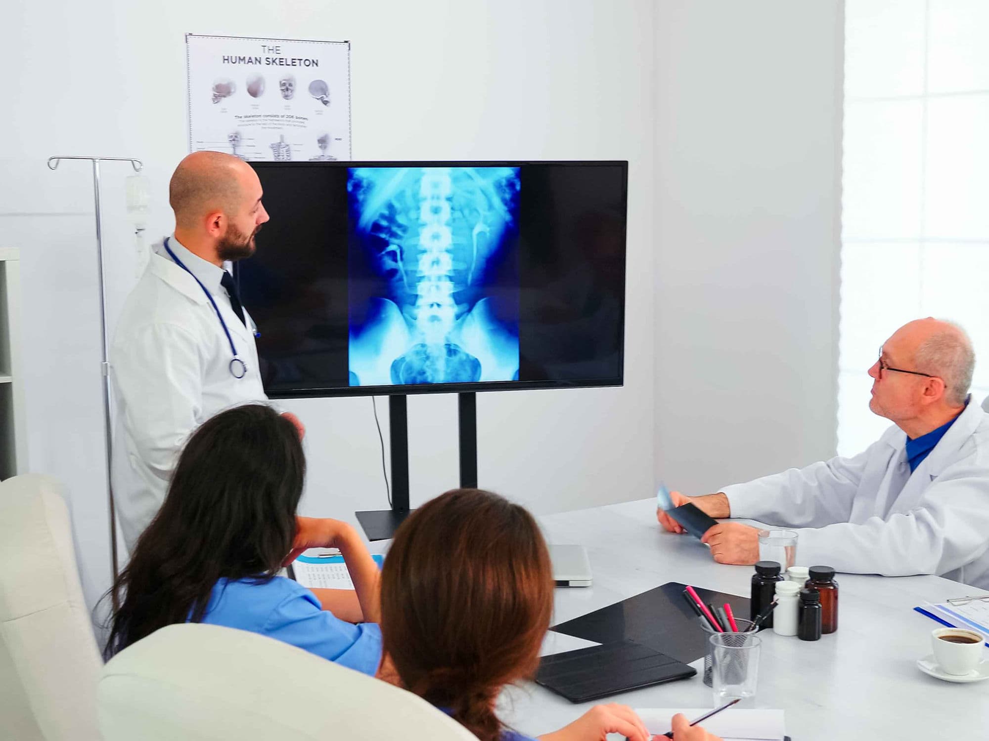
Confirmation of the specific disc level (most commonly C5-C6 or C6-C7) using high-resolution MRI and X-ray imaging.
Smoking cessation is mandatory for 4–6 weeks prior to surgery; nicotine significantly prevents the bone from fusing and increases the risk of complications.
Fasting (NPO) for at least 8 hours prior to the procedure to ensure safety under general anesthesia.
Pre-operative screening to ensure the patient can safely tolerate the retraction of the esophagus and neck tissues.
Tests Before Cervical Discectomy
Cervical MRI: The gold standard for identifying disc herniations and the degree of spinal cord or nerve root compression.
X-rays (Static and Dynamic): Used to assess overall spinal alignment and check for any abnormal movement (instability) between vertebrae.
CT Scan: Sometimes required to better visualize "hard" bone spurs (osteophytes) that may be contributing to the compression.
Electromyography (EMG): Performed to confirm that the arm symptoms are originating from the neck and not from other sites like the elbow or wrist.
Life After Cervical Discectomy
Many patients undergo the procedure as a same-day surgery or require only a single overnight stay for observation.
Depending on the surgeon’s preference and the complexity of the case, a soft or hard neck brace may be worn for 2 to 6 weeks.
Walking is encouraged immediately after surgery; however, lifting is strictly limited to less than 2–3 kg for the first 6 weeks.
Temporary hoarseness or a "lump in the throat" sensation when swallowing is common and usually fades within 2–4 weeks.
Driving is typically restricted for 2 weeks or until the patient can comfortably turn their head to check blind spots without pain.
Benefits of Cervical Discectomy
Extremely high success rates (over 90–95%) for the permanent relief of radiating arm pain and "electric shock" sensations.
Prevents the progression of permanent spinal cord damage and neurological deficits in patients with myelopathy.
Restores the ability to perform daily tasks, such as writing, buttoning clothes, and walking, by decompressing the neural pathways.
Provides significant stabilization to the neck, reducing the chronic "deep" ache associated with degenerative disc disease.