Diagnostic Hysteroscopy
Diagnostic Hysteroscopy
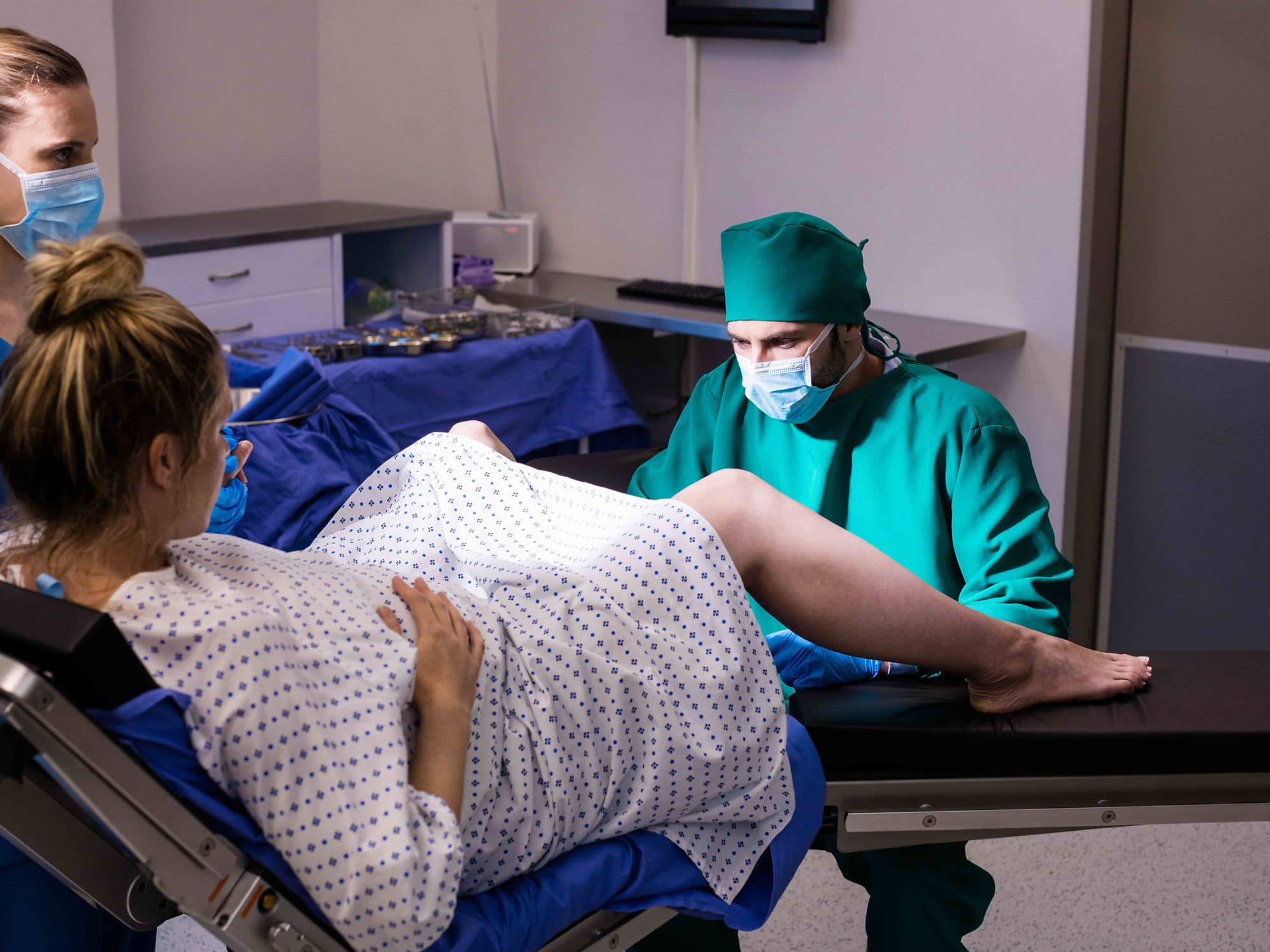
Diagnostic hysteroscopy is a minimally invasive procedure used to look inside the uterus to diagnose the cause of abnormal bleeding, infertility, or repeated miscarriages. It involves inserting a thin, lighted tube with a camera (hysteroscope) through the vagina and cervix, requiring no surgical incisions. This procedure allows for direct visualization of the uterine cavity, providing more accuracy than an ultrasound alone for certain conditions.
When You Should Consider a Diagnostic Hysteroscopy
Abnormal Uterine Bleeding: To investigate heavy, prolonged, or unexpected post-menopausal bleeding.
Infertility Evaluation: To check for structural abnormalities, such as polyps or a uterine septum, that might prevent an embryo from implanting.
Recurrent Miscarriage: Looking for internal scarring (Asherman’s Syndrome) or congenital uterine defects that could affect a pregnancy.
Displaced IUD: To locate and potentially retrieve an intrauterine device that has moved out of its proper position.
Abnormal Imaging Results: If a prior ultrasound or saline sonogram showed a suspicious shadow or growth within the uterine lining.
How It Is Performed
Anesthesia: Depending on your comfort level and the doctor's preference, it can be done with no anesthesia, local anesthesia (numbing the cervix), or light sedation.
Duration: The procedure is typically performed in an outpatient setting or doctor’s office and takes only 5 to 15 minutes.
Preparation: You are positioned similarly to a routine pelvic exam. No abdominal cuts are required as the hysteroscope uses the natural opening of the cervix.
Expansion: A saline liquid or carbon dioxide gas is gently introduced to expand the uterus, providing the doctor with a clear, panoramic view of the uterine walls and the openings of the fallopian tubes.
Inspection: The doctor moves the camera to examine the lining for fibroids, polyps, or structural defects.
Biopsy/Sampling: If a suspicious area is identified, a small tissue sample (biopsy) can be taken at the same time for laboratory testing.
Pre-Procedure Preparation
Timing Your Cycle: The procedure is best performed during the first week after your period ends, when the uterine lining is at its thinnest.
Pregnancy Test: A mandatory check to ensure you are not pregnant, as the procedure cannot be performed during pregnancy.
Medication Audit: You may be advised to take an over-the-counter pain reliever, like ibuprofen, about an hour before the procedure to minimize cramping.
Cervical Ripening (Optional): In some cases, medication may be inserted vaginally a few hours prior to help soften and slightly open the cervix.
Fasting: If you are receiving light sedation, you must follow "nothing by mouth" instructions for 6–8 hours before the appointment.
Tests Before Diagnostic Hysteroscopy
Pelvic Ultrasound: To provide a baseline view of the uterus and ovaries and to identify the location of any known fibroids.
Infection Screening: Testing for active pelvic infections or STIs to ensure it is safe to pass instruments into the uterus.
Blood Panels: A routine check of your blood count, especially if you have been experiencing heavy bleeding and anemia.
Hysterogram (HSG): Sometimes performed prior to hysteroscopy to check the patency of the fallopian tubes.
Life After Diagnostic Hysteroscopy
Recovery Time: Most patients can return to work and normal daily activities the same day or the following morning.
Short-term Symptoms: It is normal to experience mild cramping and light vaginal spotting for 1 to 2 days.
Gas/Shoulder Pain: If carbon dioxide was used for expansion, you may feel temporary referred pain in your shoulder for 24 hours.
Activity Restrictions: Most doctors recommend avoiding sexual intercourse and the use of tampons for a few days to allow the cervix to close and reduce the risk of infection.
Follow-up: Your doctor will discuss the visual findings immediately, though biopsy results typically take 5–7 days.
Why Specialized Treatment Is Highly Effective
Direct Visualization: Provides a "real-time" view that is often more definitive than X-rays or ultrasounds for identifying small polyps or adhesions.
Incision-Free: The lack of surgical cuts means there is no external scarring and a near-instantaneous recovery period.
Immediate Transition: If a problem is found, it can often be treated during the same session by switching to an operative hysteroscopy to remove the growth.
High Safety Profile: Complications like uterine perforation or infection are extremely rare when performed by experienced specialists.
Fertility Friendly: It is the "gold standard" for evaluating the uterine environment before proceeding with expensive fertility treatments like IVF.