Ectopic Pregnancy Surgery
Surgery for Ectopic Pregnancy
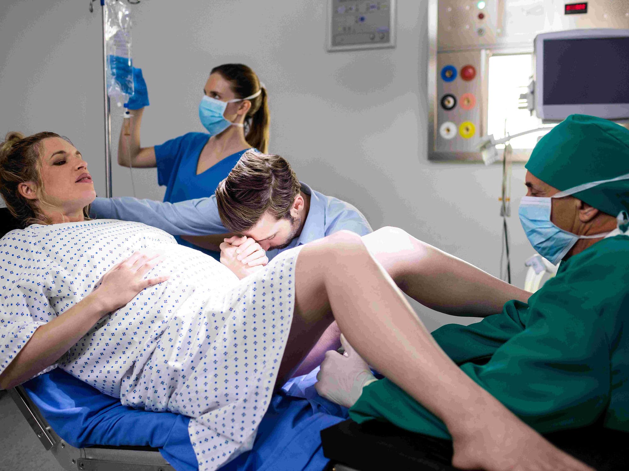
Surgery for an ectopic pregnancy is an emergency procedure performed when a fertilized egg implants outside the uterus, most commonly in a fallopian tube. Because an ectopic pregnancy cannot survive and poses a life-threatening risk of internal hemorrhage, surgical intervention is often necessary to protect the mother’s health and future fertility.
When You Should Consider Surgery for Ectopic Pregnancy
Confirmed Ectopic Pregnancy: When ultrasound and blood tests confirm the embryo has implanted outside the uterine cavity.
Severe Abdominal Pain: Sharp, stabbing pelvic pain, often on one side, that may come and go or vary in intensity.
Signs of Rupture: If you experience extreme lightheadedness, fainting, or shoulder tip pain, which indicates internal bleeding.
Failed Medical Management: When medication (such as methotrexate) has not successfully dissolved the pregnancy tissue.
High hCG Levels: If pregnancy hormone levels are too high for medical treatment to be effective or safe.
Unstable Vital Signs: An emergency situation where low blood pressure or a rapid heart rate suggests an active tubal rupture.
Surgical Approaches
Laparoscopic Surgery (Keyhole): The most common and preferred approach. The surgeon makes 2–3 tiny incisions in the abdomen to insert a high-definition camera and specialized micro-instruments.
Salpingectomy: The complete removal of the fallopian tube containing the pregnancy. This is often the safest choice if the tube is severely damaged or if the other tube is healthy.
Salpingostomy: A small, precise slit is made in the fallopian tube to remove the pregnancy tissue while leaving the tube intact. This is typically considered if the other tube is already damaged or missing.
Laparotomy (Open Surgery): A larger abdominal incision used in critical emergencies where a tube has ruptured and severe internal bleeding requires immediate, direct control.
How Is Performed
Anesthesia: The procedure is performed under general anesthesia and typically takes between 30 to 60 minutes, though emergency cases may require more time.
Access: Depending on the stability of the patient, the surgeon accesses the pelvic area through small laparoscopic ports or a traditional abdominal incision.
Tissue Removal: All pregnancy tissue is meticulously removed to prevent it from continuing to grow or causing further internal damage.
Hemostasis: The surgeon carefully seals all blood vessels to stop internal bleeding and ensure the pelvic cavity is clear of blood and debris.
Closure: Small laparoscopic incisions are closed with dissolvable stitches or surgical glue, while open incisions are secured with standard sutures or staples.
Pre-Procedure Preparation
Emergency Ultrasound: A final scan to locate the pregnancy and assess whether the fallopian tube has already ruptured.
Serial hCG Testing: Monitoring the levels of the pregnancy hormone to determine the urgency and type of surgical approach needed.
Blood Type and Cross-match: Immediate testing to ensure compatible blood is available in the event a transfusion is necessary due to blood loss.
Intravenous Access: Starting IV fluids and potentially medications to stabilize blood pressure before entering the operating room.
Fasting: In non-emergency cases, following "nothing by mouth" instructions; however, in emergencies, the surgical team proceeds immediately for patient safety.
Tests Before Surgery for Ectopic Pregnancy
Transvaginal Ultrasound: The primary diagnostic tool used to visualize the empty uterus and the mass in the fallopian tube.
Quantitative Beta-hCG: A precise blood test to measure the exact amount of pregnancy hormone in the system.
Complete Blood Count (CBC): To check for signs of anemia or internal blood loss through hemoglobin and hematocrit levels.
Coagulation Profile: Ensuring the blood’s ability to clot is normal prior to making surgical incisions.
Life After Surgery for Ectopic Pregnancy
Hospital Stay: Most laparoscopic patients are discharged the same day or after one night. Laparotomy patients typically stay for 2–3 days for observation.
Immediate Recovery: It is normal to experience temporary shoulder pain (from the gas used in laparoscopy), abdominal soreness, and light vaginal bleeding for 1–2 weeks.
Activity Restrictions: Avoid heavy lifting and strenuous exercise for 2 to 4 weeks (laparoscopic) or 6 weeks (open surgery) to allow internal healing.
Hormone Monitoring: Weekly blood tests to monitor hCG levels are often required until they reach zero to ensure no pregnancy tissue remains.
Future Pregnancy: If one healthy tube remains, the chances of a successful future pregnancy remain high. Most doctors recommend waiting at least two menstrual cycles before trying to conceive again.
Why Specialized Treatment Is Highly Effective
Life-Saving Intervention: Rapidly stops internal bleeding and removes the risk of a life-threatening tubal rupture.
Preserves Future Fertility: Specialized techniques like salpingostomy or careful laparoscopic salpingectomy protect the remaining reproductive anatomy.
Minimally Invasive Success: Laparoscopic methods result in less post-operative pain, smaller scars, and a much faster return to normal life.
Definitive Resolution: Unlike medical management, surgery provides an immediate solution for cases where the pregnancy tissue is large or the tube is compromised.
Comprehensive Emergency Care: Operating in a specialized surgical environment ensures that advanced monitoring and blood replacement are available if complications arise.