Oral Cancer Surgery
Oral Cancer Surgery
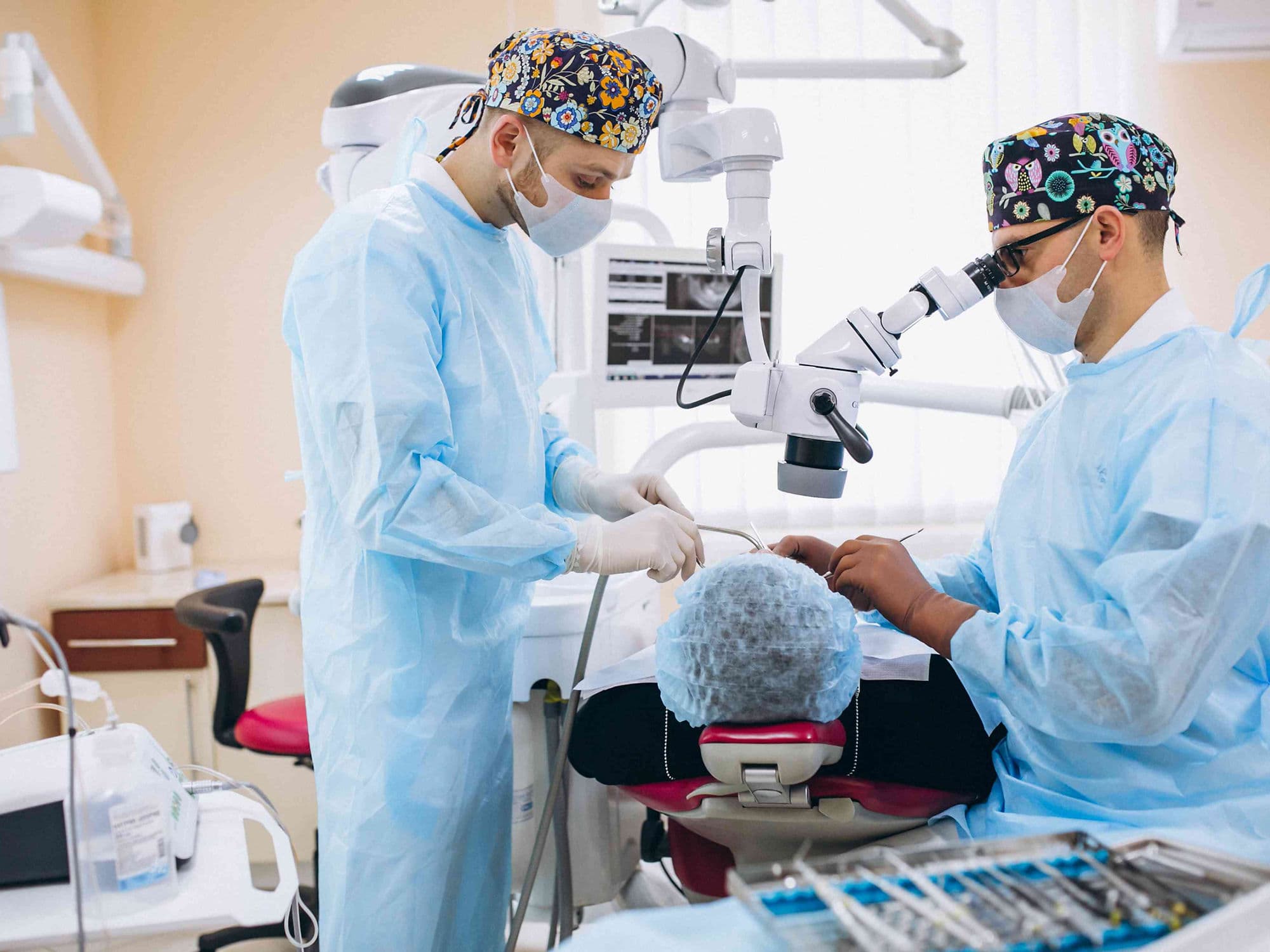
Oral Cancer Surgery (also known as head and neck surgery) is the primary treatment for cancers of the lips, tongue, inner cheeks, gums, and the floor or roof of the mouth. The goal is to remove the entire tumor while preserving as much function (speaking and swallowing) and appearance as possible. Many of these procedures are integrated with microvascular reconstruction in a single session to ensure the best functional outcomes.
When You Should Consider Oral Cancer Surgery
Tongue Malignancy: When a biopsy confirms squamous cell carcinoma on the lateral borders or base of the tongue.
Hard Palate or Gum Tumors: When cancer involves the roof of the mouth or the bony structures supporting the teeth.
Lip Cancer: For lesions that do not respond to topical treatments or show signs of deep invasion.
Floor of Mouth Lesions: When a tumor is located under the tongue, often requiring a "pull-through" resection.
Buccal Mucosa Cancer: For malignancies on the inner lining of the cheeks that may involve the underlying muscle.
Types of Primary Tumor Resection
Glossectomy: Removal of part or all of the tongue. A partial glossectomy removes only the cancerous edge, while a total glossectomy requires extensive reconstruction.
Mandiblectomy: Removal of a portion of the jawbone. A "marginal" resection removes the bone surface, while a "segmental" resection removes a full section if the cancer has invaded the marrow.
Maxillectomy: Removal of part or all of the hard palate (the roof of the mouth).
Mohs Surgery: Often utilized for lip cancer; thin layers of tissue are removed and examined microscopically in real-time until no cancer cells remain.
Wide Local Excision: Removing the tumor along with a 1-cm to 2-cm "clear margin" of healthy tissue to prevent local recurrence.
Neck Dissection (Lymph Node Removal)
Selective Neck Dissection: Removing only the lymph nodes in specific "levels" most likely to contain microscopic spread.
Radical Neck Dissection: Removing nearly all lymph nodes on one side of the neck; reserved for advanced disease where cancer involves the surrounding muscle or veins.
Sentinel Node Biopsy: Injecting a radioactive tracer or dye to identify and remove only the "first" node in the drainage path.
Level-Specific Clearance: Surgeons use precise mapping to clear Level I, II, and III nodes, which are the primary sites for oral cancer metastasis.
Reconstructive Surgery
Free Flap Transfer: The "gold standard." Surgeons transfer tissue (skin, muscle, or bone) from the forearm or leg and sew the tiny blood vessels to the neck vessels using a microscope.
Fibula Free Flap: Taking a piece of the lower leg bone to reconstruct a segment of the jawbone (mandible).
Radial Forearm Free Flap: Using skin from the inner wrist to reconstruct the tongue or the floor of the mouth.
Skin Grafts: Utilizing a thin layer of skin from the thigh to cover smaller defects within the oral cavity.
Local Flaps: Rotating nearby tissue from the neck or forehead to fill gaps in the cheek or palate.
How Is Performed
Anesthesia: Performed under general anesthesia, often involving a specialized tube to keep the mouth clear for the surgeon.
Tracheostomy: A temporary hole is made in the windpipe to ensure a safe airway while post-operative swelling subsides.
Micro-dissection: Using high-powered magnification to identify and preserve the nerves responsible for tongue movement and facial expression.
Feeding Tube Placement: A temporary tube is placed to provide nutrition while the oral tissues heal.
Frozen Section Analysis: Real-time pathology checks during surgery to confirm that all margins are negative for cancer before the reconstruction begins.
Pre-Procedure Preparation
Dental Clearance: A thorough dental exam to remove any decayed teeth in the radiation field or surgical site.
Speech and Swallow Baseline: Meeting with a therapist to evaluate your current function and plan for post-operative rehabilitation.
Allen’s Test: If a forearm flap is planned, this test ensures the hand has adequate blood supply from other arteries.
Nutritional Loading: High-protein supplementation to prevent weight loss, as eating will be difficult immediately following surgery.
Imaging Correlation: Reviewing 3D reconstructions of CT or MRI scans to plan exact bone cuts for jaw reconstruction.
Tests Before Oral Cancer Surgery
CT/MRI Head and Neck: To determine the depth of invasion and whether the tumor is attached to the jawbone.
PET-CT Scan: To rule out distant spread to the lungs or liver before committing to a major reconstructive surgery.
Angiography/Doppler: To check the blood vessels in the "donor site" (arm or leg) to ensure they are healthy enough for a free flap.
Panendoscopy: A visual inspection of the throat and esophagus under anesthesia to rule out a second primary tumor.
Biopsy Confirmation: Confirming the histological type and grade of the cancer to determine the extent of neck dissection required.
Life After Oral Surgery (Recovery & Risks)
Hospital Stay: Typically 7 to 14 days, with the first few days often spent in an ICU or High Dependency Unit for flap monitoring.
Flap Monitoring: A rare but serious risk where the blood supply to the new tissue fails, requiring an immediate return to the operating room.
Fistula: An abnormal opening where saliva leaks from the mouth into the neck; usually managed with specialized dressings.
Lymphedema: Swelling of the neck and face that may require specialized massage therapy after the lymph nodes are removed.
Rehabilitation: Daily sessions with speech and language pathologists to relearn how to swallow safely and speak clearly.
Why Specialized Treatment Is Highly Effective
Functional Restoration: Modern microvascular surgery allows patients to maintain the ability to eat and speak even after extensive resections.
High Cure Rates: For early-stage oral cancer, surgery offers a high probability of complete cure and long-term survival.
3D Precision: The use of surgical guides ensures that jaw reconstructions match the patient's original facial structure perfectly.
Integrated Care: Combining surgery with adjuvant radiation ensures that any remaining microscopic cells are eliminated.
Quality of Life: Dedicated head and neck teams focus on both removing cancer and the aesthetic and social reintegration of the patient.