Parotid Gland Surgery (Cancer)
Parotid Gland Surgery
Parotid Gland Surgery, or Parotidectomy, is the surgical removal of part or all of the parotid gland—the largest salivary gland, located just in front of the ear. When performed for cancer, the surgery is highly complex because the facial nerve, which controls all facial expressions (smiling, blinking, frowning), passes directly through the middle of the gland. The use of continuous intraoperative nerve monitoring is the standard of care to ensure the highest level of nerve preservation.
When You Should Consider a Parotidectomy
Parotid Tumors: For any growth in the parotid gland, as about 20% of these are malignant (cancerous).
Mucoepidermoid Carcinoma: The most common primary parotid cancer requiring surgical intervention.
Adenoid Cystic Carcinoma: A slow-growing but aggressive cancer known for traveling along nerve fibers.
Metastatic Skin Cancer: When skin cancer from the scalp or face spreads to the parotid lymph nodes.
Recurrent Pleomorphic Adenoma: When a previously removed benign tumor returns, requiring a more extensive resection.
Types of Parotidectomy
Superficial Parotidectomy: Removal of the portion of the gland "outside" the facial nerve. This is the most common approach for tumors that have not invaded the deep lobe.
Total Parotidectomy: Removal of the entire gland, including the deep lobe. The surgeon carefully "unfolds" the gland to peel it away from the facial nerve fibers.
Radical Parotidectomy: Removal of the entire gland and the facial nerve. This is reserved for cases where the cancer has physically encased the nerve, causing paralysis before surgery.
Extended Parotidectomy: Removal of the gland plus surrounding structures like the skin, ear canal, or jawbone if the cancer has spread beyond the gland boundaries.
Enucleation/Extracapsular Dissection: A more limited removal used only for very small, superficial, and low-grade tumors.
How Is Performed
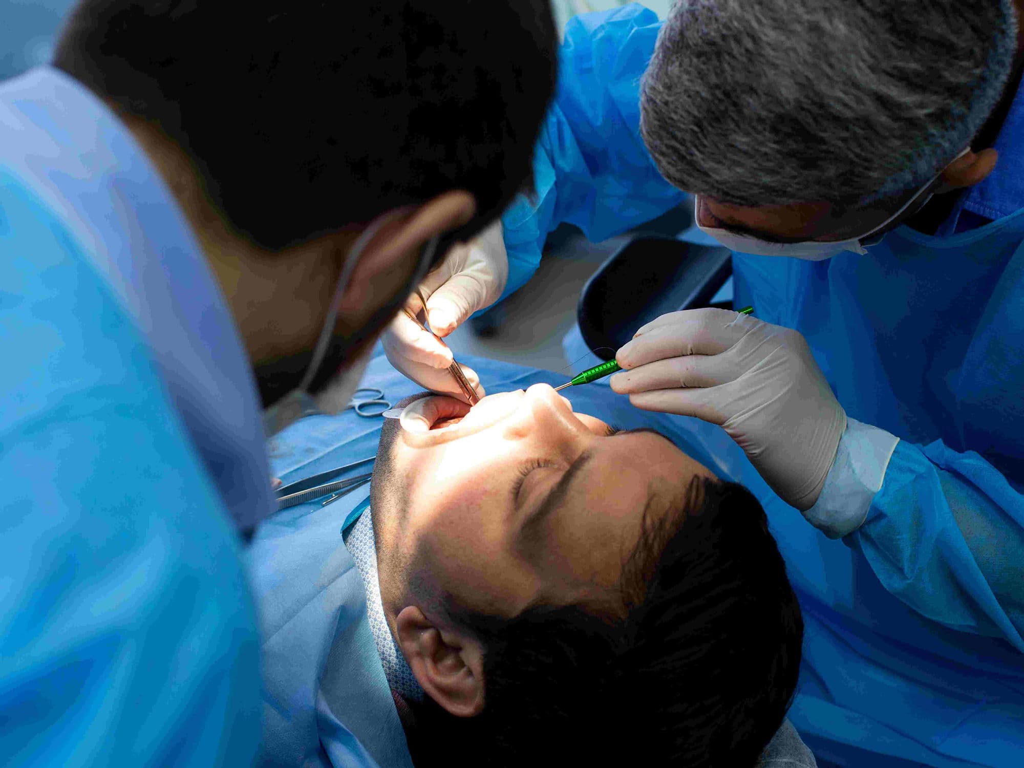
Anesthesia: Performed under general anesthesia. Surgeons avoid long-acting muscle relaxants to ensure the facial nerve can still be stimulated and monitored.
The Incision: The incision usually starts in front of the ear and curves down into the neck (Blair or Face-lift incision), often hidden in natural skin creases.
Facial Nerve Identification: The surgeon identifies the "trunk" of the facial nerve as it exits the skull and then meticulously follows its five branches.
Nerve Monitoring: Small electrodes in the facial muscles alert the surgical team if the nerve is touched or stimulated, preventing accidental injury.
Micro-dissection: Using high-power magnification or a microscope to separate the tumor from the delicate nerve fibers.
Neck Dissection: If the cancer is high-grade, the surgeon removes lymph nodes in Levels I, II, and III of the neck during the same operation.
Pre-Procedure Preparation
Facial Nerve Baseline: A thorough examination of facial movements to document any pre-existing weakness caused by the tumor.
Fine Needle Aspiration (FNA): A biopsy to determine the type and grade of the cancer, which helps plan the extent of the surgery.
Dental Check: Ensuring there are no active oral infections that could complicate the surgical site.
Tobacco Cessation: Stopping smoking at least 4 weeks prior to improve skin healing and reduce the risk of a salivary fistula.
Medication Audit: Pausing any blood thinners or supplements that increase the risk of a hematoma (blood clot) under the facial skin.
Tests Before Parotid Surgery
Contrast-Enhanced MRI: The preferred imaging to visualize the facial nerve's relationship to the tumor and check for spread along nerves.
CT Scan: Useful for evaluating whether the cancer has invaded the nearby jawbone or the base of the skull.
PET-CT Scan: Used for high-grade parotid cancers to rule out spread to the lungs or other distant sites.
Ultrasound-Guided Biopsy: To obtain a tissue sample from the tumor or suspicious neck lymph nodes.
Audiogram: Occasionally performed if the surgery involves the ear canal to establish a baseline for hearing.
Life After Parotid Surgery (Recovery & Risks)
Hospital Stay: Usually 1 to 2 nights. A small plastic drain is often left in the neck for 24 hours to prevent fluid buildup.
Facial Nerve Paresis: Temporary weakness of the face (e.g., a crooked smile or difficulty closing the eye) due to nerve manipulation. This usually resolves within 3–6 months.
Frey’s Syndrome: A long-term complication where the cheek sweats or flushes while eating; treatments include Botox injections or specialized skin barriers.
Numbness: Permanent numbness of the earlobe is common because a sensory nerve (greater auricular nerve) is often divided to reach the gland.
Salivary Fistula: Saliva may leak from the remaining gland tissue under the skin, often managed with temporary pressure dressings.
Why Specialized Treatment Is Highly Effective
Nerve Preservation: Intraoperative monitoring has significantly reduced the rates of permanent facial paralysis in parotid surgery.
Aesthetic Focus: Modern incisions ensure that surgical scars are nearly invisible once fully healed.
Advanced Reconstruction: If the nerve must be removed, "cable grafting" techniques can often restore facial movement over 6–12 months.
Targeted Adjuvant Therapy: Following surgery with precision radiation ensures that any microscopic cells near the facial nerve are eliminated.
Multidisciplinary Excellence: Combining the skills of head and neck surgeons with reconstructive experts provides the best balance of cancer clearance and functional preservation.