Spinal Fusion Surgery
Spinal Fusion Surgery
Spinal Fusion is a major surgical procedure designed to permanently connect two or more vertebrae, eliminating painful motion between them. The procedure is characterized by Minimally Invasive Spine Surgery (MISS) and robotic assistance, utilizing bone grafts and high-precision hardware to create a solid bone mass. This approach aims to stabilize the structural integrity of the spine while protecting the surrounding nerves and musculature.
When You Should Consider Spinal Fusion
Chronic Pain: Debilitating back or neck pain that has not responded to physical therapy, medications, or injections.
Neurological Symptoms: Persistent numbness, tingling, or weakness in the arms or legs caused by sustained nerve compression.
Mechanical Instability: Significant pain that worsens with specific movements, such as bending, twisting, or lifting.
Spinal Deformity: Visible curvature or a sensation of the spine "slipping," often associated with structural instability.
Functional Limitation: Difficulty standing or walking for extended periods due to structural spinal narrowing or collapse.
Trauma or Tumor: Severe pain or instability following a spinal fracture or the surgical removal of a spinal tumor.
Conditions That Require Specialized Care
Degenerative Disc Disease: Where worn-out discs cause painful friction and micro-motion between vertebrae.
Spondylolisthesis: A condition where one vertebra slips forward over the one below it, potentially pinching nerves.
Spinal Stenosis: Resulting in the narrowing of the spinal canal and significant nerve pressure.
Scoliosis or Kyphosis: Involving abnormal curvatures of the spine that require corrective alignment and stabilization.
Pseudoarthrosis: A condition where a previous fusion attempt failed to heal into a solid bone mass.
Methods of Spinal Fusion
Minimally Invasive Spine Surgery (MISS): Techniques that use tubular retractors to spread muscles rather than cutting them, reducing blood loss and recovery time.
Robotic-Assisted Fusion: The use of advanced guidance systems to ensure screws and rods are placed with sub-millimeter accuracy.
Anterior Lumbar Interbody Fusion (ALIF): Accessing the spine through the abdomen to provide a large surface area for the fusion cage.
Lateral Interbody Fusion (XLIF/LLIF): A side-access approach that avoids major back muscles and the spinal canal, often allowing for faster mobilization.
Posterior Lumbar Interbody Fusion (PLIF): The traditional approach from the back, offering the most direct access to compressed nerves and the spinal canal.
Transforaminal Lumbar Interbody Fusion (TLIF): An evolution of the posterior approach that accesses the disc space from a more lateral angle to reduce nerve retraction.
How Spinal Fusion Is Performed
Surgical Mapping: Digital mapping or Augmented Reality (AR) is used to project the patient's internal anatomy for the surgeon.
Access: Minimally invasive incisions are made to reach the spine from the most appropriate clinical angle (front, back, or side).
Disc Removal: The intervertebral disc or damaged bone is removed to decompress nerves and create space for the fusion.
Cage Insertion: A "cage" or spacer filled with bone graft material is inserted between the vertebrae to stimulate bone growth.
Hardware Stabilization: Robotic arms often assist in the precise placement of pedicle screws and rods to hold the vertebrae steady while they fuse.
Biological Stimulation: Bone Morphogenetic Proteins (BMP) or specialized bone grafts are applied to accelerate the natural bone-healing process.
Pre-Procedure Preparation
Smoking Cessation: Patients must commit to a strict no-nicotine program, as smoking significantly increases the risk of fusion failure (non-union).
Pre-habilitation: Strengthening "core" stabilizer muscles through directed physical therapy to support the spine post-operatively.
Home Setup: Coordinate a "home recovery station" to avoid the need for bending, lifting, or twisting during the initial healing phase.
Bone Health Optimization: Undergo a bone density scan (DEXA) to ensure the vertebrae are strong enough to support surgical hardware.
Bracing Consultation: Discuss the use of a post-operative back brace with the surgical team to ensure proper fitting and sizing.
Tests Before Spinal Fusion Surgery
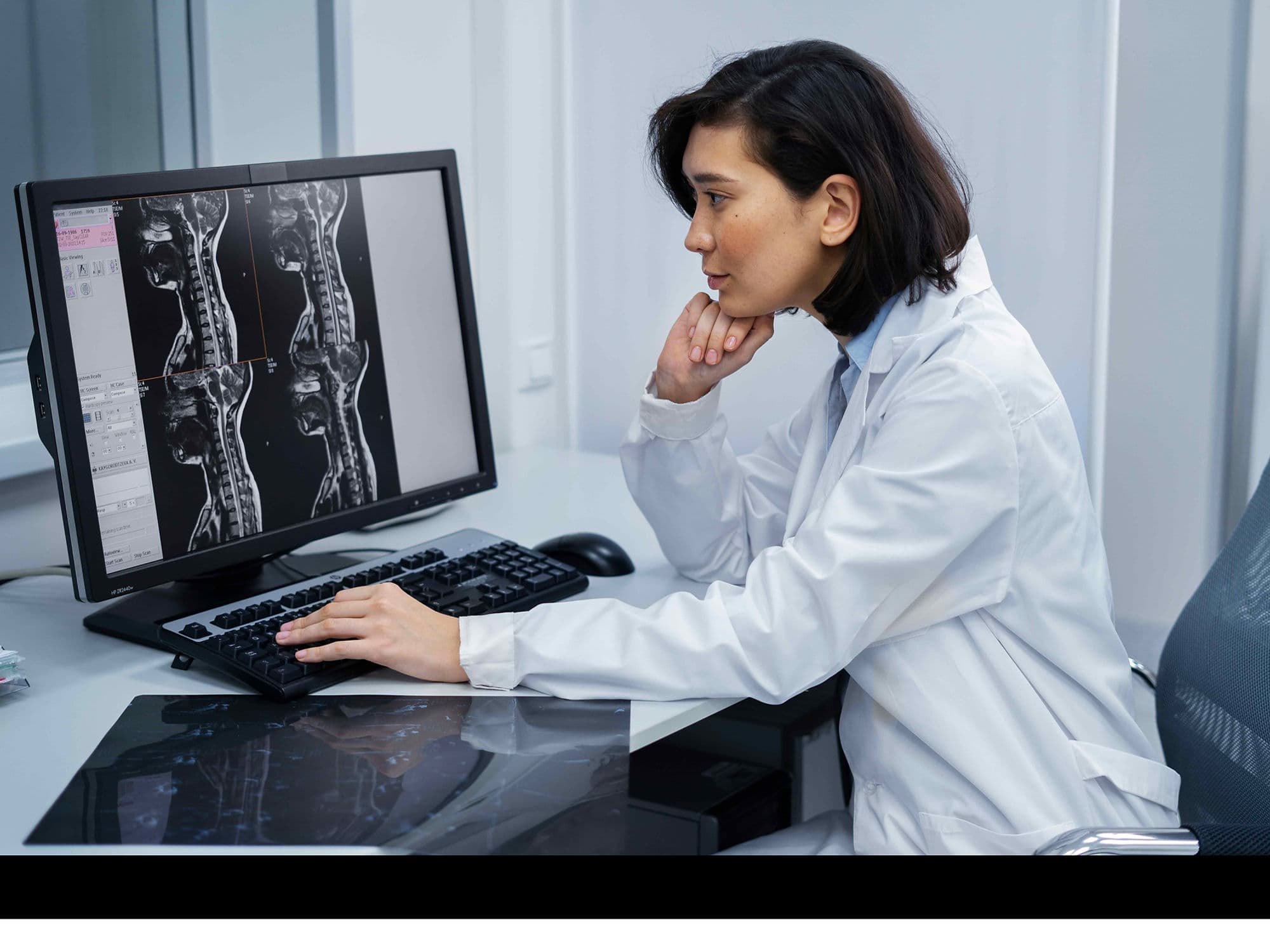
Standing X-rays and MRI: Used to identify the exact source of nerve compression and the degree of mechanical instability.
High-resolution CT Scan: Essential for 3D surgical planning and robotic navigation mapping.
Electrocardiogram (EKG): Along with comprehensive blood panels to confirm cardiovascular readiness for a major procedure.
Neurological Baseline Testing: Measuring nerve conduction and muscle strength to provide a comparison for post-operative recovery.
DEXA Scan: To evaluate the quality of the "host bone" for successful graft integration and hardware stability.
Life After Spinal Fusion Surgery
Immediate Recovery: Hospital stays typically last 1 to 3 days, with an emphasis on early, assisted walking to prevent blood clots.
The "No BLT" Rule: For the first 3 to 6 months, patients must strictly avoid Bending at the waist, Lifting over 3kg, and Twisting the spine.
Bracing: Wear a customized back brace as prescribed to maintain spinal alignment during the critical fusion window.
Phased Physical Therapy: Focusing on core stabilization and safe movement patterns once the initial bone healing is confirmed.
Long-term Monitoring: Routine follow-up imaging (X-rays or CT) is required to confirm the success of the bone bridge across the joint.
Activity Resumption: Gradual return to a more active lifestyle once the vertebrae have fused into a single, solid, and stable bone mass.
Benefits of Spinal Fusion Surgery
Significant Pain Reduction: Achieves a 70% to 90% success rate for patients with chronic instability-related pain.
Enhanced Precision: Utilizes AR and robotic technology to make surgery safer and more accurate than traditional "freehand" methods.
Neurological Protection: Stabilizes the spine to prevent further nerve injury or progressive physical deformity.
Accelerated Healing: Stimulates the body's natural recovery using bio-engineered proteins for faster bone growth.
Permanent Stability: Eliminates the painful micro-motion that causes chronic inflammation and structural wear.