Tongue Resection (Cancer)
Tongue Resection
Tongue Resection, clinically termed a glossectomy, is the surgical removal of all or part of the tongue to treat oral cancer. The primary goal is to excise the malignant tumor with a 1–2 cm "clear margin" of healthy tissue to prevent recurrence. Advanced microvascular reconstruction is now the standard for maintaining speech and swallowing functions after a resection.
When You Should Consider a Glossectomy
Squamous Cell Carcinoma (SCC): The most common form of tongue cancer, often appearing as a persistent ulcer or growth on the lateral (side) border.
Deep Invasion: When a tumor has grown into the underlying intrinsic muscles of the tongue.
Leukoplakia with Dysplasia: When precancerous white patches show high-grade changes that are likely to become invasive.
Recurrent Disease: When cancer returns in a previously treated area of the mouth.
Base of Tongue Tumors: When the malignancy is located at the very back of the tongue, near the throat.
Types of Tongue Resection
Partial Glossectomy: Removal of a small portion of the tongue. Usually, the remaining tissue is sewn together, and speech and swallowing remain near normal.
Hemiglossectomy: Removal of one full side of the tongue. This typically requires reconstruction using tissue from another part of the body to maintain volume and mobility.
Total Glossectomy: Removal of the entire tongue. This is a life-altering procedure reserved for advanced cancers and requires extensive microvascular reconstruction.
Base of Tongue Resection: A specialized procedure for tumors at the back of the tongue, often performed robotically (TORS) to avoid large external incisions.
Compartmental Resection: Removing the tumor along with the entire anatomical compartment of muscles to ensure no microscopic cells remain.
How Is Performed
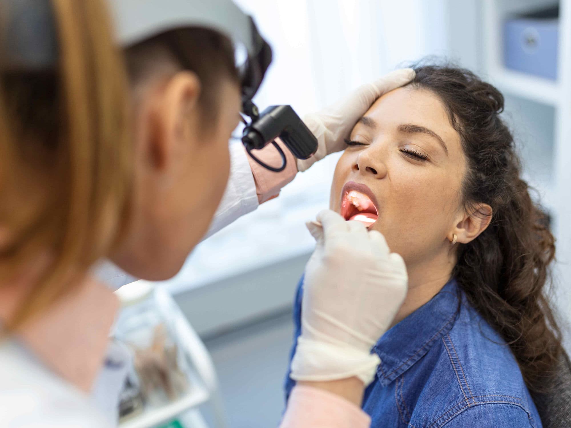
Anesthesia: Performed under general anesthesia, often with a "nasotracheal" tube to provide the surgeon with a clear view of the oral cavity.
Neck Dissection: A concurrent procedure where lymph nodes are removed from the neck to check for microscopic cancer spread.
Resection with Margins: The surgeon uses specialized tools to cut 1–2 cm away from the visible tumor to ensure a "pathologically clear" margin.
Microvascular Reconstruction (Free Flap): For larger defects, tissue (skin, fat, or muscle) is taken from the forearm or thigh, and its blood vessels are sewn to vessels in the neck using a microscope.
Tracheostomy: A temporary breathing hole is made in the neck because postoperative swelling can block the airway; it is usually removed after 5–10 days.
Feeding Tube Placement: Since the patient cannot swallow while the sutures heal, a temporary NG (nose-to-stomach) or PEG tube provides nutrition for 1–2 weeks.
Pre-Procedure Preparation
Speech and Swallow Baseline: Meeting with a specialist to assess current function and plan for intensive rehabilitation after surgery.
Dental Evaluation: Removing any decayed teeth that might cause infection during healing or interfere with future radiation therapy.
Allen’s Test: If a forearm flap is planned, this test ensures the hand has a sufficient secondary blood supply.
Nutritional Optimization: Starting high-protein supplements to ensure the body has the resources to heal complex microvascular connections.
Imaging Correlation: Reviewing 3D CT or MRI scans to map the tumor's depth and its proximity to the lingual artery and nerve.
Tests Before Tongue Resection
Contrast-Enhanced MRI: The "gold standard" for determining the exact depth of invasion (DOI) into the tongue muscle.
PET-CT Scan: To rule out any spread to the lungs or distant lymph nodes before committing to a major reconstructive procedure.
Biopsy Verification: Confirming the histological grade of the cancer to determine the necessary extent of the neck dissection.
Doppler Ultrasound: To map the blood vessels in the donor site (arm or leg) to ensure they are suitable for a "free flap" transfer.
Coagulation Profile: To ensure blood clots properly at the resection site but remains fluid enough for microscopic vascular connections.
Life After Tongue Surgery (Recovery & Risks)
Hospital Stay: Typically 7 to 14 days, with the first few days spent in a specialized unit for frequent "flap checks" to ensure blood flow.
Flap Failure: A rare but critical risk where the microscopic blood vessel connection clots, requiring immediate emergency re-operation.
Aspiration Risk: If the new tongue cannot protect the airway during swallowing, food or saliva may enter the lungs, potentially causing pneumonia.
Fistula: An abnormal leak of saliva from the mouth into the neck tissues, which usually requires specialized wound care to heal.
Sensory Changes: Permanent numbness in the resected area or a loss of taste is common, though the other side of the tongue often compensates.
Why Specialized Treatment Is Highly Effective
Microvascular Precision: Modern "free flap" techniques allow surgeons to rebuild a tongue that can still move, speak, and push food to the back of the throat.
Comprehensive Staging: Performing a neck dissection during the same surgery ensures that any microscopic spread is caught and treated early.
Robotic (TORS) Advancements: For base-of-tongue cancers, robotic surgery allows for removal through the mouth, avoiding the need to "split" the jawbone.
Intensive Rehabilitation: Standardized speech and swallow therapy significantly improves quality of life, helping patients return to a normal diet.
Multidisciplinary Success: When surgery is followed by modern adjuvant radiation, local control rates for tongue cancer are at an all-time high.