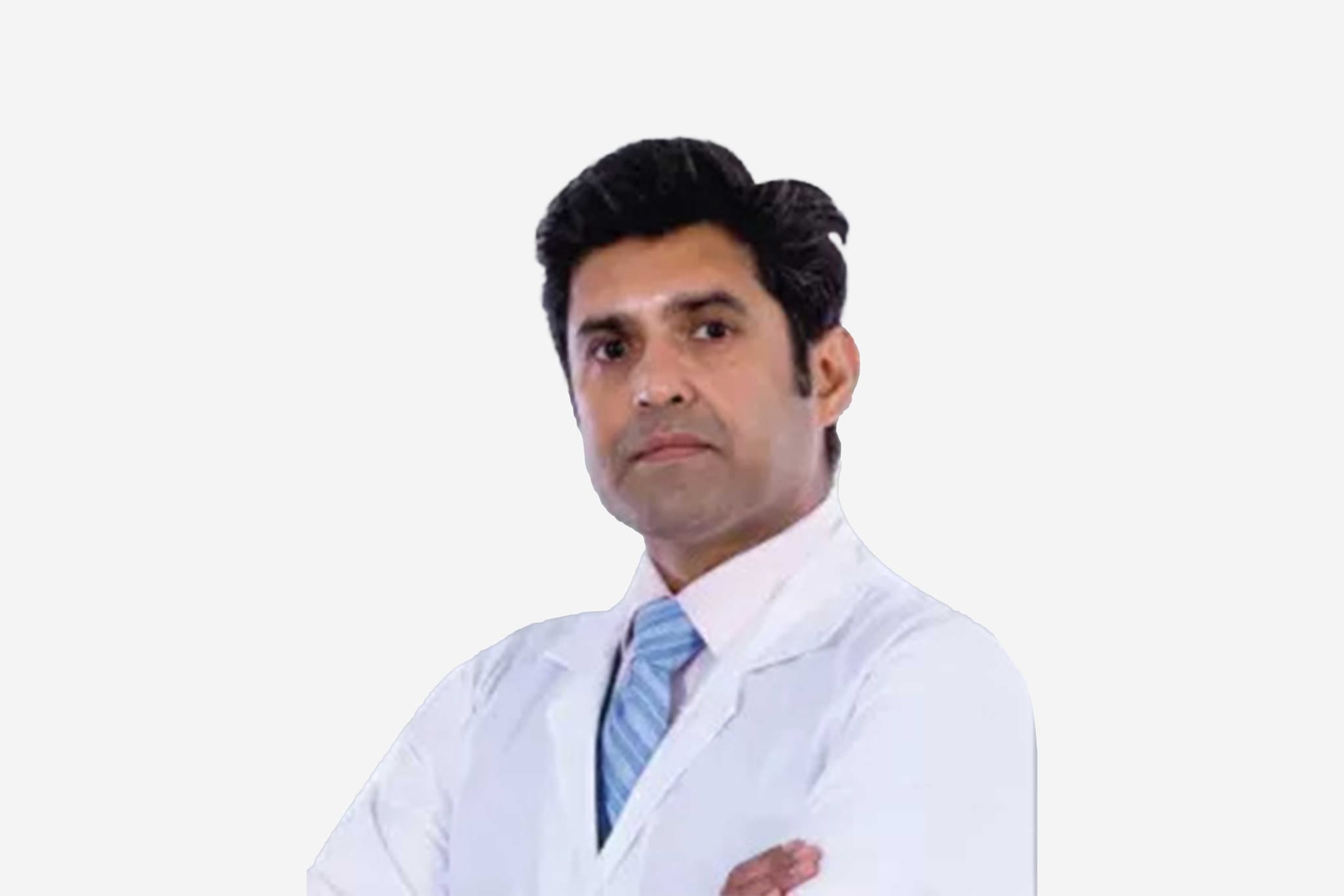
Medivisor India Treatment Logo
  • Home
              • Become a Partner

              +91 83407 80250

              Medivisor Logo

              Medivisor India Treatment is a trusted New Delhi–based medical travel partner connecting global patients to advanced, affordable, high-quality healthcare in India.

              • Medivisor House 359, Sector 1, Vaishali, Ghaziabad, (Delhi/NCR) India
              • +91 8340 780 250
              • info@medivisorhealth.com

              About Us

              • About Us
              • Our Services
              • Our Team
              • Our Medical Advisors
              • Our Safety Measures
              • Join Our Network
              • Contact Us

              India Treatment

              • Hospitals
              • Treatments
              • Treatment Cost
              • Treatment Process
              • Visa Process
              • Travel Guide
              • FAQs
              • Why Medivisor

              Gallery

              • Patient Testimonials
              • Patient Activities
              • News Coverage
              • Blog

              Get in Touch

              © 2026 Medivisor India Treatment. All rights reserved.

              Dr. Dr. Sudhansu Bhattacharyya

              Dr. Sudhansu Bhattacharyya

              50+ years experience

              M.B.B.S., M.S. (General Surgery), M.Ch. (Cardio-Thoracic Surgery)
              1. Home
              2. Doctors
              3. Dr. Sudhansu Bhattacharyya

              About Dr. Sudhansu Bhattacharyya

              Dr. Sudhansu Bhattacharya is a highly revered Consultant in the Cardiac Surgery Department at Sir H. N. Reliance Foundation Hospital and Research Centre, Mumbai. With a career spanning several decades, he is recognized not only for his surgical expertise but also for his significant contributions to academic medicine and the development of specialized surgical instrumentation.

              Before moving into full-time clinical practice in 1983, Dr. Bhattacharya dedicated over a decade to medical education and research. He served as a Professor of Cardio-Thoracic Surgery at the prestigious Seth GS Medical College and KEM Hospital in Mumbai. This strong academic foundation has defined his career, characterized by a commitment to teaching and the continuous advancement of cardiac surgical techniques.

              Mentorship and Global Training

              Dr. Bhattacharya holds a Fellowship in Cardiothoracic Surgery and was mentored by the world-renowned cardiac surgeon, Dr. Dudley Johnson. Under this mentorship, he refined his skills in complex coronary and valvular procedures, bringing advanced surgical perspectives back to the Indian healthcare landscape. His extensive clinical experience includes tenures at several of Mumbai’s premier medical institutions, such as Jaslok Hospital and Breach Candy Hospital.

              Innovation and Patented Surgical Instruments

              One of Dr. Bhattacharya’s most unique contributions to the field of cardiac surgery is his work as an inventor. He has designed and patented several surgical instruments that are used to improve visibility and precision during delicate operations. Notable inventions include:

              • Atrial Retractor: Specially designed for use during mitral valve replacement surgeries.

              • Internal Mammary Artery Retractor: A specialized tool utilized for LIMA-RIMA (Left/Right Internal Mammary Artery) grafting procedures, facilitating easier access for arterial revascularization.

              Awards and Academic Contributions

              Throughout his distinguished career, Dr. Bhattacharya has received numerous national and international awards in recognition of his clinical excellence and innovative spirit. He remains an active voice in the medical community, having authored several journal publications and presented numerous papers at global conferences. His work continues to influence modern surgical protocols in the domain of cardiothoracic medicine.

              Dr. Sudhansu Bhattacharya at a Glance

              • Consultant Cardiac Surgeon at Sir H. N. Reliance Foundation Hospital and Research Centre.

              • Over 10 years of experience as a Professor of Cardio-Thoracic Surgery at KEM Hospital.

              • Mentored by the pioneering cardiac surgeon Dr. Dudley Johnson.

              • Inventor and patent-holder of specialized instruments for mitral valve and arterial grafting.

              • Extensive clinical history with premier institutions like Jaslok and Breach Candy Hospitals.

              • Recognized globally with numerous awards for his career accomplishments.

              • Prolific author and contributor to international cardiac surgery journals and conferences.

              M.B.B.S., M.S. (General Surgery), M.Ch. (Cardio-Thoracic Surgery)

              No awards & achievements available

              Affiliated Hospitals

              Hiranandani Fortis Hospital, Vashi, Navi Mumbai
              Hiranandani Fortis Hospital, Vashi, Navi Mumbai

              Multi Specialty

              Mumbai, Maharashtra

              2007

              Estd.

              150+

              Beds

              115+

              Doctors

              May we help you?

              Enter digits only (6-15 digits). We'll add your country code.

              May we help you?

              Enter digits only (6-15 digits). We'll add your country code.

              Related Treatments

              VSD Device Closure
              VSD Device Closure

              VSD Device Closure VSD (Ventricular Septal Defect) device closure is a minimally invasive, non-surgical procedure used to seal a "hole in the heart" between the two lower chambers (ventricles). Unlike traditional open-heart surgery, this procedure is performed entirely through a catheter, resulting in no chest scars and a significantly faster recovery. This advanced technique allows for the permanent repair of the heart's internal wall without the need for a heart-lung bypass machine. When You Should Consider VSD Device Closure Muscular VSDs: This is the primary treatment for holes located in the muscular portion of the ventricular septum. Symptom Management: For children or adults experiencing poor weight gain, frequent lung infections, or persistent shortness of breath. Heart Protection: To prevent the left side of the heart from overworking, which can lead to an enlarged heart (cardiomegaly). Pulmonary Hypertension Prevention: To reduce the risk of developing dangerously high blood pressure in the lung arteries. Heart Failure Prevention: Correcting the defect before it leads to more serious long-term cardiac complications. How It Is Performed Access: A small incision is made in the groin to access the femoral vein or artery. No large incisions are made on the chest. Anesthesia: The procedure is performed in a specialized Cardiac Catheterization Lab (Cath Lab) under general anesthesia or heavy sedation, typically taking 1 to 2 hours. Guidance: A thin, flexible tube (catheter) is threaded through the blood vessels into the heart, guided by real-time X-ray (Fluoroscopy) and detailed ultrasound (Transesophageal Echo). Measurement: The specialist measures the exact size and location of the hole to select a custom-sized Nitinol mesh device. Deployment: A folded, umbrella-like device is pushed through the catheter. Once it reaches the hole, it is carefully unfolded to "sandwich" the defect from both sides. Verification: Once the device is securely in place and the hole is confirmed to be sealed, the catheter is removed and the small puncture in the groin is closed. Pre-Procedure Preparation Echocardiogram: A detailed ultrasound of the heart to map the VSD's size and its proximity to the heart's valves. Transesophageal Echo (TEE): A specialized ultrasound performed through the esophagus for high-resolution images of the defect. Dental Clearance: Ensuring there are no active dental infections, which could increase the risk of heart infection (endocarditis) after the device is placed. Fasting: Following "nothing by mouth" instructions for 8 hours prior to the procedure. Medication Audit: You may be asked to adjust or stop certain medications, particularly blood thinners, a few days before the procedure. Tests Before VSD Device Closure Chest X-ray: To evaluate the current size of the heart and check for any fluid in the lungs. Electrocardiogram (ECG): A baseline check of the heart's electrical system to identify any pre-existing arrhythmias. Blood Panels: A routine check of your blood count, electrolytes, and kidney function. Cardiac MRI or CT: Occasionally used to provide a 3D model of the heart for complex or multiple VSDs. Life After VSD Device Closure Hospital Stay: Most patients stay for one night for observation and are discharged the next day. Medication: You will typically take blood-thinning medication (usually Aspirin) for 6 months to prevent clots from forming on the device while the heart lining grows over it. Activity Restrictions: Most patients can return to school or light work within 3 to 5 days. You should avoid strenuous exercise and heavy lifting for at least 2 weeks. Dental Care Precautions: For the first 6 months post-procedure, you must take preventive antibiotics before any dental work to prevent heart infections. Long-term Integration: Over 3–6 months, the heart's natural lining (endocardium) grows completely over the device, making it a permanent and seamless part of your heart. Why Specialized Treatment Is Highly Effective Scar-Free Recovery: By avoiding a sternotomy (opening the chest), patients experience much less pain and have no permanent surgical scars. Rapid Return to Normalcy: Recovery is measured in days rather than the months required for open-heart surgery. High Success Rates: Device closure is a highly reliable method for sealing muscular VSDs with a very low risk of the hole reopening. Protects Electrical System: Advanced imaging ensures the device is positioned to minimize pressure on the heart's natural "wiring." Permanent Solution: The Nitinol mesh is designed to last a lifetime, providing a durable repair that grows with the patient.

              Aortic Valve Replacement AVR
              Aortic Valve Replacement AVR

              Aortic Valve Replacement (AVR) Aortic Valve Replacement (AVR) is an advanced cardiac procedure that replaces a damaged, stiff, or leaking aortic valve with a new mechanical or tissue valve. This restores healthy blood flow, improves heart pumping capacity, reduces symptoms, and prevents long-term heart failure or life-threatening complications. When You Should Consider AVR Severe or persistent shortness of breath that limits walking, climbing stairs, or daily activity. Chest pain, pressure, or heaviness due to the heart struggling to push blood through a narrowed valve. Extreme tiredness or low energy even during simple tasks. Dizziness or fainting episodes, especially during exertion. Irregular heartbeat or noticeable palpitations, indicating the heart is under stress. Swelling in the feet, legs, or ankles, a sign of poor blood circulation or early heart failure. Conditions That Require AVR Severe Aortic Stenosis – the valve becomes narrowed and heavily calcified, restricting blood flow. Severe Aortic Regurgitation – the valve leaks and allows blood to flow backward into the heart. Congenital valve abnormalities, including bicuspid valves. Infection-related valve damage (endocarditis) that weakens or destroys the valve. Aged, stiff, or heavily calcified aortic valve due to long-term wear and tear. How Aortic Valve Replacement Is Performed General anesthesia is given to ensure a pain-free and comfortable procedure. The surgeon makes either a full chest incision or a minimally invasive cut depending on your case. The damaged aortic valve is carefully removed. A new mechanical or biological valve is implanted to restore proper blood flow. The heart is restarted, and valve function is tested to ensure smooth operation. You are shifted to the ICU for continuous monitoring and early recovery. Types of Aortic Valve Replacement Mechanical Valve Replacement Long-lasting artificial valve; ideal for younger patients. Requires lifelong blood thinners to prevent clots. Biological (Tissue) Valve Replacement Made from natural tissue. Offers natural blood flow and usually requires minimal blood thinner use. Minimally Invasive AVR Smaller incisions, less pain, reduced blood loss, and faster healing. Robotic AVR Performed using robotic precision tools for high accuracy, minimal scars, and quicker recovery. TAVR (Transcatheter Aortic Valve Replacement) A non-surgical, catheter-based procedure performed through the groin. Ideal for elderly or high-risk patients. Pre-Surgery Preparation Quit smoking at least 2–3 weeks before surgery for better lung function. Keep blood pressure, diabetes, and heart rate well controlled. Follow fasting instructions before the procedure. Stop blood thinners only if your cardiologist advises. Complete all required heart and blood tests before the surgery date. Pre-Surgery Tests ECG to check heart rhythm. 2D Echocardiography to evaluate valve structure and pumping strength. CT scan or MRI for detailed imaging when needed. Coronary Angiography to detect any artery blockages. Chest X-ray to assess lung health. Routine blood tests including CBC, kidney/liver function, and clotting profile. Why AVR Is Highly Effective Restores normal forward blood flow from the heart. Reduces breathlessness and chest discomfort. Prevents the heart from becoming enlarged or weak. Improves daily stamina, energy levels, and activity tolerance. Provides long-lasting results with modern valve technology. Recovery After AVR ICU stay: Usually 1–2 days for close monitoring. Early walking begins within 24 hours. Tubes and drains are removed in 48–72 hours. Home recovery: Typically 4–8 weeks depending on the surgery type. Return to work: Usually within 6–10 weeks. Life After AVR Avoid smoking permanently to protect the new valve. Follow a heart-healthy, low-salt diet for lifelong cardiac wellness. Exercise daily with light walking, avoid heavy lifting initially. Take medications regularly, especially blood thinners if you have a mechanical valve. Join a cardiac rehabilitation program for guided recovery and long-term heart strength.

              Mitral Valve Replacement (MVR)
              Mitral Valve Replacement (MVR)

              Mitral Valve Replacement (MVR) (Cardiology) Mitral Valve Replacement (MVR) is a specialized heart procedure that restores healthy blood flow by replacing a diseased mitral valve with a mechanical or biological valve. This improves heart efficiency, reduces symptoms like breathlessness and fatigue, and prevents long-term complications such as heart failure. When You Should Consider MVR Shortness of breath during daily activities or while lying down. Chest discomfort or pressure caused by poor blood flow through the heart. Fatigue or low energy during simple tasks. Irregular heartbeat or palpitations due to valve dysfunction. Swelling in feet, legs, or ankles from fluid retention. Fainting or dizziness, especially during physical activity. Conditions That Require MVR Severe Mitral Stenosis – narrowing of the mitral valve restricting blood flow. Severe Mitral Regurgitation – leaking mitral valve causing backward blood flow. Congenital mitral valve defects present from birth. Valve damage from infection (endocarditis). Calcified or thickened mitral valve leading to poor heart function. How Mitral Valve Replacement Is Performed General anesthesia is administered for a safe, painless procedure. A chest or minimally invasive incision is made based on patient suitability. The damaged mitral valve is carefully removed. A mechanical or biological replacement valve is implanted. Heart function is tested before closing the incision. Patient is moved to the ICU for monitored recovery. Types of Mitral Valve Replacement Mechanical Valve Replacement Long-lasting artificial valve; requires lifelong blood thinners. Biological (Tissue) Valve Replacement Natural tissue valve; usually requires minimal blood thinner use. Minimally Invasive MVR Smaller incisions, less pain, quicker healing, and reduced scarring. Robotic MVR Uses robotic precision for high accuracy, minimal scarring, and faster recovery. Transcatheter Mitral Valve Replacement (TMVR) Non-surgical, catheter-based procedure for high-risk or elderly patients. Pre-Surgery Preparation Stop smoking 2–3 weeks before surgery. Maintain blood pressure, diabetes, and heart rate within target range. Follow fasting instructions as advised. Pause blood thinners only if instructed by your cardiologist. Complete all cardiac and routine blood tests prior to surgery. Pre-Surgery Tests ECG to check heart rhythm. Echocardiography (2D/3D) to evaluate mitral valve function. CT or MRI scans for detailed imaging if required. Coronary angiography to detect any blocked arteries. Chest X-ray to assess lung and heart health. Routine blood tests including CBC, kidney/liver function, and clotting profile. Why MVR Is Highly Effective Restores normal blood flow through the heart. Reduces shortness of breath, fatigue, and chest discomfort. Prevents heart enlargement and failure. Improves daily activity tolerance and quality of life. Provides long-lasting results with modern valve options. Recovery After MVR ICU stay: 1–2 days for close monitoring. Walking usually begins within 24 hours. Tubes and drains are removed in 48–72 hours. Home recovery: 4–8 weeks depending on the procedure type. Return to work: Typically 6–10 weeks, gradually increasing activity. Life After MVR Avoid smoking permanently. Follow a heart-healthy, low-salt diet. Engage in daily light exercise; avoid heavy lifting initially. Take prescribed medications regularly, especially blood thinners for mechanical valves. Join a cardiac rehabilitation program for optimal long-term recovery.

              Heart Bypass Surgery (CABG)
              Heart Bypass Surgery (CABG)

              Coronary Artery Bypass Grafting (CABG) Coronary Artery Bypass Grafting (CABG), commonly called "heart bypass surgery," is a major surgical procedure used to treat severe coronary artery disease. It creates new pathways for blood to flow to the heart muscle by bypassing clogged or narrowed sections of the coronary arteries. By using healthy blood vessels from elsewhere in the body to "reroute" blood, CABG restores vital oxygen supply to the heart muscle and reduces the risk of a heart attack. When You Should Consider CABG Left Main Disease: A severe blockage in the main artery supplying the left side of the heart, which is considered high-risk. Triple Vessel Disease: Significant blockages in all three major coronary arteries. Diabetes: Patients with diabetes and multi-vessel disease often have better long-term outcomes with surgery than with stenting. Complex Anatomy: Blockages that are too long, heavily calcified (hardened), or located in areas where a stent cannot be safely placed. Failed Angioplasty: When previous attempts to open arteries with balloons or stents have not been successful or the artery has narrowed again. Surgical Techniques On-Pump CABG: The traditional method where a heart-lung bypass machine takes over the work of the heart and lungs, allowing the surgeon to operate on a still, non-beating heart. Off-Pump (Beating Heart) CABG: The surgeon uses specialized stabilizers to operate while the heart continues to beat, avoiding the bypass machine. This is often preferred for patients at high risk for stroke. Minimally Invasive (MIDCAB): Small incisions are made between the ribs rather than through the breastbone. This is typically used for bypassing one or two arteries on the front of the heart. Endoscopic Vessel Harvesting (EVH): A 2026 standard where grafts from the leg or arm are removed through tiny incisions using a camera, reducing scarring and pain. How CABG Is Performed Incision: A midline incision is made, and the breastbone (sternum) is divided to access the heart. Harvesting: Simultaneously, healthy vessels are harvested: the Internal Mammary Artery (chest), Saphenous Vein (leg), or Radial Artery (arm). Bypass: One end of the graft is attached to the aorta (the main artery) and the other end below the blockage, creating a permanent "detour." Restarting: Once the connections are tested for leaks, the heart is restarted (if it was stopped), and the bypass machine is disconnected. Closing: The sternum is secured with permanent stainless steel wires, and the skin is closed with stitches or staples. Pre-Procedure Preparation Fasting for at least 8–12 hours before surgery, as it is performed under general anesthesia. Extensive blood work, chest X-rays, and an ECG to ensure you are fit for major surgery. Dental clearance is often required to ensure no hidden infections could travel to the heart. Stopping or adjusting certain medications, especially blood thinners like Clopidogrel or Aspirin, as directed. Shaving and surgical scrubbing of the chest, legs, and arms to prevent infection. Tests Before CABG Coronary Angiogram: The "roadmap" that shows exactly where the blockages are located. Echocardiogram: To assess the heart's pumping strength (Ejection Fraction) and valve function. Carotid Doppler: To check for blockages in the neck arteries that might increase the risk of stroke during surgery. Pulmonary Function Test (PFT): To ensure the lungs are strong enough to handle anesthesia and recovery. Vein Mapping: Ultrasound of the legs or arms to ensure the vessels are healthy enough to be used as grafts. Life After CABG ICU Stay: Expect to spend the first 24 hours in the Intensive Care Unit for close monitoring of heart rhythm and blood pressure. Hospital Stay: Total recovery in the hospital usually lasts 5 to 7 days. Sternal Precautions: For the first 6 weeks, you must avoid lifting anything heavier than 2–3 kg to allow the breastbone to heal properly. Cardiac Rehabilitation: Starting around week 6, supervised exercise programs are highly recommended to rebuild strength. Long-term Meds: You will likely remain on Aspirin and cholesterol-lowering medications (statins) indefinitely to keep the new grafts clear. Benefits of CABG Superior Longevity: Provides a long-term solution for complex multi-vessel disease, often outlasting stents. Symptom Relief: Significant reduction or total elimination of chest pain (angina) and shortness of breath. Reduced Heart Attack Risk: By restoring blood flow to large areas of the heart, the risk of a future major cardiac event is lowered. Improved Quality of Life: Most patients return to an active lifestyle and can exercise more effectively than before surgery. 2026 Success Rates: Elective CABG has a high survival rate (approx. 98–99%) due to advanced surgical and anesthesia protocols.

              ASD Device Closure
              ASD Device Closure

              Atrial Septal Defect (ASD) Closure Atrial Septal Defect (ASD) closure is a specialized cardiac procedure performed to repair a hole in the septum, which is the wall separating the heart's upper chambers. This treatment is essential for restoring normal blood flow, preventing the heart from overworking, and reducing the risk of long term complications such as pulmonary hypertension or stroke. When You Should Consider ASD Closure Persistent shortness of breath, especially during exercise or physical activity. Frequent respiratory infections or lung issues. Chronic fatigue or low energy levels during simple daily tasks. Heart palpitations or the sensation of a skipped heartbeat. Swelling in the legs, feet, or abdomen caused by fluid buildup. Detection of a heart murmur during a routine physical checkup. Conditions That Require ASD Closure Secundum ASD which is the most common form located in the middle of the atrial wall. Primum ASD which occurs in the lower part of the septum and may affect heart valves. Sinus Venosus ASD located near the entry points of the large veins into the right atrium. Coronary Sinus ASD which involves a defect in the wall between the coronary sinus and the left atrium. Large defects that cause significant blood shunting and heart chamber enlargement. How ASD Closure Is Performed General anesthesia is administered to ensure the patient is comfortable and pain free. For transcatheter closure, a thin tube is guided through a vein in the groin to the heart. For surgical repair, a chest incision is made to provide direct access to the heart wall. A specialized mesh device or a surgical patch is placed to permanently seal the hole. The heart function is tested using real time imaging to ensure the defect is fully closed. Patients are moved to a specialized recovery unit for continuous monitoring. Types of ASD Closure Transcatheter Device Closure A minimally invasive method using a catheter to deliver a permanent sealing device to the heart. Open Heart ASD Repair The traditional surgical approach used for very large or complex defects involving a chest incision. Minimally Invasive ASD Surgery Performed through small incisions between the ribs to minimize scarring and speed up healing. Robotic Assisted Repair Uses advanced robotic systems for high precision closure with the smallest possible incisions. Pre Surgery Preparation Stop smoking at least two to three weeks before the procedure for better lung recovery. Ensure blood pressure and blood sugar levels are well controlled. Follow specific fasting instructions provided by your Medivisor India Treatment coordinator. Adjust or pause blood thinning medications only as advised by your cardiologist. Complete all required cardiac imaging and blood work before the scheduled surgery date. Pre Surgery Tests ECG to monitor the electrical activity and rhythm of the heart. 2D or 3D Echocardiography to visualize the size and location of the defect. Transesophageal Echo (TEE) for a more detailed view of the heart structures. Chest X ray to evaluate the size of the heart and the condition of the lungs. Routine blood panels including CBC, liver function, and clotting profiles. Why ASD Closure Is Highly Effective Restores normal blood circulation and prevents oxygen rich blood from mixing with poor blood. Eliminates symptoms like breathlessness and chronic fatigue within weeks. Prevents the right side of the heart from becoming enlarged or failing. Significantly improves daily stamina and long term quality of life. Provides a permanent solution with high success rates in both children and adults. Recovery After ASD Closure ICU or recovery room stay for one to two days for close observation. Early mobilization and walking are encouraged within twenty four hours. For transcatheter patients, discharge is often possible within forty eight hours. Surgical patients typically require four to seven days of hospital care. Most patients return to school or work within one to four weeks depending on the method. Life After ASD Closure Exercise tolerance often improves significantly within two to three months of the repair. Follow a heart healthy diet and stay hydrated to support the healing process. Take daily aspirin or blood thinners for six months as prescribed to prevent clots. Use antibiotics before dental procedures for six months to prevent heart infections. Attend regular follow up appointments with a cardiologist to monitor heart health.

              Tetralogy of Fallot Repair
              Tetralogy of Fallot Repair

              Tetralogy of Fallot (ToF) Repair Tetralogy of Fallot (ToF) Repair is a major open-heart surgery performed to correct a combination of four specific heart defects present at birth. The goal of the procedure is to restore normal blood flow to the lungs and ensure that oxygen-rich blood is pumped effectively to the rest of the body. Most infants undergo this definitive correction within their first year of life, typically between 3 to 6 months of age, to prevent long-term damage to the heart muscle and lungs. When You Should Consider ToF Repair Cyanosis ("Blue Baby" Syndrome): When a newborn has noticeably blue or purple-tinted skin, lips, or nails due to low oxygen levels in the blood. "Tet" Spells: Sudden episodes of profound cyanosis and shortness of breath, often triggered by crying or feeding, which are medical emergencies. Failure to Thrive: When a baby is not gaining weight or growing at a normal rate because the heart is working too hard to circulate oxygen. Heart Murmur: The discovery of a loud, harsh heart murmur during a newborn exam, which often indicates turbulent blood flow through a narrowed pulmonary valve. Low Oxygen Saturation: If pulse oximetry readings consistently show oxygen levels below the normal range, indicating an intracardiac shunt. Methods Of ToF Repair Complete Intracardiac Repair: The definitive surgical correction involving patching the VSD and widening the pulmonary outflow tract in a single operation. Blalock-Thomas-Taussig (BTT) Shunt: A temporary "palliative" procedure where a small synthetic tube is sewn between a major artery and the pulmonary artery to increase blood flow to the lungs in very small or weak infants. Transannular Patching: A specialized technique used when the pulmonary valve ring is too small, involving a patch that extends across the valve to significantly enlarge the opening. Pulmonary Valve Sparing Repair: A method that focuses on preserving the patient's own pulmonary valve to prevent "leaking" later in life. Monocusp Valve Reconstruction: Using a piece of the patient's own tissue (pericardium) to create a temporary valve leaf to help regulate blood flow immediately after surgery. How Is Performed Surgical Access: Under general anesthesia, a midline incision is made through the breastbone (median sternotomy) to provide the surgeon with direct access to the heart. Cardiopulmonary Bypass: The child is connected to a heart-lung machine, which takes over the job of circulating and oxygenating the blood so the surgeon can work on a still heart. VSD Patching: The surgeon identifies the large hole between the lower chambers (the Ventricular Septal Defect) and sews a synthetic patch—usually made of Dacron or the patient’s own pericardium—to close it. Relieving Obstruction: Thickened muscle bundles in the right ventricle that block the path to the lungs are carefully cut away. Pulmonary Valve Widening: If the pulmonary valve is narrowed, the surgeon opens it or uses a patch to enlarge the pathway (the pulmonary outflow tract) to ensure easy blood flow to the lungs. Weaning from Bypass: Once the repairs are complete, the heart is restarted, and the heart-lung machine is gradually removed as the heart takes over its new, corrected circulation. Pre-Procedure Preparation Echocardiogram (Echo): A detailed ultrasound of the heart is mandatory to map the exact size of the VSD and the degree of pulmonary narrowing. Cardiac Catheterization: Occasionally performed to measure the pressures inside the heart chambers and check for any additional abnormal blood vessels. Nutritional Optimization: Many infants are placed on high-calorie formulas or fortified breast milk to ensure they are strong enough for the major surgery. Infection Screening: Ensuring the baby has no signs of a cold, fever, or respiratory infection, which could delay the procedure. Fasting (NPO): Infants must stop feeding several hours before the surgery according to strict hospital guidelines to ensure safety during anesthesia. Tests Before ToF Repair Chest X-ray: To evaluate the size and shape of the heart (often appearing "boot-shaped" in ToF) and the blood flow patterns in the lungs. Electrocardiogram (EKG): To record the heart's electrical activity and establish a baseline before the VSD patch is placed near the heart’s conduction system. Complete Blood Count (CBC): To check for polycythemia (an abnormally high red blood cell count), which is the body's way of compensating for low oxygen. Cross-match Blood Work: To ensure that appropriately typed blood is available in the operating room for a potential transfusion. Life After ToF Repair ICU Recovery: Patients usually spend 2 to 4 days in the Pediatric Cardiac ICU for intensive monitoring of heart rhythm, blood pressure, and oxygen levels. Hospital Stay: The typical total stay is 7 to 10 days, depending on how quickly the child transitions back to normal feeding and breathing on their own. Wound Care: The chest incision is closed with dissolvable stitches under the skin; parents are taught how to keep the site clean and dry during the first weeks at home. Activity: Most children recover quickly and are back to their normal baseline activity within a few weeks, though "tummy time" may be restricted to protect the breastbone. Lifelong Follow-up: Regular visits with a Congenital Heart Specialist are mandatory, as some patients may need a pulmonary valve replacement 20–30 years later. Benefits Of ToF Repair Normal Oxygen Levels: Immediately corrects the "blueness" and allows the child to have normal energy levels and pink skin and lips. Restores Growth: Once the heart is working efficiently, most children experience a "catch-up" period of rapid growth and weight gain. Protects the Heart Muscle: Closing the VSD and relieving the pressure on the right ventricle prevents the heart from becoming dangerously thickened or weak. High Success Rate: With modern surgical techniques, the survival rate for this complex repair is excellent, typically exceeding 95%. Full Active Life: Most children who undergo ToF repair grow up to lead completely normal lives, participating in school, sports, and all regular childhood activities.

              PDA Surgical Ligation
              PDA Surgical Ligation

              Patent Ductus Arteriosus (PDA) Surgical Ligation Surgical Ligation of a Patent Ductus Arteriosus (PDA) is a definitive procedure to manually close an abnormal, persistent connection between the aorta and the pulmonary artery. While many PDAs are now closed using minimally invasive catheters, surgery remains the primary choice for premature infants, very small babies, or patients with a ductal shape that cannot safely hold a synthetic plug or coil. Closing this "extra" vessel prevents blood from flooding the lungs, which can lead to heart failure and respiratory distress. When You Should Consider PDA Surgical Ligation Symptomatic Prematurity: For extremely low-birth-weight infants who experience difficulty breathing or feeding and have not responded to medical treatments like Ibuprofen or Indomethacin. Large Ductal Shunt: When the PDA is large enough to cause "volume overload," leading to an enlarged heart and high blood pressure in the lungs (pulmonary hypertension). Anatomical Constraints: If the PDA is too short, wide, or "window-shaped," making it technically difficult or dangerous to place a transcatheter device. Failure of Catheter Closure: When a previous attempt to close the ductus using a catheter-based plug has failed or the device was unable to stay in a stable position. Recurrent Infections: For patients who develop endocarditis (an infection of the heart lining) specifically related to the turbulent blood flow through the PDA. Methods Of PDA Surgical Ligation Left Posterolateral Thoracotomy: The traditional surgical approach involving a small incision on the left side of the chest, usually between the 4th and 5th ribs. Surgical Clipping: Using a small, permanent titanium clip to pinch the ductus vessel shut, which is often faster and less traumatic than traditional stitching. Suture Ligation: The surgeon uses two thick silk threads to tie the vessel tightly in two places, ensuring no blood can pass through the connection. Ductal Division: A more extensive method where the surgeon ties the vessel in two spots and then cuts the tissue in the middle to ensure it can never reopen. VATS (Video-Assisted) Ligation: A minimally invasive surgical option using a camera and small instruments for older children or larger infants to avoid a full thoracotomy. How Is Performed Surgical Access: Under general anesthesia, the surgeon makes a small incision on the left side of the chest, reaching the heart from the side rather than through the breastbone. Lung Retraction: The left lung is gently moved aside and protected to provide the surgeon with a clear, direct view of the aorta and the pulmonary artery. Vessel Identification: The surgeon carefully isolates the ductus arteriosus, taking extreme care to identify the nearby nerves that control the voice box and diaphragm. The Closure: Depending on the anatomy, the surgeon either applies a titanium clip or ties two heavy silk sutures around the vessel to "ligate" it. Flow Confirmation: The surgeon confirms that the vessel is completely flattened and that there is no residual "thrill" or vibration, indicating the shunt is closed. Chest Tube Placement: A small drainage tube is often placed in the chest cavity to remove any air or fluid and ensure the left lung re-expands fully after the procedure. Pre-Procedure Preparation Echocardiogram (Echo): A detailed ultrasound is mandatory to measure the exact diameter of the PDA and assess how much blood is shunting into the lungs. Respiratory Support Optimization: For premature infants in the NICU, ventilator settings are adjusted to ensure the baby is stable enough for the move to the operating room. Infection Screening: Ensuring the patient is free from active pneumonia or other infections that could complicate the surgical recovery. Blood Cross-match: Ensuring that appropriately typed blood is available, as the ductal tissue in premature babies can be extremely fragile and prone to bleeding. Fasting (NPO): Infants must follow strict fasting guidelines before surgery to ensure safety under general anesthesia. Tests Before PDA Surgical Ligation Chest X-ray: To evaluate the degree of heart enlargement and see how much fluid or "congestion" is present in the lung fields. Electrocardiogram (EKG): To check the heart’s electrical rhythm and look for signs of strain on the left side of the heart caused by the extra blood flow. Complete Blood Count (CBC): To check for adequate hemoglobin and ensures there is no underlying infection before the sterile procedure. Coagulation Profile: To confirm the blood's ability to clot normally, which is vital when working on major blood vessels like the aorta. Life After PDA Surgical Ligation Chest Tube Removal: The drainage tube is typically removed within 24 to 48 hours once the surgeon confirms the lung is fully expanded and there is no fluid buildup. NICU/Hospital Monitoring: Full-term babies typically stay 2 to 4 days, while premature infants return to the NICU until they reach their original growth and respiratory goals. Pain Management: Discomfort at the rib incision is managed with local nerve blocks and IV medications, transitioning to oral pain relief as the baby begins feeding. Vocal Assessment: Doctors and nurses monitor the baby's cry or voice, as the nerve controlling the left vocal cord is located very close to the ligation site. Activity: Most older children return to normal play and activity within 1 to 2 weeks, with the heart usually returning to its normal size shortly after. Benefits Of PDA Surgical Ligation Permanent Cure: Surgical ligation has a success rate of nearly 100%; once the vessel is tied or clipped, it is considered permanently closed. Immediate Respiratory Relief: Removing the "flood" of blood to the lungs often allows premature babies to be weaned off ventilators much faster. Protects the Heart: By stopping the volume overload, the surgery prevents the left side of the heart from becoming stretched or weakened. Prevents Lung Damage: Closing the PDA early prevents permanent damage to the small blood vessels in the lungs (pulmonary hypertension). Enables Growth: Many infants experience a rapid improvement in their ability to feed and gain weight once the heart and lungs are no longer struggling.

              Complex Congenital Heart Surgery
              Complex Congenital Heart Surgery

              Complex Congenital Heart Surgery Complex Congenital Heart Surgery refers to a group of highly specialized operations performed to treat severe, often life-threatening structural heart defects present from birth. Unlike "simple" repairs, such as closing a small hole, complex surgeries often involve rearranging the entire circulatory system. These procedures are frequently performed in multiple stages over several years to allow the heart and lungs to adapt to new blood flow patterns. When You Should Consider Complex Heart Surgery Hypoplastic Left Heart Syndrome (HLHS): When the left side of the heart is severely underdeveloped and cannot pump enough blood to the body. Transposition of the Great Arteries (TGA): A critical condition where the two main arteries leaving the heart are "switched," sending oxygen-poor blood to the body. Tricuspid Atresia: When a missing heart valve prevents blood from flowing from the right atrium to the right ventricle, resulting in a "single ventricle" circulation. Total Anomalous Pulmonary Venous Return (TAPVR): A defect where the veins bringing blood from the lungs attach to the wrong place in the heart. Truncus Arteriosus: When a single large blood vessel stems from the heart instead of the separate aorta and pulmonary artery. Common Complex Procedures The Norwood Procedure (Stage 1 of 3): The first step in treating HLHS; the right ventricle is converted into the main pumping chamber, and the aorta is reconstructed to ensure the body receives blood. Arterial Switch Operation (ASO): Performed for TGA; the aorta and pulmonary artery are disconnected and reattached to the correct ventricles, including the delicate transfer of coronary arteries. The Fontan Procedure (Stage 3 of 3): The final stage for single-ventricle defects; oxygen-poor blood from the lower body is connected directly to the pulmonary artery, bypassing the heart. The Glenn Procedure (Stage 2 of 3): Connects the large vein from the upper body (SVC) directly to the pulmonary artery to reduce the workload on a single working ventricle. Ross Procedure: A sophisticated valve replacement where the patient’s own healthy pulmonary valve is moved to the aortic position, allowing it to grow as the child grows. How Is Performed Median Sternotomy: Under general anesthesia, a midline incision is made through the breastbone to allow the surgical team full access to the heart and great vessels. Advanced Cardiopulmonary Bypass: The patient is connected to a heart-lung machine designed to manage the tiny blood volumes of newborns while maintaining oxygenation to the brain and organs. Deep Hypothermic Circulatory Arrest (DHCA): For the most intricate repairs, the body temperature is lowered to approximately 18°C, and circulation is briefly stopped to provide a still, bloodless field for the surgeon. Anatomical Reconstruction: Using the patient's own tissue or synthetic patches (Dacron/Gore-Tex), the surgeon "re-plumbs" the heart, enlarging vessels and closing internal defects. Coronary Re-implantation: In "switch" procedures, the tiny coronary arteries—often the size of a needle—are meticulously moved to the new aortic root to ensure the heart muscle receives blood. Delayed Chest Closure: In some newborn cases, the chest is left "open" for 2–3 days with a sterile covering to allow the heart to recover from swelling before the final closure. Pre-Procedure Preparation 3D Anatomical Modeling: Surgeons often use 3D-printed models of the patient's specific heart anatomy to "rehearse" and plan the complex reconstruction before surgery. Prostaglandin Infusion: Many newborns are kept on a continuous IV medication (Alprostadil) to keep the ductus arteriosus open, ensuring survival until surgery can be performed. Nutritional Optimization: Infants may require specialized high-calorie feeding or TPN (IV nutrition) to reach a stable weight and strength for the operation. Cardiac Catheterization: A detailed study to measure internal heart pressures and resistance in the lung vessels, which is critical for planning "staged" procedures. Fasting (NPO): Strict adherence to fasting guidelines is required to ensure safety during the induction of general anesthesia. Tests Before Complex Heart Surgery Fetal and Neonatal Echocardiogram: The primary diagnostic tool used to visualize the internal structures of the heart and the origin of the great vessels. Cardiac MRI or CT: Provides high-resolution, three-dimensional images of the heart's relationship to the lungs and chest wall. Genetic Screening: To check for associated syndromes (such as DiGeorge Syndrome) that may impact the child's overall surgical risk and recovery. Cross-match Blood Work: Because these surgeries involve significant blood volumes, several units of specifically typed and screened blood are prepared in advance. Life After Complex Heart Surgery Cardiac ICU (CICU): Patients spend 7 to 21 days in a specialized ICU where heart function, rhythm, and oxygen levels are monitored second-by-second. Inotropic Support: High doses of IV medications are often used for several days to help the "re-plumbed" heart pump effectively as it adapts to the new circulation. Neurological Monitoring: Given the use of bypass and circulatory arrest, the medical team closely monitors for seizures or developmental milestones during recovery. Wound and Bone Healing: For children, the breastbone typically heals within 6 to 8 weeks; parents are taught specific "lifting" techniques to protect the chest. Lifelong CHD Specialist Care: These patients are considered "repaired" rather than "cured" and require lifelong surveillance to monitor for valve issues or rhythm changes. Benefits Of Complex Heart Surgery Life-Saving Intervention: Provides a definitive chance at survival for infants born with defects that would otherwise be fatal within days or weeks. Improved Oxygenation: Corrects "cyanosis" (blueness), allowing the child’s brain and organs to receive the oxygen necessary for normal development. Restores Physical Potential: Many children grow up to lead active lives, attend school, and participate in sports that would have been impossible without repair. Growth and Development: Relieving the heart's workload allows the body to redirect energy toward physical growth and cognitive milestones. Staged Success: The multi-stage approach (Norwood/Glenn/Fontan) allows the heart to grow and the lungs to mature, leading to better long-term outcomes in single-ventricle patients.

              VSD Device Closure
              VSD Device Closure

              VSD Device Closure VSD (Ventricular Septal Defect) device closure is a minimally invasive, non-surgical procedure used to seal a "hole in the heart" between the two lower chambers (ventricles). Unlike traditional open-heart surgery, this procedure is performed entirely through a catheter, resulting in no chest scars and a significantly faster recovery. This advanced technique allows for the permanent repair of the heart's internal wall without the need for a heart-lung bypass machine. When You Should Consider VSD Device Closure Muscular VSDs: This is the primary treatment for holes located in the muscular portion of the ventricular septum. Symptom Management: For children or adults experiencing poor weight gain, frequent lung infections, or persistent shortness of breath. Heart Protection: To prevent the left side of the heart from overworking, which can lead to an enlarged heart (cardiomegaly). Pulmonary Hypertension Prevention: To reduce the risk of developing dangerously high blood pressure in the lung arteries. Heart Failure Prevention: Correcting the defect before it leads to more serious long-term cardiac complications. How It Is Performed Access: A small incision is made in the groin to access the femoral vein or artery. No large incisions are made on the chest. Anesthesia: The procedure is performed in a specialized Cardiac Catheterization Lab (Cath Lab) under general anesthesia or heavy sedation, typically taking 1 to 2 hours. Guidance: A thin, flexible tube (catheter) is threaded through the blood vessels into the heart, guided by real-time X-ray (Fluoroscopy) and detailed ultrasound (Transesophageal Echo). Measurement: The specialist measures the exact size and location of the hole to select a custom-sized Nitinol mesh device. Deployment: A folded, umbrella-like device is pushed through the catheter. Once it reaches the hole, it is carefully unfolded to "sandwich" the defect from both sides. Verification: Once the device is securely in place and the hole is confirmed to be sealed, the catheter is removed and the small puncture in the groin is closed. Pre-Procedure Preparation Echocardiogram: A detailed ultrasound of the heart to map the VSD's size and its proximity to the heart's valves. Transesophageal Echo (TEE): A specialized ultrasound performed through the esophagus for high-resolution images of the defect. Dental Clearance: Ensuring there are no active dental infections, which could increase the risk of heart infection (endocarditis) after the device is placed. Fasting: Following "nothing by mouth" instructions for 8 hours prior to the procedure. Medication Audit: You may be asked to adjust or stop certain medications, particularly blood thinners, a few days before the procedure. Tests Before VSD Device Closure Chest X-ray: To evaluate the current size of the heart and check for any fluid in the lungs. Electrocardiogram (ECG): A baseline check of the heart's electrical system to identify any pre-existing arrhythmias. Blood Panels: A routine check of your blood count, electrolytes, and kidney function. Cardiac MRI or CT: Occasionally used to provide a 3D model of the heart for complex or multiple VSDs. Life After VSD Device Closure Hospital Stay: Most patients stay for one night for observation and are discharged the next day. Medication: You will typically take blood-thinning medication (usually Aspirin) for 6 months to prevent clots from forming on the device while the heart lining grows over it. Activity Restrictions: Most patients can return to school or light work within 3 to 5 days. You should avoid strenuous exercise and heavy lifting for at least 2 weeks. Dental Care Precautions: For the first 6 months post-procedure, you must take preventive antibiotics before any dental work to prevent heart infections. Long-term Integration: Over 3–6 months, the heart's natural lining (endocardium) grows completely over the device, making it a permanent and seamless part of your heart. Why Specialized Treatment Is Highly Effective Scar-Free Recovery: By avoiding a sternotomy (opening the chest), patients experience much less pain and have no permanent surgical scars. Rapid Return to Normalcy: Recovery is measured in days rather than the months required for open-heart surgery. High Success Rates: Device closure is a highly reliable method for sealing muscular VSDs with a very low risk of the hole reopening. Protects Electrical System: Advanced imaging ensures the device is positioned to minimize pressure on the heart's natural "wiring." Permanent Solution: The Nitinol mesh is designed to last a lifetime, providing a durable repair that grows with the patient.

              Aortic Valve Replacement AVR
              Aortic Valve Replacement AVR

              Aortic Valve Replacement (AVR) Aortic Valve Replacement (AVR) is an advanced cardiac procedure that replaces a damaged, stiff, or leaking aortic valve with a new mechanical or tissue valve. This restores healthy blood flow, improves heart pumping capacity, reduces symptoms, and prevents long-term heart failure or life-threatening complications. When You Should Consider AVR Severe or persistent shortness of breath that limits walking, climbing stairs, or daily activity. Chest pain, pressure, or heaviness due to the heart struggling to push blood through a narrowed valve. Extreme tiredness or low energy even during simple tasks. Dizziness or fainting episodes, especially during exertion. Irregular heartbeat or noticeable palpitations, indicating the heart is under stress. Swelling in the feet, legs, or ankles, a sign of poor blood circulation or early heart failure. Conditions That Require AVR Severe Aortic Stenosis – the valve becomes narrowed and heavily calcified, restricting blood flow. Severe Aortic Regurgitation – the valve leaks and allows blood to flow backward into the heart. Congenital valve abnormalities, including bicuspid valves. Infection-related valve damage (endocarditis) that weakens or destroys the valve. Aged, stiff, or heavily calcified aortic valve due to long-term wear and tear. How Aortic Valve Replacement Is Performed General anesthesia is given to ensure a pain-free and comfortable procedure. The surgeon makes either a full chest incision or a minimally invasive cut depending on your case. The damaged aortic valve is carefully removed. A new mechanical or biological valve is implanted to restore proper blood flow. The heart is restarted, and valve function is tested to ensure smooth operation. You are shifted to the ICU for continuous monitoring and early recovery. Types of Aortic Valve Replacement Mechanical Valve Replacement Long-lasting artificial valve; ideal for younger patients. Requires lifelong blood thinners to prevent clots. Biological (Tissue) Valve Replacement Made from natural tissue. Offers natural blood flow and usually requires minimal blood thinner use. Minimally Invasive AVR Smaller incisions, less pain, reduced blood loss, and faster healing. Robotic AVR Performed using robotic precision tools for high accuracy, minimal scars, and quicker recovery. TAVR (Transcatheter Aortic Valve Replacement) A non-surgical, catheter-based procedure performed through the groin. Ideal for elderly or high-risk patients. Pre-Surgery Preparation Quit smoking at least 2–3 weeks before surgery for better lung function. Keep blood pressure, diabetes, and heart rate well controlled. Follow fasting instructions before the procedure. Stop blood thinners only if your cardiologist advises. Complete all required heart and blood tests before the surgery date. Pre-Surgery Tests ECG to check heart rhythm. 2D Echocardiography to evaluate valve structure and pumping strength. CT scan or MRI for detailed imaging when needed. Coronary Angiography to detect any artery blockages. Chest X-ray to assess lung health. Routine blood tests including CBC, kidney/liver function, and clotting profile. Why AVR Is Highly Effective Restores normal forward blood flow from the heart. Reduces breathlessness and chest discomfort. Prevents the heart from becoming enlarged or weak. Improves daily stamina, energy levels, and activity tolerance. Provides long-lasting results with modern valve technology. Recovery After AVR ICU stay: Usually 1–2 days for close monitoring. Early walking begins within 24 hours. Tubes and drains are removed in 48–72 hours. Home recovery: Typically 4–8 weeks depending on the surgery type. Return to work: Usually within 6–10 weeks. Life After AVR Avoid smoking permanently to protect the new valve. Follow a heart-healthy, low-salt diet for lifelong cardiac wellness. Exercise daily with light walking, avoid heavy lifting initially. Take medications regularly, especially blood thinners if you have a mechanical valve. Join a cardiac rehabilitation program for guided recovery and long-term heart strength.

              Mitral Valve Replacement (MVR)
              Mitral Valve Replacement (MVR)

              Mitral Valve Replacement (MVR) (Cardiology) Mitral Valve Replacement (MVR) is a specialized heart procedure that restores healthy blood flow by replacing a diseased mitral valve with a mechanical or biological valve. This improves heart efficiency, reduces symptoms like breathlessness and fatigue, and prevents long-term complications such as heart failure. When You Should Consider MVR Shortness of breath during daily activities or while lying down. Chest discomfort or pressure caused by poor blood flow through the heart. Fatigue or low energy during simple tasks. Irregular heartbeat or palpitations due to valve dysfunction. Swelling in feet, legs, or ankles from fluid retention. Fainting or dizziness, especially during physical activity. Conditions That Require MVR Severe Mitral Stenosis – narrowing of the mitral valve restricting blood flow. Severe Mitral Regurgitation – leaking mitral valve causing backward blood flow. Congenital mitral valve defects present from birth. Valve damage from infection (endocarditis). Calcified or thickened mitral valve leading to poor heart function. How Mitral Valve Replacement Is Performed General anesthesia is administered for a safe, painless procedure. A chest or minimally invasive incision is made based on patient suitability. The damaged mitral valve is carefully removed. A mechanical or biological replacement valve is implanted. Heart function is tested before closing the incision. Patient is moved to the ICU for monitored recovery. Types of Mitral Valve Replacement Mechanical Valve Replacement Long-lasting artificial valve; requires lifelong blood thinners. Biological (Tissue) Valve Replacement Natural tissue valve; usually requires minimal blood thinner use. Minimally Invasive MVR Smaller incisions, less pain, quicker healing, and reduced scarring. Robotic MVR Uses robotic precision for high accuracy, minimal scarring, and faster recovery. Transcatheter Mitral Valve Replacement (TMVR) Non-surgical, catheter-based procedure for high-risk or elderly patients. Pre-Surgery Preparation Stop smoking 2–3 weeks before surgery. Maintain blood pressure, diabetes, and heart rate within target range. Follow fasting instructions as advised. Pause blood thinners only if instructed by your cardiologist. Complete all cardiac and routine blood tests prior to surgery. Pre-Surgery Tests ECG to check heart rhythm. Echocardiography (2D/3D) to evaluate mitral valve function. CT or MRI scans for detailed imaging if required. Coronary angiography to detect any blocked arteries. Chest X-ray to assess lung and heart health. Routine blood tests including CBC, kidney/liver function, and clotting profile. Why MVR Is Highly Effective Restores normal blood flow through the heart. Reduces shortness of breath, fatigue, and chest discomfort. Prevents heart enlargement and failure. Improves daily activity tolerance and quality of life. Provides long-lasting results with modern valve options. Recovery After MVR ICU stay: 1–2 days for close monitoring. Walking usually begins within 24 hours. Tubes and drains are removed in 48–72 hours. Home recovery: 4–8 weeks depending on the procedure type. Return to work: Typically 6–10 weeks, gradually increasing activity. Life After MVR Avoid smoking permanently. Follow a heart-healthy, low-salt diet. Engage in daily light exercise; avoid heavy lifting initially. Take prescribed medications regularly, especially blood thinners for mechanical valves. Join a cardiac rehabilitation program for optimal long-term recovery.

              Heart Bypass Surgery (CABG)
              Heart Bypass Surgery (CABG)

              Coronary Artery Bypass Grafting (CABG) Coronary Artery Bypass Grafting (CABG), commonly called "heart bypass surgery," is a major surgical procedure used to treat severe coronary artery disease. It creates new pathways for blood to flow to the heart muscle by bypassing clogged or narrowed sections of the coronary arteries. By using healthy blood vessels from elsewhere in the body to "reroute" blood, CABG restores vital oxygen supply to the heart muscle and reduces the risk of a heart attack. When You Should Consider CABG Left Main Disease: A severe blockage in the main artery supplying the left side of the heart, which is considered high-risk. Triple Vessel Disease: Significant blockages in all three major coronary arteries. Diabetes: Patients with diabetes and multi-vessel disease often have better long-term outcomes with surgery than with stenting. Complex Anatomy: Blockages that are too long, heavily calcified (hardened), or located in areas where a stent cannot be safely placed. Failed Angioplasty: When previous attempts to open arteries with balloons or stents have not been successful or the artery has narrowed again. Surgical Techniques On-Pump CABG: The traditional method where a heart-lung bypass machine takes over the work of the heart and lungs, allowing the surgeon to operate on a still, non-beating heart. Off-Pump (Beating Heart) CABG: The surgeon uses specialized stabilizers to operate while the heart continues to beat, avoiding the bypass machine. This is often preferred for patients at high risk for stroke. Minimally Invasive (MIDCAB): Small incisions are made between the ribs rather than through the breastbone. This is typically used for bypassing one or two arteries on the front of the heart. Endoscopic Vessel Harvesting (EVH): A 2026 standard where grafts from the leg or arm are removed through tiny incisions using a camera, reducing scarring and pain. How CABG Is Performed Incision: A midline incision is made, and the breastbone (sternum) is divided to access the heart. Harvesting: Simultaneously, healthy vessels are harvested: the Internal Mammary Artery (chest), Saphenous Vein (leg), or Radial Artery (arm). Bypass: One end of the graft is attached to the aorta (the main artery) and the other end below the blockage, creating a permanent "detour." Restarting: Once the connections are tested for leaks, the heart is restarted (if it was stopped), and the bypass machine is disconnected. Closing: The sternum is secured with permanent stainless steel wires, and the skin is closed with stitches or staples. Pre-Procedure Preparation Fasting for at least 8–12 hours before surgery, as it is performed under general anesthesia. Extensive blood work, chest X-rays, and an ECG to ensure you are fit for major surgery. Dental clearance is often required to ensure no hidden infections could travel to the heart. Stopping or adjusting certain medications, especially blood thinners like Clopidogrel or Aspirin, as directed. Shaving and surgical scrubbing of the chest, legs, and arms to prevent infection. Tests Before CABG Coronary Angiogram: The "roadmap" that shows exactly where the blockages are located. Echocardiogram: To assess the heart's pumping strength (Ejection Fraction) and valve function. Carotid Doppler: To check for blockages in the neck arteries that might increase the risk of stroke during surgery. Pulmonary Function Test (PFT): To ensure the lungs are strong enough to handle anesthesia and recovery. Vein Mapping: Ultrasound of the legs or arms to ensure the vessels are healthy enough to be used as grafts. Life After CABG ICU Stay: Expect to spend the first 24 hours in the Intensive Care Unit for close monitoring of heart rhythm and blood pressure. Hospital Stay: Total recovery in the hospital usually lasts 5 to 7 days. Sternal Precautions: For the first 6 weeks, you must avoid lifting anything heavier than 2–3 kg to allow the breastbone to heal properly. Cardiac Rehabilitation: Starting around week 6, supervised exercise programs are highly recommended to rebuild strength. Long-term Meds: You will likely remain on Aspirin and cholesterol-lowering medications (statins) indefinitely to keep the new grafts clear. Benefits of CABG Superior Longevity: Provides a long-term solution for complex multi-vessel disease, often outlasting stents. Symptom Relief: Significant reduction or total elimination of chest pain (angina) and shortness of breath. Reduced Heart Attack Risk: By restoring blood flow to large areas of the heart, the risk of a future major cardiac event is lowered. Improved Quality of Life: Most patients return to an active lifestyle and can exercise more effectively than before surgery. 2026 Success Rates: Elective CABG has a high survival rate (approx. 98–99%) due to advanced surgical and anesthesia protocols.

              ASD Device Closure
              ASD Device Closure

              Atrial Septal Defect (ASD) Closure Atrial Septal Defect (ASD) closure is a specialized cardiac procedure performed to repair a hole in the septum, which is the wall separating the heart's upper chambers. This treatment is essential for restoring normal blood flow, preventing the heart from overworking, and reducing the risk of long term complications such as pulmonary hypertension or stroke. When You Should Consider ASD Closure Persistent shortness of breath, especially during exercise or physical activity. Frequent respiratory infections or lung issues. Chronic fatigue or low energy levels during simple daily tasks. Heart palpitations or the sensation of a skipped heartbeat. Swelling in the legs, feet, or abdomen caused by fluid buildup. Detection of a heart murmur during a routine physical checkup. Conditions That Require ASD Closure Secundum ASD which is the most common form located in the middle of the atrial wall. Primum ASD which occurs in the lower part of the septum and may affect heart valves. Sinus Venosus ASD located near the entry points of the large veins into the right atrium. Coronary Sinus ASD which involves a defect in the wall between the coronary sinus and the left atrium. Large defects that cause significant blood shunting and heart chamber enlargement. How ASD Closure Is Performed General anesthesia is administered to ensure the patient is comfortable and pain free. For transcatheter closure, a thin tube is guided through a vein in the groin to the heart. For surgical repair, a chest incision is made to provide direct access to the heart wall. A specialized mesh device or a surgical patch is placed to permanently seal the hole. The heart function is tested using real time imaging to ensure the defect is fully closed. Patients are moved to a specialized recovery unit for continuous monitoring. Types of ASD Closure Transcatheter Device Closure A minimally invasive method using a catheter to deliver a permanent sealing device to the heart. Open Heart ASD Repair The traditional surgical approach used for very large or complex defects involving a chest incision. Minimally Invasive ASD Surgery Performed through small incisions between the ribs to minimize scarring and speed up healing. Robotic Assisted Repair Uses advanced robotic systems for high precision closure with the smallest possible incisions. Pre Surgery Preparation Stop smoking at least two to three weeks before the procedure for better lung recovery. Ensure blood pressure and blood sugar levels are well controlled. Follow specific fasting instructions provided by your Medivisor India Treatment coordinator. Adjust or pause blood thinning medications only as advised by your cardiologist. Complete all required cardiac imaging and blood work before the scheduled surgery date. Pre Surgery Tests ECG to monitor the electrical activity and rhythm of the heart. 2D or 3D Echocardiography to visualize the size and location of the defect. Transesophageal Echo (TEE) for a more detailed view of the heart structures. Chest X ray to evaluate the size of the heart and the condition of the lungs. Routine blood panels including CBC, liver function, and clotting profiles. Why ASD Closure Is Highly Effective Restores normal blood circulation and prevents oxygen rich blood from mixing with poor blood. Eliminates symptoms like breathlessness and chronic fatigue within weeks. Prevents the right side of the heart from becoming enlarged or failing. Significantly improves daily stamina and long term quality of life. Provides a permanent solution with high success rates in both children and adults. Recovery After ASD Closure ICU or recovery room stay for one to two days for close observation. Early mobilization and walking are encouraged within twenty four hours. For transcatheter patients, discharge is often possible within forty eight hours. Surgical patients typically require four to seven days of hospital care. Most patients return to school or work within one to four weeks depending on the method. Life After ASD Closure Exercise tolerance often improves significantly within two to three months of the repair. Follow a heart healthy diet and stay hydrated to support the healing process. Take daily aspirin or blood thinners for six months as prescribed to prevent clots. Use antibiotics before dental procedures for six months to prevent heart infections. Attend regular follow up appointments with a cardiologist to monitor heart health.

              Tetralogy of Fallot Repair
              Tetralogy of Fallot Repair

              Tetralogy of Fallot (ToF) Repair Tetralogy of Fallot (ToF) Repair is a major open-heart surgery performed to correct a combination of four specific heart defects present at birth. The goal of the procedure is to restore normal blood flow to the lungs and ensure that oxygen-rich blood is pumped effectively to the rest of the body. Most infants undergo this definitive correction within their first year of life, typically between 3 to 6 months of age, to prevent long-term damage to the heart muscle and lungs. When You Should Consider ToF Repair Cyanosis ("Blue Baby" Syndrome): When a newborn has noticeably blue or purple-tinted skin, lips, or nails due to low oxygen levels in the blood. "Tet" Spells: Sudden episodes of profound cyanosis and shortness of breath, often triggered by crying or feeding, which are medical emergencies. Failure to Thrive: When a baby is not gaining weight or growing at a normal rate because the heart is working too hard to circulate oxygen. Heart Murmur: The discovery of a loud, harsh heart murmur during a newborn exam, which often indicates turbulent blood flow through a narrowed pulmonary valve. Low Oxygen Saturation: If pulse oximetry readings consistently show oxygen levels below the normal range, indicating an intracardiac shunt. Methods Of ToF Repair Complete Intracardiac Repair: The definitive surgical correction involving patching the VSD and widening the pulmonary outflow tract in a single operation. Blalock-Thomas-Taussig (BTT) Shunt: A temporary "palliative" procedure where a small synthetic tube is sewn between a major artery and the pulmonary artery to increase blood flow to the lungs in very small or weak infants. Transannular Patching: A specialized technique used when the pulmonary valve ring is too small, involving a patch that extends across the valve to significantly enlarge the opening. Pulmonary Valve Sparing Repair: A method that focuses on preserving the patient's own pulmonary valve to prevent "leaking" later in life. Monocusp Valve Reconstruction: Using a piece of the patient's own tissue (pericardium) to create a temporary valve leaf to help regulate blood flow immediately after surgery. How Is Performed Surgical Access: Under general anesthesia, a midline incision is made through the breastbone (median sternotomy) to provide the surgeon with direct access to the heart. Cardiopulmonary Bypass: The child is connected to a heart-lung machine, which takes over the job of circulating and oxygenating the blood so the surgeon can work on a still heart. VSD Patching: The surgeon identifies the large hole between the lower chambers (the Ventricular Septal Defect) and sews a synthetic patch—usually made of Dacron or the patient’s own pericardium—to close it. Relieving Obstruction: Thickened muscle bundles in the right ventricle that block the path to the lungs are carefully cut away. Pulmonary Valve Widening: If the pulmonary valve is narrowed, the surgeon opens it or uses a patch to enlarge the pathway (the pulmonary outflow tract) to ensure easy blood flow to the lungs. Weaning from Bypass: Once the repairs are complete, the heart is restarted, and the heart-lung machine is gradually removed as the heart takes over its new, corrected circulation. Pre-Procedure Preparation Echocardiogram (Echo): A detailed ultrasound of the heart is mandatory to map the exact size of the VSD and the degree of pulmonary narrowing. Cardiac Catheterization: Occasionally performed to measure the pressures inside the heart chambers and check for any additional abnormal blood vessels. Nutritional Optimization: Many infants are placed on high-calorie formulas or fortified breast milk to ensure they are strong enough for the major surgery. Infection Screening: Ensuring the baby has no signs of a cold, fever, or respiratory infection, which could delay the procedure. Fasting (NPO): Infants must stop feeding several hours before the surgery according to strict hospital guidelines to ensure safety during anesthesia. Tests Before ToF Repair Chest X-ray: To evaluate the size and shape of the heart (often appearing "boot-shaped" in ToF) and the blood flow patterns in the lungs. Electrocardiogram (EKG): To record the heart's electrical activity and establish a baseline before the VSD patch is placed near the heart’s conduction system. Complete Blood Count (CBC): To check for polycythemia (an abnormally high red blood cell count), which is the body's way of compensating for low oxygen. Cross-match Blood Work: To ensure that appropriately typed blood is available in the operating room for a potential transfusion. Life After ToF Repair ICU Recovery: Patients usually spend 2 to 4 days in the Pediatric Cardiac ICU for intensive monitoring of heart rhythm, blood pressure, and oxygen levels. Hospital Stay: The typical total stay is 7 to 10 days, depending on how quickly the child transitions back to normal feeding and breathing on their own. Wound Care: The chest incision is closed with dissolvable stitches under the skin; parents are taught how to keep the site clean and dry during the first weeks at home. Activity: Most children recover quickly and are back to their normal baseline activity within a few weeks, though "tummy time" may be restricted to protect the breastbone. Lifelong Follow-up: Regular visits with a Congenital Heart Specialist are mandatory, as some patients may need a pulmonary valve replacement 20–30 years later. Benefits Of ToF Repair Normal Oxygen Levels: Immediately corrects the "blueness" and allows the child to have normal energy levels and pink skin and lips. Restores Growth: Once the heart is working efficiently, most children experience a "catch-up" period of rapid growth and weight gain. Protects the Heart Muscle: Closing the VSD and relieving the pressure on the right ventricle prevents the heart from becoming dangerously thickened or weak. High Success Rate: With modern surgical techniques, the survival rate for this complex repair is excellent, typically exceeding 95%. Full Active Life: Most children who undergo ToF repair grow up to lead completely normal lives, participating in school, sports, and all regular childhood activities.

              PDA Surgical Ligation
              PDA Surgical Ligation

              Patent Ductus Arteriosus (PDA) Surgical Ligation Surgical Ligation of a Patent Ductus Arteriosus (PDA) is a definitive procedure to manually close an abnormal, persistent connection between the aorta and the pulmonary artery. While many PDAs are now closed using minimally invasive catheters, surgery remains the primary choice for premature infants, very small babies, or patients with a ductal shape that cannot safely hold a synthetic plug or coil. Closing this "extra" vessel prevents blood from flooding the lungs, which can lead to heart failure and respiratory distress. When You Should Consider PDA Surgical Ligation Symptomatic Prematurity: For extremely low-birth-weight infants who experience difficulty breathing or feeding and have not responded to medical treatments like Ibuprofen or Indomethacin. Large Ductal Shunt: When the PDA is large enough to cause "volume overload," leading to an enlarged heart and high blood pressure in the lungs (pulmonary hypertension). Anatomical Constraints: If the PDA is too short, wide, or "window-shaped," making it technically difficult or dangerous to place a transcatheter device. Failure of Catheter Closure: When a previous attempt to close the ductus using a catheter-based plug has failed or the device was unable to stay in a stable position. Recurrent Infections: For patients who develop endocarditis (an infection of the heart lining) specifically related to the turbulent blood flow through the PDA. Methods Of PDA Surgical Ligation Left Posterolateral Thoracotomy: The traditional surgical approach involving a small incision on the left side of the chest, usually between the 4th and 5th ribs. Surgical Clipping: Using a small, permanent titanium clip to pinch the ductus vessel shut, which is often faster and less traumatic than traditional stitching. Suture Ligation: The surgeon uses two thick silk threads to tie the vessel tightly in two places, ensuring no blood can pass through the connection. Ductal Division: A more extensive method where the surgeon ties the vessel in two spots and then cuts the tissue in the middle to ensure it can never reopen. VATS (Video-Assisted) Ligation: A minimally invasive surgical option using a camera and small instruments for older children or larger infants to avoid a full thoracotomy. How Is Performed Surgical Access: Under general anesthesia, the surgeon makes a small incision on the left side of the chest, reaching the heart from the side rather than through the breastbone. Lung Retraction: The left lung is gently moved aside and protected to provide the surgeon with a clear, direct view of the aorta and the pulmonary artery. Vessel Identification: The surgeon carefully isolates the ductus arteriosus, taking extreme care to identify the nearby nerves that control the voice box and diaphragm. The Closure: Depending on the anatomy, the surgeon either applies a titanium clip or ties two heavy silk sutures around the vessel to "ligate" it. Flow Confirmation: The surgeon confirms that the vessel is completely flattened and that there is no residual "thrill" or vibration, indicating the shunt is closed. Chest Tube Placement: A small drainage tube is often placed in the chest cavity to remove any air or fluid and ensure the left lung re-expands fully after the procedure. Pre-Procedure Preparation Echocardiogram (Echo): A detailed ultrasound is mandatory to measure the exact diameter of the PDA and assess how much blood is shunting into the lungs. Respiratory Support Optimization: For premature infants in the NICU, ventilator settings are adjusted to ensure the baby is stable enough for the move to the operating room. Infection Screening: Ensuring the patient is free from active pneumonia or other infections that could complicate the surgical recovery. Blood Cross-match: Ensuring that appropriately typed blood is available, as the ductal tissue in premature babies can be extremely fragile and prone to bleeding. Fasting (NPO): Infants must follow strict fasting guidelines before surgery to ensure safety under general anesthesia. Tests Before PDA Surgical Ligation Chest X-ray: To evaluate the degree of heart enlargement and see how much fluid or "congestion" is present in the lung fields. Electrocardiogram (EKG): To check the heart’s electrical rhythm and look for signs of strain on the left side of the heart caused by the extra blood flow. Complete Blood Count (CBC): To check for adequate hemoglobin and ensures there is no underlying infection before the sterile procedure. Coagulation Profile: To confirm the blood's ability to clot normally, which is vital when working on major blood vessels like the aorta. Life After PDA Surgical Ligation Chest Tube Removal: The drainage tube is typically removed within 24 to 48 hours once the surgeon confirms the lung is fully expanded and there is no fluid buildup. NICU/Hospital Monitoring: Full-term babies typically stay 2 to 4 days, while premature infants return to the NICU until they reach their original growth and respiratory goals. Pain Management: Discomfort at the rib incision is managed with local nerve blocks and IV medications, transitioning to oral pain relief as the baby begins feeding. Vocal Assessment: Doctors and nurses monitor the baby's cry or voice, as the nerve controlling the left vocal cord is located very close to the ligation site. Activity: Most older children return to normal play and activity within 1 to 2 weeks, with the heart usually returning to its normal size shortly after. Benefits Of PDA Surgical Ligation Permanent Cure: Surgical ligation has a success rate of nearly 100%; once the vessel is tied or clipped, it is considered permanently closed. Immediate Respiratory Relief: Removing the "flood" of blood to the lungs often allows premature babies to be weaned off ventilators much faster. Protects the Heart: By stopping the volume overload, the surgery prevents the left side of the heart from becoming stretched or weakened. Prevents Lung Damage: Closing the PDA early prevents permanent damage to the small blood vessels in the lungs (pulmonary hypertension). Enables Growth: Many infants experience a rapid improvement in their ability to feed and gain weight once the heart and lungs are no longer struggling.

              Complex Congenital Heart Surgery
              Complex Congenital Heart Surgery

              Complex Congenital Heart Surgery Complex Congenital Heart Surgery refers to a group of highly specialized operations performed to treat severe, often life-threatening structural heart defects present from birth. Unlike "simple" repairs, such as closing a small hole, complex surgeries often involve rearranging the entire circulatory system. These procedures are frequently performed in multiple stages over several years to allow the heart and lungs to adapt to new blood flow patterns. When You Should Consider Complex Heart Surgery Hypoplastic Left Heart Syndrome (HLHS): When the left side of the heart is severely underdeveloped and cannot pump enough blood to the body. Transposition of the Great Arteries (TGA): A critical condition where the two main arteries leaving the heart are "switched," sending oxygen-poor blood to the body. Tricuspid Atresia: When a missing heart valve prevents blood from flowing from the right atrium to the right ventricle, resulting in a "single ventricle" circulation. Total Anomalous Pulmonary Venous Return (TAPVR): A defect where the veins bringing blood from the lungs attach to the wrong place in the heart. Truncus Arteriosus: When a single large blood vessel stems from the heart instead of the separate aorta and pulmonary artery. Common Complex Procedures The Norwood Procedure (Stage 1 of 3): The first step in treating HLHS; the right ventricle is converted into the main pumping chamber, and the aorta is reconstructed to ensure the body receives blood. Arterial Switch Operation (ASO): Performed for TGA; the aorta and pulmonary artery are disconnected and reattached to the correct ventricles, including the delicate transfer of coronary arteries. The Fontan Procedure (Stage 3 of 3): The final stage for single-ventricle defects; oxygen-poor blood from the lower body is connected directly to the pulmonary artery, bypassing the heart. The Glenn Procedure (Stage 2 of 3): Connects the large vein from the upper body (SVC) directly to the pulmonary artery to reduce the workload on a single working ventricle. Ross Procedure: A sophisticated valve replacement where the patient’s own healthy pulmonary valve is moved to the aortic position, allowing it to grow as the child grows. How Is Performed Median Sternotomy: Under general anesthesia, a midline incision is made through the breastbone to allow the surgical team full access to the heart and great vessels. Advanced Cardiopulmonary Bypass: The patient is connected to a heart-lung machine designed to manage the tiny blood volumes of newborns while maintaining oxygenation to the brain and organs. Deep Hypothermic Circulatory Arrest (DHCA): For the most intricate repairs, the body temperature is lowered to approximately 18°C, and circulation is briefly stopped to provide a still, bloodless field for the surgeon. Anatomical Reconstruction: Using the patient's own tissue or synthetic patches (Dacron/Gore-Tex), the surgeon "re-plumbs" the heart, enlarging vessels and closing internal defects. Coronary Re-implantation: In "switch" procedures, the tiny coronary arteries—often the size of a needle—are meticulously moved to the new aortic root to ensure the heart muscle receives blood. Delayed Chest Closure: In some newborn cases, the chest is left "open" for 2–3 days with a sterile covering to allow the heart to recover from swelling before the final closure. Pre-Procedure Preparation 3D Anatomical Modeling: Surgeons often use 3D-printed models of the patient's specific heart anatomy to "rehearse" and plan the complex reconstruction before surgery. Prostaglandin Infusion: Many newborns are kept on a continuous IV medication (Alprostadil) to keep the ductus arteriosus open, ensuring survival until surgery can be performed. Nutritional Optimization: Infants may require specialized high-calorie feeding or TPN (IV nutrition) to reach a stable weight and strength for the operation. Cardiac Catheterization: A detailed study to measure internal heart pressures and resistance in the lung vessels, which is critical for planning "staged" procedures. Fasting (NPO): Strict adherence to fasting guidelines is required to ensure safety during the induction of general anesthesia. Tests Before Complex Heart Surgery Fetal and Neonatal Echocardiogram: The primary diagnostic tool used to visualize the internal structures of the heart and the origin of the great vessels. Cardiac MRI or CT: Provides high-resolution, three-dimensional images of the heart's relationship to the lungs and chest wall. Genetic Screening: To check for associated syndromes (such as DiGeorge Syndrome) that may impact the child's overall surgical risk and recovery. Cross-match Blood Work: Because these surgeries involve significant blood volumes, several units of specifically typed and screened blood are prepared in advance. Life After Complex Heart Surgery Cardiac ICU (CICU): Patients spend 7 to 21 days in a specialized ICU where heart function, rhythm, and oxygen levels are monitored second-by-second. Inotropic Support: High doses of IV medications are often used for several days to help the "re-plumbed" heart pump effectively as it adapts to the new circulation. Neurological Monitoring: Given the use of bypass and circulatory arrest, the medical team closely monitors for seizures or developmental milestones during recovery. Wound and Bone Healing: For children, the breastbone typically heals within 6 to 8 weeks; parents are taught specific "lifting" techniques to protect the chest. Lifelong CHD Specialist Care: These patients are considered "repaired" rather than "cured" and require lifelong surveillance to monitor for valve issues or rhythm changes. Benefits Of Complex Heart Surgery Life-Saving Intervention: Provides a definitive chance at survival for infants born with defects that would otherwise be fatal within days or weeks. Improved Oxygenation: Corrects "cyanosis" (blueness), allowing the child’s brain and organs to receive the oxygen necessary for normal development. Restores Physical Potential: Many children grow up to lead active lives, attend school, and participate in sports that would have been impossible without repair. Growth and Development: Relieving the heart's workload allows the body to redirect energy toward physical growth and cognitive milestones. Staged Success: The multi-stage approach (Norwood/Glenn/Fontan) allows the heart to grow and the lungs to mature, leading to better long-term outcomes in single-ventricle patients.

              Heart Bypass Surgery (CABG)
              Heart Bypass Surgery (CABG)

              Coronary Artery Bypass Grafting (CABG) Coronary Artery Bypass Grafting (CABG), commonly called "heart bypass surgery," is a major surgical procedure used to treat severe coronary artery disease. It creates new pathways for blood to flow to the heart muscle by bypassing clogged or narrowed sections of the coronary arteries. By using healthy blood vessels from elsewhere in the body to "reroute" blood, CABG restores vital oxygen supply to the heart muscle and reduces the risk of a heart attack. When You Should Consider CABG Left Main Disease: A severe blockage in the main artery supplying the left side of the heart, which is considered high-risk. Triple Vessel Disease: Significant blockages in all three major coronary arteries. Diabetes: Patients with diabetes and multi-vessel disease often have better long-term outcomes with surgery than with stenting. Complex Anatomy: Blockages that are too long, heavily calcified (hardened), or located in areas where a stent cannot be safely placed. Failed Angioplasty: When previous attempts to open arteries with balloons or stents have not been successful or the artery has narrowed again. Surgical Techniques On-Pump CABG: The traditional method where a heart-lung bypass machine takes over the work of the heart and lungs, allowing the surgeon to operate on a still, non-beating heart. Off-Pump (Beating Heart) CABG: The surgeon uses specialized stabilizers to operate while the heart continues to beat, avoiding the bypass machine. This is often preferred for patients at high risk for stroke. Minimally Invasive (MIDCAB): Small incisions are made between the ribs rather than through the breastbone. This is typically used for bypassing one or two arteries on the front of the heart. Endoscopic Vessel Harvesting (EVH): A 2026 standard where grafts from the leg or arm are removed through tiny incisions using a camera, reducing scarring and pain. How CABG Is Performed Incision: A midline incision is made, and the breastbone (sternum) is divided to access the heart. Harvesting: Simultaneously, healthy vessels are harvested: the Internal Mammary Artery (chest), Saphenous Vein (leg), or Radial Artery (arm). Bypass: One end of the graft is attached to the aorta (the main artery) and the other end below the blockage, creating a permanent "detour." Restarting: Once the connections are tested for leaks, the heart is restarted (if it was stopped), and the bypass machine is disconnected. Closing: The sternum is secured with permanent stainless steel wires, and the skin is closed with stitches or staples. Pre-Procedure Preparation Fasting for at least 8–12 hours before surgery, as it is performed under general anesthesia. Extensive blood work, chest X-rays, and an ECG to ensure you are fit for major surgery. Dental clearance is often required to ensure no hidden infections could travel to the heart. Stopping or adjusting certain medications, especially blood thinners like Clopidogrel or Aspirin, as directed. Shaving and surgical scrubbing of the chest, legs, and arms to prevent infection. Tests Before CABG Coronary Angiogram: The "roadmap" that shows exactly where the blockages are located. Echocardiogram: To assess the heart's pumping strength (Ejection Fraction) and valve function. Carotid Doppler: To check for blockages in the neck arteries that might increase the risk of stroke during surgery. Pulmonary Function Test (PFT): To ensure the lungs are strong enough to handle anesthesia and recovery. Vein Mapping: Ultrasound of the legs or arms to ensure the vessels are healthy enough to be used as grafts. Life After CABG ICU Stay: Expect to spend the first 24 hours in the Intensive Care Unit for close monitoring of heart rhythm and blood pressure. Hospital Stay: Total recovery in the hospital usually lasts 5 to 7 days. Sternal Precautions: For the first 6 weeks, you must avoid lifting anything heavier than 2–3 kg to allow the breastbone to heal properly. Cardiac Rehabilitation: Starting around week 6, supervised exercise programs are highly recommended to rebuild strength. Long-term Meds: You will likely remain on Aspirin and cholesterol-lowering medications (statins) indefinitely to keep the new grafts clear. Benefits of CABG Superior Longevity: Provides a long-term solution for complex multi-vessel disease, often outlasting stents. Symptom Relief: Significant reduction or total elimination of chest pain (angina) and shortness of breath. Reduced Heart Attack Risk: By restoring blood flow to large areas of the heart, the risk of a future major cardiac event is lowered. Improved Quality of Life: Most patients return to an active lifestyle and can exercise more effectively than before surgery. 2026 Success Rates: Elective CABG has a high survival rate (approx. 98–99%) due to advanced surgical and anesthesia protocols.

              ASD Device Closure
              ASD Device Closure

              Atrial Septal Defect (ASD) Closure Atrial Septal Defect (ASD) closure is a specialized cardiac procedure performed to repair a hole in the septum, which is the wall separating the heart's upper chambers. This treatment is essential for restoring normal blood flow, preventing the heart from overworking, and reducing the risk of long term complications such as pulmonary hypertension or stroke. When You Should Consider ASD Closure Persistent shortness of breath, especially during exercise or physical activity. Frequent respiratory infections or lung issues. Chronic fatigue or low energy levels during simple daily tasks. Heart palpitations or the sensation of a skipped heartbeat. Swelling in the legs, feet, or abdomen caused by fluid buildup. Detection of a heart murmur during a routine physical checkup. Conditions That Require ASD Closure Secundum ASD which is the most common form located in the middle of the atrial wall. Primum ASD which occurs in the lower part of the septum and may affect heart valves. Sinus Venosus ASD located near the entry points of the large veins into the right atrium. Coronary Sinus ASD which involves a defect in the wall between the coronary sinus and the left atrium. Large defects that cause significant blood shunting and heart chamber enlargement. How ASD Closure Is Performed General anesthesia is administered to ensure the patient is comfortable and pain free. For transcatheter closure, a thin tube is guided through a vein in the groin to the heart. For surgical repair, a chest incision is made to provide direct access to the heart wall. A specialized mesh device or a surgical patch is placed to permanently seal the hole. The heart function is tested using real time imaging to ensure the defect is fully closed. Patients are moved to a specialized recovery unit for continuous monitoring. Types of ASD Closure Transcatheter Device Closure A minimally invasive method using a catheter to deliver a permanent sealing device to the heart. Open Heart ASD Repair The traditional surgical approach used for very large or complex defects involving a chest incision. Minimally Invasive ASD Surgery Performed through small incisions between the ribs to minimize scarring and speed up healing. Robotic Assisted Repair Uses advanced robotic systems for high precision closure with the smallest possible incisions. Pre Surgery Preparation Stop smoking at least two to three weeks before the procedure for better lung recovery. Ensure blood pressure and blood sugar levels are well controlled. Follow specific fasting instructions provided by your Medivisor India Treatment coordinator. Adjust or pause blood thinning medications only as advised by your cardiologist. Complete all required cardiac imaging and blood work before the scheduled surgery date. Pre Surgery Tests ECG to monitor the electrical activity and rhythm of the heart. 2D or 3D Echocardiography to visualize the size and location of the defect. Transesophageal Echo (TEE) for a more detailed view of the heart structures. Chest X ray to evaluate the size of the heart and the condition of the lungs. Routine blood panels including CBC, liver function, and clotting profiles. Why ASD Closure Is Highly Effective Restores normal blood circulation and prevents oxygen rich blood from mixing with poor blood. Eliminates symptoms like breathlessness and chronic fatigue within weeks. Prevents the right side of the heart from becoming enlarged or failing. Significantly improves daily stamina and long term quality of life. Provides a permanent solution with high success rates in both children and adults. Recovery After ASD Closure ICU or recovery room stay for one to two days for close observation. Early mobilization and walking are encouraged within twenty four hours. For transcatheter patients, discharge is often possible within forty eight hours. Surgical patients typically require four to seven days of hospital care. Most patients return to school or work within one to four weeks depending on the method. Life After ASD Closure Exercise tolerance often improves significantly within two to three months of the repair. Follow a heart healthy diet and stay hydrated to support the healing process. Take daily aspirin or blood thinners for six months as prescribed to prevent clots. Use antibiotics before dental procedures for six months to prevent heart infections. Attend regular follow up appointments with a cardiologist to monitor heart health.

              Tetralogy of Fallot Repair
              Tetralogy of Fallot Repair

              Tetralogy of Fallot (ToF) Repair Tetralogy of Fallot (ToF) Repair is a major open-heart surgery performed to correct a combination of four specific heart defects present at birth. The goal of the procedure is to restore normal blood flow to the lungs and ensure that oxygen-rich blood is pumped effectively to the rest of the body. Most infants undergo this definitive correction within their first year of life, typically between 3 to 6 months of age, to prevent long-term damage to the heart muscle and lungs. When You Should Consider ToF Repair Cyanosis ("Blue Baby" Syndrome): When a newborn has noticeably blue or purple-tinted skin, lips, or nails due to low oxygen levels in the blood. "Tet" Spells: Sudden episodes of profound cyanosis and shortness of breath, often triggered by crying or feeding, which are medical emergencies. Failure to Thrive: When a baby is not gaining weight or growing at a normal rate because the heart is working too hard to circulate oxygen. Heart Murmur: The discovery of a loud, harsh heart murmur during a newborn exam, which often indicates turbulent blood flow through a narrowed pulmonary valve. Low Oxygen Saturation: If pulse oximetry readings consistently show oxygen levels below the normal range, indicating an intracardiac shunt. Methods Of ToF Repair Complete Intracardiac Repair: The definitive surgical correction involving patching the VSD and widening the pulmonary outflow tract in a single operation. Blalock-Thomas-Taussig (BTT) Shunt: A temporary "palliative" procedure where a small synthetic tube is sewn between a major artery and the pulmonary artery to increase blood flow to the lungs in very small or weak infants. Transannular Patching: A specialized technique used when the pulmonary valve ring is too small, involving a patch that extends across the valve to significantly enlarge the opening. Pulmonary Valve Sparing Repair: A method that focuses on preserving the patient's own pulmonary valve to prevent "leaking" later in life. Monocusp Valve Reconstruction: Using a piece of the patient's own tissue (pericardium) to create a temporary valve leaf to help regulate blood flow immediately after surgery. How Is Performed Surgical Access: Under general anesthesia, a midline incision is made through the breastbone (median sternotomy) to provide the surgeon with direct access to the heart. Cardiopulmonary Bypass: The child is connected to a heart-lung machine, which takes over the job of circulating and oxygenating the blood so the surgeon can work on a still heart. VSD Patching: The surgeon identifies the large hole between the lower chambers (the Ventricular Septal Defect) and sews a synthetic patch—usually made of Dacron or the patient’s own pericardium—to close it. Relieving Obstruction: Thickened muscle bundles in the right ventricle that block the path to the lungs are carefully cut away. Pulmonary Valve Widening: If the pulmonary valve is narrowed, the surgeon opens it or uses a patch to enlarge the pathway (the pulmonary outflow tract) to ensure easy blood flow to the lungs. Weaning from Bypass: Once the repairs are complete, the heart is restarted, and the heart-lung machine is gradually removed as the heart takes over its new, corrected circulation. Pre-Procedure Preparation Echocardiogram (Echo): A detailed ultrasound of the heart is mandatory to map the exact size of the VSD and the degree of pulmonary narrowing. Cardiac Catheterization: Occasionally performed to measure the pressures inside the heart chambers and check for any additional abnormal blood vessels. Nutritional Optimization: Many infants are placed on high-calorie formulas or fortified breast milk to ensure they are strong enough for the major surgery. Infection Screening: Ensuring the baby has no signs of a cold, fever, or respiratory infection, which could delay the procedure. Fasting (NPO): Infants must stop feeding several hours before the surgery according to strict hospital guidelines to ensure safety during anesthesia. Tests Before ToF Repair Chest X-ray: To evaluate the size and shape of the heart (often appearing "boot-shaped" in ToF) and the blood flow patterns in the lungs. Electrocardiogram (EKG): To record the heart's electrical activity and establish a baseline before the VSD patch is placed near the heart’s conduction system. Complete Blood Count (CBC): To check for polycythemia (an abnormally high red blood cell count), which is the body's way of compensating for low oxygen. Cross-match Blood Work: To ensure that appropriately typed blood is available in the operating room for a potential transfusion. Life After ToF Repair ICU Recovery: Patients usually spend 2 to 4 days in the Pediatric Cardiac ICU for intensive monitoring of heart rhythm, blood pressure, and oxygen levels. Hospital Stay: The typical total stay is 7 to 10 days, depending on how quickly the child transitions back to normal feeding and breathing on their own. Wound Care: The chest incision is closed with dissolvable stitches under the skin; parents are taught how to keep the site clean and dry during the first weeks at home. Activity: Most children recover quickly and are back to their normal baseline activity within a few weeks, though "tummy time" may be restricted to protect the breastbone. Lifelong Follow-up: Regular visits with a Congenital Heart Specialist are mandatory, as some patients may need a pulmonary valve replacement 20–30 years later. Benefits Of ToF Repair Normal Oxygen Levels: Immediately corrects the "blueness" and allows the child to have normal energy levels and pink skin and lips. Restores Growth: Once the heart is working efficiently, most children experience a "catch-up" period of rapid growth and weight gain. Protects the Heart Muscle: Closing the VSD and relieving the pressure on the right ventricle prevents the heart from becoming dangerously thickened or weak. High Success Rate: With modern surgical techniques, the survival rate for this complex repair is excellent, typically exceeding 95%. Full Active Life: Most children who undergo ToF repair grow up to lead completely normal lives, participating in school, sports, and all regular childhood activities.

              PDA Surgical Ligation
              PDA Surgical Ligation

              Patent Ductus Arteriosus (PDA) Surgical Ligation Surgical Ligation of a Patent Ductus Arteriosus (PDA) is a definitive procedure to manually close an abnormal, persistent connection between the aorta and the pulmonary artery. While many PDAs are now closed using minimally invasive catheters, surgery remains the primary choice for premature infants, very small babies, or patients with a ductal shape that cannot safely hold a synthetic plug or coil. Closing this "extra" vessel prevents blood from flooding the lungs, which can lead to heart failure and respiratory distress. When You Should Consider PDA Surgical Ligation Symptomatic Prematurity: For extremely low-birth-weight infants who experience difficulty breathing or feeding and have not responded to medical treatments like Ibuprofen or Indomethacin. Large Ductal Shunt: When the PDA is large enough to cause "volume overload," leading to an enlarged heart and high blood pressure in the lungs (pulmonary hypertension). Anatomical Constraints: If the PDA is too short, wide, or "window-shaped," making it technically difficult or dangerous to place a transcatheter device. Failure of Catheter Closure: When a previous attempt to close the ductus using a catheter-based plug has failed or the device was unable to stay in a stable position. Recurrent Infections: For patients who develop endocarditis (an infection of the heart lining) specifically related to the turbulent blood flow through the PDA. Methods Of PDA Surgical Ligation Left Posterolateral Thoracotomy: The traditional surgical approach involving a small incision on the left side of the chest, usually between the 4th and 5th ribs. Surgical Clipping: Using a small, permanent titanium clip to pinch the ductus vessel shut, which is often faster and less traumatic than traditional stitching. Suture Ligation: The surgeon uses two thick silk threads to tie the vessel tightly in two places, ensuring no blood can pass through the connection. Ductal Division: A more extensive method where the surgeon ties the vessel in two spots and then cuts the tissue in the middle to ensure it can never reopen. VATS (Video-Assisted) Ligation: A minimally invasive surgical option using a camera and small instruments for older children or larger infants to avoid a full thoracotomy. How Is Performed Surgical Access: Under general anesthesia, the surgeon makes a small incision on the left side of the chest, reaching the heart from the side rather than through the breastbone. Lung Retraction: The left lung is gently moved aside and protected to provide the surgeon with a clear, direct view of the aorta and the pulmonary artery. Vessel Identification: The surgeon carefully isolates the ductus arteriosus, taking extreme care to identify the nearby nerves that control the voice box and diaphragm. The Closure: Depending on the anatomy, the surgeon either applies a titanium clip or ties two heavy silk sutures around the vessel to "ligate" it. Flow Confirmation: The surgeon confirms that the vessel is completely flattened and that there is no residual "thrill" or vibration, indicating the shunt is closed. Chest Tube Placement: A small drainage tube is often placed in the chest cavity to remove any air or fluid and ensure the left lung re-expands fully after the procedure. Pre-Procedure Preparation Echocardiogram (Echo): A detailed ultrasound is mandatory to measure the exact diameter of the PDA and assess how much blood is shunting into the lungs. Respiratory Support Optimization: For premature infants in the NICU, ventilator settings are adjusted to ensure the baby is stable enough for the move to the operating room. Infection Screening: Ensuring the patient is free from active pneumonia or other infections that could complicate the surgical recovery. Blood Cross-match: Ensuring that appropriately typed blood is available, as the ductal tissue in premature babies can be extremely fragile and prone to bleeding. Fasting (NPO): Infants must follow strict fasting guidelines before surgery to ensure safety under general anesthesia. Tests Before PDA Surgical Ligation Chest X-ray: To evaluate the degree of heart enlargement and see how much fluid or "congestion" is present in the lung fields. Electrocardiogram (EKG): To check the heart’s electrical rhythm and look for signs of strain on the left side of the heart caused by the extra blood flow. Complete Blood Count (CBC): To check for adequate hemoglobin and ensures there is no underlying infection before the sterile procedure. Coagulation Profile: To confirm the blood's ability to clot normally, which is vital when working on major blood vessels like the aorta. Life After PDA Surgical Ligation Chest Tube Removal: The drainage tube is typically removed within 24 to 48 hours once the surgeon confirms the lung is fully expanded and there is no fluid buildup. NICU/Hospital Monitoring: Full-term babies typically stay 2 to 4 days, while premature infants return to the NICU until they reach their original growth and respiratory goals. Pain Management: Discomfort at the rib incision is managed with local nerve blocks and IV medications, transitioning to oral pain relief as the baby begins feeding. Vocal Assessment: Doctors and nurses monitor the baby's cry or voice, as the nerve controlling the left vocal cord is located very close to the ligation site. Activity: Most older children return to normal play and activity within 1 to 2 weeks, with the heart usually returning to its normal size shortly after. Benefits Of PDA Surgical Ligation Permanent Cure: Surgical ligation has a success rate of nearly 100%; once the vessel is tied or clipped, it is considered permanently closed. Immediate Respiratory Relief: Removing the "flood" of blood to the lungs often allows premature babies to be weaned off ventilators much faster. Protects the Heart: By stopping the volume overload, the surgery prevents the left side of the heart from becoming stretched or weakened. Prevents Lung Damage: Closing the PDA early prevents permanent damage to the small blood vessels in the lungs (pulmonary hypertension). Enables Growth: Many infants experience a rapid improvement in their ability to feed and gain weight once the heart and lungs are no longer struggling.

              Complex Congenital Heart Surgery
              Complex Congenital Heart Surgery

              Complex Congenital Heart Surgery Complex Congenital Heart Surgery refers to a group of highly specialized operations performed to treat severe, often life-threatening structural heart defects present from birth. Unlike "simple" repairs, such as closing a small hole, complex surgeries often involve rearranging the entire circulatory system. These procedures are frequently performed in multiple stages over several years to allow the heart and lungs to adapt to new blood flow patterns. When You Should Consider Complex Heart Surgery Hypoplastic Left Heart Syndrome (HLHS): When the left side of the heart is severely underdeveloped and cannot pump enough blood to the body. Transposition of the Great Arteries (TGA): A critical condition where the two main arteries leaving the heart are "switched," sending oxygen-poor blood to the body. Tricuspid Atresia: When a missing heart valve prevents blood from flowing from the right atrium to the right ventricle, resulting in a "single ventricle" circulation. Total Anomalous Pulmonary Venous Return (TAPVR): A defect where the veins bringing blood from the lungs attach to the wrong place in the heart. Truncus Arteriosus: When a single large blood vessel stems from the heart instead of the separate aorta and pulmonary artery. Common Complex Procedures The Norwood Procedure (Stage 1 of 3): The first step in treating HLHS; the right ventricle is converted into the main pumping chamber, and the aorta is reconstructed to ensure the body receives blood. Arterial Switch Operation (ASO): Performed for TGA; the aorta and pulmonary artery are disconnected and reattached to the correct ventricles, including the delicate transfer of coronary arteries. The Fontan Procedure (Stage 3 of 3): The final stage for single-ventricle defects; oxygen-poor blood from the lower body is connected directly to the pulmonary artery, bypassing the heart. The Glenn Procedure (Stage 2 of 3): Connects the large vein from the upper body (SVC) directly to the pulmonary artery to reduce the workload on a single working ventricle. Ross Procedure: A sophisticated valve replacement where the patient’s own healthy pulmonary valve is moved to the aortic position, allowing it to grow as the child grows. How Is Performed Median Sternotomy: Under general anesthesia, a midline incision is made through the breastbone to allow the surgical team full access to the heart and great vessels. Advanced Cardiopulmonary Bypass: The patient is connected to a heart-lung machine designed to manage the tiny blood volumes of newborns while maintaining oxygenation to the brain and organs. Deep Hypothermic Circulatory Arrest (DHCA): For the most intricate repairs, the body temperature is lowered to approximately 18°C, and circulation is briefly stopped to provide a still, bloodless field for the surgeon. Anatomical Reconstruction: Using the patient's own tissue or synthetic patches (Dacron/Gore-Tex), the surgeon "re-plumbs" the heart, enlarging vessels and closing internal defects. Coronary Re-implantation: In "switch" procedures, the tiny coronary arteries—often the size of a needle—are meticulously moved to the new aortic root to ensure the heart muscle receives blood. Delayed Chest Closure: In some newborn cases, the chest is left "open" for 2–3 days with a sterile covering to allow the heart to recover from swelling before the final closure. Pre-Procedure Preparation 3D Anatomical Modeling: Surgeons often use 3D-printed models of the patient's specific heart anatomy to "rehearse" and plan the complex reconstruction before surgery. Prostaglandin Infusion: Many newborns are kept on a continuous IV medication (Alprostadil) to keep the ductus arteriosus open, ensuring survival until surgery can be performed. Nutritional Optimization: Infants may require specialized high-calorie feeding or TPN (IV nutrition) to reach a stable weight and strength for the operation. Cardiac Catheterization: A detailed study to measure internal heart pressures and resistance in the lung vessels, which is critical for planning "staged" procedures. Fasting (NPO): Strict adherence to fasting guidelines is required to ensure safety during the induction of general anesthesia. Tests Before Complex Heart Surgery Fetal and Neonatal Echocardiogram: The primary diagnostic tool used to visualize the internal structures of the heart and the origin of the great vessels. Cardiac MRI or CT: Provides high-resolution, three-dimensional images of the heart's relationship to the lungs and chest wall. Genetic Screening: To check for associated syndromes (such as DiGeorge Syndrome) that may impact the child's overall surgical risk and recovery. Cross-match Blood Work: Because these surgeries involve significant blood volumes, several units of specifically typed and screened blood are prepared in advance. Life After Complex Heart Surgery Cardiac ICU (CICU): Patients spend 7 to 21 days in a specialized ICU where heart function, rhythm, and oxygen levels are monitored second-by-second. Inotropic Support: High doses of IV medications are often used for several days to help the "re-plumbed" heart pump effectively as it adapts to the new circulation. Neurological Monitoring: Given the use of bypass and circulatory arrest, the medical team closely monitors for seizures or developmental milestones during recovery. Wound and Bone Healing: For children, the breastbone typically heals within 6 to 8 weeks; parents are taught specific "lifting" techniques to protect the chest. Lifelong CHD Specialist Care: These patients are considered "repaired" rather than "cured" and require lifelong surveillance to monitor for valve issues or rhythm changes. Benefits Of Complex Heart Surgery Life-Saving Intervention: Provides a definitive chance at survival for infants born with defects that would otherwise be fatal within days or weeks. Improved Oxygenation: Corrects "cyanosis" (blueness), allowing the child’s brain and organs to receive the oxygen necessary for normal development. Restores Physical Potential: Many children grow up to lead active lives, attend school, and participate in sports that would have been impossible without repair. Growth and Development: Relieving the heart's workload allows the body to redirect energy toward physical growth and cognitive milestones. Staged Success: The multi-stage approach (Norwood/Glenn/Fontan) allows the heart to grow and the lungs to mature, leading to better long-term outcomes in single-ventricle patients.

              VSD Device Closure
              VSD Device Closure

              VSD Device Closure VSD (Ventricular Septal Defect) device closure is a minimally invasive, non-surgical procedure used to seal a "hole in the heart" between the two lower chambers (ventricles). Unlike traditional open-heart surgery, this procedure is performed entirely through a catheter, resulting in no chest scars and a significantly faster recovery. This advanced technique allows for the permanent repair of the heart's internal wall without the need for a heart-lung bypass machine. When You Should Consider VSD Device Closure Muscular VSDs: This is the primary treatment for holes located in the muscular portion of the ventricular septum. Symptom Management: For children or adults experiencing poor weight gain, frequent lung infections, or persistent shortness of breath. Heart Protection: To prevent the left side of the heart from overworking, which can lead to an enlarged heart (cardiomegaly). Pulmonary Hypertension Prevention: To reduce the risk of developing dangerously high blood pressure in the lung arteries. Heart Failure Prevention: Correcting the defect before it leads to more serious long-term cardiac complications. How It Is Performed Access: A small incision is made in the groin to access the femoral vein or artery. No large incisions are made on the chest. Anesthesia: The procedure is performed in a specialized Cardiac Catheterization Lab (Cath Lab) under general anesthesia or heavy sedation, typically taking 1 to 2 hours. Guidance: A thin, flexible tube (catheter) is threaded through the blood vessels into the heart, guided by real-time X-ray (Fluoroscopy) and detailed ultrasound (Transesophageal Echo). Measurement: The specialist measures the exact size and location of the hole to select a custom-sized Nitinol mesh device. Deployment: A folded, umbrella-like device is pushed through the catheter. Once it reaches the hole, it is carefully unfolded to "sandwich" the defect from both sides. Verification: Once the device is securely in place and the hole is confirmed to be sealed, the catheter is removed and the small puncture in the groin is closed. Pre-Procedure Preparation Echocardiogram: A detailed ultrasound of the heart to map the VSD's size and its proximity to the heart's valves. Transesophageal Echo (TEE): A specialized ultrasound performed through the esophagus for high-resolution images of the defect. Dental Clearance: Ensuring there are no active dental infections, which could increase the risk of heart infection (endocarditis) after the device is placed. Fasting: Following "nothing by mouth" instructions for 8 hours prior to the procedure. Medication Audit: You may be asked to adjust or stop certain medications, particularly blood thinners, a few days before the procedure. Tests Before VSD Device Closure Chest X-ray: To evaluate the current size of the heart and check for any fluid in the lungs. Electrocardiogram (ECG): A baseline check of the heart's electrical system to identify any pre-existing arrhythmias. Blood Panels: A routine check of your blood count, electrolytes, and kidney function. Cardiac MRI or CT: Occasionally used to provide a 3D model of the heart for complex or multiple VSDs. Life After VSD Device Closure Hospital Stay: Most patients stay for one night for observation and are discharged the next day. Medication: You will typically take blood-thinning medication (usually Aspirin) for 6 months to prevent clots from forming on the device while the heart lining grows over it. Activity Restrictions: Most patients can return to school or light work within 3 to 5 days. You should avoid strenuous exercise and heavy lifting for at least 2 weeks. Dental Care Precautions: For the first 6 months post-procedure, you must take preventive antibiotics before any dental work to prevent heart infections. Long-term Integration: Over 3–6 months, the heart's natural lining (endocardium) grows completely over the device, making it a permanent and seamless part of your heart. Why Specialized Treatment Is Highly Effective Scar-Free Recovery: By avoiding a sternotomy (opening the chest), patients experience much less pain and have no permanent surgical scars. Rapid Return to Normalcy: Recovery is measured in days rather than the months required for open-heart surgery. High Success Rates: Device closure is a highly reliable method for sealing muscular VSDs with a very low risk of the hole reopening. Protects Electrical System: Advanced imaging ensures the device is positioned to minimize pressure on the heart's natural "wiring." Permanent Solution: The Nitinol mesh is designed to last a lifetime, providing a durable repair that grows with the patient.

              Aortic Valve Replacement AVR
              Aortic Valve Replacement AVR

              Aortic Valve Replacement (AVR) Aortic Valve Replacement (AVR) is an advanced cardiac procedure that replaces a damaged, stiff, or leaking aortic valve with a new mechanical or tissue valve. This restores healthy blood flow, improves heart pumping capacity, reduces symptoms, and prevents long-term heart failure or life-threatening complications. When You Should Consider AVR Severe or persistent shortness of breath that limits walking, climbing stairs, or daily activity. Chest pain, pressure, or heaviness due to the heart struggling to push blood through a narrowed valve. Extreme tiredness or low energy even during simple tasks. Dizziness or fainting episodes, especially during exertion. Irregular heartbeat or noticeable palpitations, indicating the heart is under stress. Swelling in the feet, legs, or ankles, a sign of poor blood circulation or early heart failure. Conditions That Require AVR Severe Aortic Stenosis – the valve becomes narrowed and heavily calcified, restricting blood flow. Severe Aortic Regurgitation – the valve leaks and allows blood to flow backward into the heart. Congenital valve abnormalities, including bicuspid valves. Infection-related valve damage (endocarditis) that weakens or destroys the valve. Aged, stiff, or heavily calcified aortic valve due to long-term wear and tear. How Aortic Valve Replacement Is Performed General anesthesia is given to ensure a pain-free and comfortable procedure. The surgeon makes either a full chest incision or a minimally invasive cut depending on your case. The damaged aortic valve is carefully removed. A new mechanical or biological valve is implanted to restore proper blood flow. The heart is restarted, and valve function is tested to ensure smooth operation. You are shifted to the ICU for continuous monitoring and early recovery. Types of Aortic Valve Replacement Mechanical Valve Replacement Long-lasting artificial valve; ideal for younger patients. Requires lifelong blood thinners to prevent clots. Biological (Tissue) Valve Replacement Made from natural tissue. Offers natural blood flow and usually requires minimal blood thinner use. Minimally Invasive AVR Smaller incisions, less pain, reduced blood loss, and faster healing. Robotic AVR Performed using robotic precision tools for high accuracy, minimal scars, and quicker recovery. TAVR (Transcatheter Aortic Valve Replacement) A non-surgical, catheter-based procedure performed through the groin. Ideal for elderly or high-risk patients. Pre-Surgery Preparation Quit smoking at least 2–3 weeks before surgery for better lung function. Keep blood pressure, diabetes, and heart rate well controlled. Follow fasting instructions before the procedure. Stop blood thinners only if your cardiologist advises. Complete all required heart and blood tests before the surgery date. Pre-Surgery Tests ECG to check heart rhythm. 2D Echocardiography to evaluate valve structure and pumping strength. CT scan or MRI for detailed imaging when needed. Coronary Angiography to detect any artery blockages. Chest X-ray to assess lung health. Routine blood tests including CBC, kidney/liver function, and clotting profile. Why AVR Is Highly Effective Restores normal forward blood flow from the heart. Reduces breathlessness and chest discomfort. Prevents the heart from becoming enlarged or weak. Improves daily stamina, energy levels, and activity tolerance. Provides long-lasting results with modern valve technology. Recovery After AVR ICU stay: Usually 1–2 days for close monitoring. Early walking begins within 24 hours. Tubes and drains are removed in 48–72 hours. Home recovery: Typically 4–8 weeks depending on the surgery type. Return to work: Usually within 6–10 weeks. Life After AVR Avoid smoking permanently to protect the new valve. Follow a heart-healthy, low-salt diet for lifelong cardiac wellness. Exercise daily with light walking, avoid heavy lifting initially. Take medications regularly, especially blood thinners if you have a mechanical valve. Join a cardiac rehabilitation program for guided recovery and long-term heart strength.

              Mitral Valve Replacement (MVR)
              Mitral Valve Replacement (MVR)

              Mitral Valve Replacement (MVR) (Cardiology) Mitral Valve Replacement (MVR) is a specialized heart procedure that restores healthy blood flow by replacing a diseased mitral valve with a mechanical or biological valve. This improves heart efficiency, reduces symptoms like breathlessness and fatigue, and prevents long-term complications such as heart failure. When You Should Consider MVR Shortness of breath during daily activities or while lying down. Chest discomfort or pressure caused by poor blood flow through the heart. Fatigue or low energy during simple tasks. Irregular heartbeat or palpitations due to valve dysfunction. Swelling in feet, legs, or ankles from fluid retention. Fainting or dizziness, especially during physical activity. Conditions That Require MVR Severe Mitral Stenosis – narrowing of the mitral valve restricting blood flow. Severe Mitral Regurgitation – leaking mitral valve causing backward blood flow. Congenital mitral valve defects present from birth. Valve damage from infection (endocarditis). Calcified or thickened mitral valve leading to poor heart function. How Mitral Valve Replacement Is Performed General anesthesia is administered for a safe, painless procedure. A chest or minimally invasive incision is made based on patient suitability. The damaged mitral valve is carefully removed. A mechanical or biological replacement valve is implanted. Heart function is tested before closing the incision. Patient is moved to the ICU for monitored recovery. Types of Mitral Valve Replacement Mechanical Valve Replacement Long-lasting artificial valve; requires lifelong blood thinners. Biological (Tissue) Valve Replacement Natural tissue valve; usually requires minimal blood thinner use. Minimally Invasive MVR Smaller incisions, less pain, quicker healing, and reduced scarring. Robotic MVR Uses robotic precision for high accuracy, minimal scarring, and faster recovery. Transcatheter Mitral Valve Replacement (TMVR) Non-surgical, catheter-based procedure for high-risk or elderly patients. Pre-Surgery Preparation Stop smoking 2–3 weeks before surgery. Maintain blood pressure, diabetes, and heart rate within target range. Follow fasting instructions as advised. Pause blood thinners only if instructed by your cardiologist. Complete all cardiac and routine blood tests prior to surgery. Pre-Surgery Tests ECG to check heart rhythm. Echocardiography (2D/3D) to evaluate mitral valve function. CT or MRI scans for detailed imaging if required. Coronary angiography to detect any blocked arteries. Chest X-ray to assess lung and heart health. Routine blood tests including CBC, kidney/liver function, and clotting profile. Why MVR Is Highly Effective Restores normal blood flow through the heart. Reduces shortness of breath, fatigue, and chest discomfort. Prevents heart enlargement and failure. Improves daily activity tolerance and quality of life. Provides long-lasting results with modern valve options. Recovery After MVR ICU stay: 1–2 days for close monitoring. Walking usually begins within 24 hours. Tubes and drains are removed in 48–72 hours. Home recovery: 4–8 weeks depending on the procedure type. Return to work: Typically 6–10 weeks, gradually increasing activity. Life After MVR Avoid smoking permanently. Follow a heart-healthy, low-salt diet. Engage in daily light exercise; avoid heavy lifting initially. Take prescribed medications regularly, especially blood thinners for mechanical valves. Join a cardiac rehabilitation program for optimal long-term recovery.

              Heart Bypass Surgery (CABG)
              Heart Bypass Surgery (CABG)

              Coronary Artery Bypass Grafting (CABG) Coronary Artery Bypass Grafting (CABG), commonly called "heart bypass surgery," is a major surgical procedure used to treat severe coronary artery disease. It creates new pathways for blood to flow to the heart muscle by bypassing clogged or narrowed sections of the coronary arteries. By using healthy blood vessels from elsewhere in the body to "reroute" blood, CABG restores vital oxygen supply to the heart muscle and reduces the risk of a heart attack. When You Should Consider CABG Left Main Disease: A severe blockage in the main artery supplying the left side of the heart, which is considered high-risk. Triple Vessel Disease: Significant blockages in all three major coronary arteries. Diabetes: Patients with diabetes and multi-vessel disease often have better long-term outcomes with surgery than with stenting. Complex Anatomy: Blockages that are too long, heavily calcified (hardened), or located in areas where a stent cannot be safely placed. Failed Angioplasty: When previous attempts to open arteries with balloons or stents have not been successful or the artery has narrowed again. Surgical Techniques On-Pump CABG: The traditional method where a heart-lung bypass machine takes over the work of the heart and lungs, allowing the surgeon to operate on a still, non-beating heart. Off-Pump (Beating Heart) CABG: The surgeon uses specialized stabilizers to operate while the heart continues to beat, avoiding the bypass machine. This is often preferred for patients at high risk for stroke. Minimally Invasive (MIDCAB): Small incisions are made between the ribs rather than through the breastbone. This is typically used for bypassing one or two arteries on the front of the heart. Endoscopic Vessel Harvesting (EVH): A 2026 standard where grafts from the leg or arm are removed through tiny incisions using a camera, reducing scarring and pain. How CABG Is Performed Incision: A midline incision is made, and the breastbone (sternum) is divided to access the heart. Harvesting: Simultaneously, healthy vessels are harvested: the Internal Mammary Artery (chest), Saphenous Vein (leg), or Radial Artery (arm). Bypass: One end of the graft is attached to the aorta (the main artery) and the other end below the blockage, creating a permanent "detour." Restarting: Once the connections are tested for leaks, the heart is restarted (if it was stopped), and the bypass machine is disconnected. Closing: The sternum is secured with permanent stainless steel wires, and the skin is closed with stitches or staples. Pre-Procedure Preparation Fasting for at least 8–12 hours before surgery, as it is performed under general anesthesia. Extensive blood work, chest X-rays, and an ECG to ensure you are fit for major surgery. Dental clearance is often required to ensure no hidden infections could travel to the heart. Stopping or adjusting certain medications, especially blood thinners like Clopidogrel or Aspirin, as directed. Shaving and surgical scrubbing of the chest, legs, and arms to prevent infection. Tests Before CABG Coronary Angiogram: The "roadmap" that shows exactly where the blockages are located. Echocardiogram: To assess the heart's pumping strength (Ejection Fraction) and valve function. Carotid Doppler: To check for blockages in the neck arteries that might increase the risk of stroke during surgery. Pulmonary Function Test (PFT): To ensure the lungs are strong enough to handle anesthesia and recovery. Vein Mapping: Ultrasound of the legs or arms to ensure the vessels are healthy enough to be used as grafts. Life After CABG ICU Stay: Expect to spend the first 24 hours in the Intensive Care Unit for close monitoring of heart rhythm and blood pressure. Hospital Stay: Total recovery in the hospital usually lasts 5 to 7 days. Sternal Precautions: For the first 6 weeks, you must avoid lifting anything heavier than 2–3 kg to allow the breastbone to heal properly. Cardiac Rehabilitation: Starting around week 6, supervised exercise programs are highly recommended to rebuild strength. Long-term Meds: You will likely remain on Aspirin and cholesterol-lowering medications (statins) indefinitely to keep the new grafts clear. Benefits of CABG Superior Longevity: Provides a long-term solution for complex multi-vessel disease, often outlasting stents. Symptom Relief: Significant reduction or total elimination of chest pain (angina) and shortness of breath. Reduced Heart Attack Risk: By restoring blood flow to large areas of the heart, the risk of a future major cardiac event is lowered. Improved Quality of Life: Most patients return to an active lifestyle and can exercise more effectively than before surgery. 2026 Success Rates: Elective CABG has a high survival rate (approx. 98–99%) due to advanced surgical and anesthesia protocols.

              ASD Device Closure
              ASD Device Closure

              Atrial Septal Defect (ASD) Closure Atrial Septal Defect (ASD) closure is a specialized cardiac procedure performed to repair a hole in the septum, which is the wall separating the heart's upper chambers. This treatment is essential for restoring normal blood flow, preventing the heart from overworking, and reducing the risk of long term complications such as pulmonary hypertension or stroke. When You Should Consider ASD Closure Persistent shortness of breath, especially during exercise or physical activity. Frequent respiratory infections or lung issues. Chronic fatigue or low energy levels during simple daily tasks. Heart palpitations or the sensation of a skipped heartbeat. Swelling in the legs, feet, or abdomen caused by fluid buildup. Detection of a heart murmur during a routine physical checkup. Conditions That Require ASD Closure Secundum ASD which is the most common form located in the middle of the atrial wall. Primum ASD which occurs in the lower part of the septum and may affect heart valves. Sinus Venosus ASD located near the entry points of the large veins into the right atrium. Coronary Sinus ASD which involves a defect in the wall between the coronary sinus and the left atrium. Large defects that cause significant blood shunting and heart chamber enlargement. How ASD Closure Is Performed General anesthesia is administered to ensure the patient is comfortable and pain free. For transcatheter closure, a thin tube is guided through a vein in the groin to the heart. For surgical repair, a chest incision is made to provide direct access to the heart wall. A specialized mesh device or a surgical patch is placed to permanently seal the hole. The heart function is tested using real time imaging to ensure the defect is fully closed. Patients are moved to a specialized recovery unit for continuous monitoring. Types of ASD Closure Transcatheter Device Closure A minimally invasive method using a catheter to deliver a permanent sealing device to the heart. Open Heart ASD Repair The traditional surgical approach used for very large or complex defects involving a chest incision. Minimally Invasive ASD Surgery Performed through small incisions between the ribs to minimize scarring and speed up healing. Robotic Assisted Repair Uses advanced robotic systems for high precision closure with the smallest possible incisions. Pre Surgery Preparation Stop smoking at least two to three weeks before the procedure for better lung recovery. Ensure blood pressure and blood sugar levels are well controlled. Follow specific fasting instructions provided by your Medivisor India Treatment coordinator. Adjust or pause blood thinning medications only as advised by your cardiologist. Complete all required cardiac imaging and blood work before the scheduled surgery date. Pre Surgery Tests ECG to monitor the electrical activity and rhythm of the heart. 2D or 3D Echocardiography to visualize the size and location of the defect. Transesophageal Echo (TEE) for a more detailed view of the heart structures. Chest X ray to evaluate the size of the heart and the condition of the lungs. Routine blood panels including CBC, liver function, and clotting profiles. Why ASD Closure Is Highly Effective Restores normal blood circulation and prevents oxygen rich blood from mixing with poor blood. Eliminates symptoms like breathlessness and chronic fatigue within weeks. Prevents the right side of the heart from becoming enlarged or failing. Significantly improves daily stamina and long term quality of life. Provides a permanent solution with high success rates in both children and adults. Recovery After ASD Closure ICU or recovery room stay for one to two days for close observation. Early mobilization and walking are encouraged within twenty four hours. For transcatheter patients, discharge is often possible within forty eight hours. Surgical patients typically require four to seven days of hospital care. Most patients return to school or work within one to four weeks depending on the method. Life After ASD Closure Exercise tolerance often improves significantly within two to three months of the repair. Follow a heart healthy diet and stay hydrated to support the healing process. Take daily aspirin or blood thinners for six months as prescribed to prevent clots. Use antibiotics before dental procedures for six months to prevent heart infections. Attend regular follow up appointments with a cardiologist to monitor heart health.

              Related Specialists

              Dr. Anvay Mulay
              Dr. Anvay Mulay

              Director - Cardiac Surgery

              Cardiothoracic & Vascular Surgeon (CTVS)

              30+ years experience

              Dr. Bhaba Nanda Das
              Dr. Bhaba Nanda Das

              Heart Transplant Surgeon

              37+ years experience

              Dr. Deepak Gowda
              Dr. Deepak Gowda

              Consultant - Cardiothoracic & Vascular Surgery

              Cardiothoracic & Vascular Surgeon (CTVS)

              16+ years experience

              Dr. Ganesh Krishnan Iyer
              Dr. Ganesh Krishnan Iyer

              Lead Consultant - Cardiothoracic & Vascular Surgery

              Cardiac Surgeon

              39+ years experience

              Dr. Ajay Kaul
              Dr. Ajay Kaul

              Chairman - Cardiac Science

              Lung Transplant Surgeon

              38+ years experience

              Dr. Devi Prasad Shetty
              Dr. Devi Prasad Shetty

              Founder, Chairman & Senior Consultant - Cardiac Surgeon

              Cardiothoracic & Vascular Surgeon (CTVS)

              43+ years experience

              Dr Mukesh Goel
              Dr Mukesh Goel

              Cardiac Surgeon

              24+ years experience

              Dr. Anil Bhan
              Dr. Anil Bhan

              Chairman - Cardiac Surgery

              Cardiac Surgeon

              45+ years experience

              Dr. Anvay Mulay
              Dr. Anvay Mulay

              Director - Cardiac Surgery

              Cardiothoracic & Vascular Surgeon (CTVS)

              30+ years experience

              Dr. Bhaba Nanda Das
              Dr. Bhaba Nanda Das

              Heart Transplant Surgeon

              37+ years experience

              Dr. Deepak Gowda
              Dr. Deepak Gowda

              Consultant - Cardiothoracic & Vascular Surgery

              Cardiothoracic & Vascular Surgeon (CTVS)

              16+ years experience

              Dr. Ganesh Krishnan Iyer
              Dr. Ganesh Krishnan Iyer

              Lead Consultant - Cardiothoracic & Vascular Surgery

              Cardiac Surgeon

              39+ years experience

              Dr. Ajay Kaul
              Dr. Ajay Kaul

              Chairman - Cardiac Science

              Lung Transplant Surgeon

              38+ years experience

              Dr. Devi Prasad Shetty
              Dr. Devi Prasad Shetty

              Founder, Chairman & Senior Consultant - Cardiac Surgeon

              Cardiothoracic & Vascular Surgeon (CTVS)

              43+ years experience

              Dr Mukesh Goel
              Dr Mukesh Goel

              Cardiac Surgeon

              24+ years experience

              Dr. Anil Bhan
              Dr. Anil Bhan

              Chairman - Cardiac Surgery

              Cardiac Surgeon

              45+ years experience

              Dr. Ganesh Krishnan Iyer
              Dr. Ganesh Krishnan Iyer

              Lead Consultant - Cardiothoracic & Vascular Surgery

              Cardiac Surgeon

              39+ years experience

              Dr. Ajay Kaul
              Dr. Ajay Kaul

              Chairman - Cardiac Science

              Lung Transplant Surgeon

              38+ years experience

              Dr. Devi Prasad Shetty
              Dr. Devi Prasad Shetty

              Founder, Chairman & Senior Consultant - Cardiac Surgeon

              Cardiothoracic & Vascular Surgeon (CTVS)

              43+ years experience

              Dr Mukesh Goel
              Dr Mukesh Goel

              Cardiac Surgeon

              24+ years experience

              Dr. Anil Bhan
              Dr. Anil Bhan

              Chairman - Cardiac Surgery

              Cardiac Surgeon

              45+ years experience

              Dr. Anvay Mulay
              Dr. Anvay Mulay

              Director - Cardiac Surgery

              Cardiothoracic & Vascular Surgeon (CTVS)

              30+ years experience

              Dr. Bhaba Nanda Das
              Dr. Bhaba Nanda Das

              Heart Transplant Surgeon

              37+ years experience

              Dr. Deepak Gowda
              Dr. Deepak Gowda

              Consultant - Cardiothoracic & Vascular Surgery

              Cardiothoracic & Vascular Surgeon (CTVS)

              16+ years experience

              Dr. Ganesh Krishnan Iyer
              Dr. Ganesh Krishnan Iyer

              Lead Consultant - Cardiothoracic & Vascular Surgery

              Cardiac Surgeon

              39+ years experience

              Dr. Ajay Kaul
              Dr. Ajay Kaul

              Chairman - Cardiac Science

              Lung Transplant Surgeon

              38+ years experience