Trabeculectomy Surgery
Trabeculectomy (Glaucoma Surgery)
Trabeculectomy is a specialized surgical procedure used to treat glaucoma by creating a new drainage pathway to lower the internal pressure of the eye (IOP). It is typically recommended when eye drops or laser treatments are no longer effective at preventing progressive optic nerve damage and vision loss.
When You Should Consider Trabeculectomy
Uncontrolled Glaucoma: When your intraocular pressure remains high despite the maximum use of eye drops or oral medications.
Progressive Vision Loss: If visual field tests show that your peripheral vision is continuing to deteriorate.
Optic Nerve Damage: When clinical examinations show worsening "cupping" or thinning of the optic nerve fibers.
Inadequate Laser Results: If previous procedures like Selective Laser Trabeculoplasty (SLT) have failed to maintain a safe pressure level.
Advanced Disease: In cases of severe glaucoma where a very low "target pressure" is required to preserve the remaining sight.
How Is Performed
Anesthesia: The surgery is usually performed as an outpatient procedure under local anesthesia and takes about 45 to 60 minutes.
Creating the Flap: The surgeon creates a microscopic "trapdoor" (flap) in the sclera—the white part of the eye—usually hidden under the upper eyelid.
Removing Tissue: A tiny piece of the eye's blocked drainage meshwork is removed from under the flap to create a new opening.
The "Bleb": The flap is loosely stitched back in place, allowing fluid (aqueous humor) to bypass the blocked natural drains and flow into a small reservoir called a Filtering Bleb.
Fluid Absorption: The fluid in the bleb is naturally reabsorbed by the surrounding blood vessels, effectively lowering the pressure inside the eye.
Anti-Scarring Medication: Medications like Mitomycin-C (MMC) are applied during surgery to prevent the new drainage hole from scarring shut.
Pre-Procedure Preparation
Pressure Mapping: Recording several pressure readings to establish the baseline and determine the "target pressure" needed for safety.
Medication Audit: Reviewing current glaucoma drops; some may need to be stopped or adjusted before surgery to reduce inflammation.
Infection Screen: Ensuring the eyelids and tear ducts are healthy and free of infection before the procedure.
Transportation: Arranging for a ride home, as the eye will be patched and vision will be temporarily blurry.
Anesthesia Discussion: Confirming the type of sedation or local numbing that will be used for your comfort.
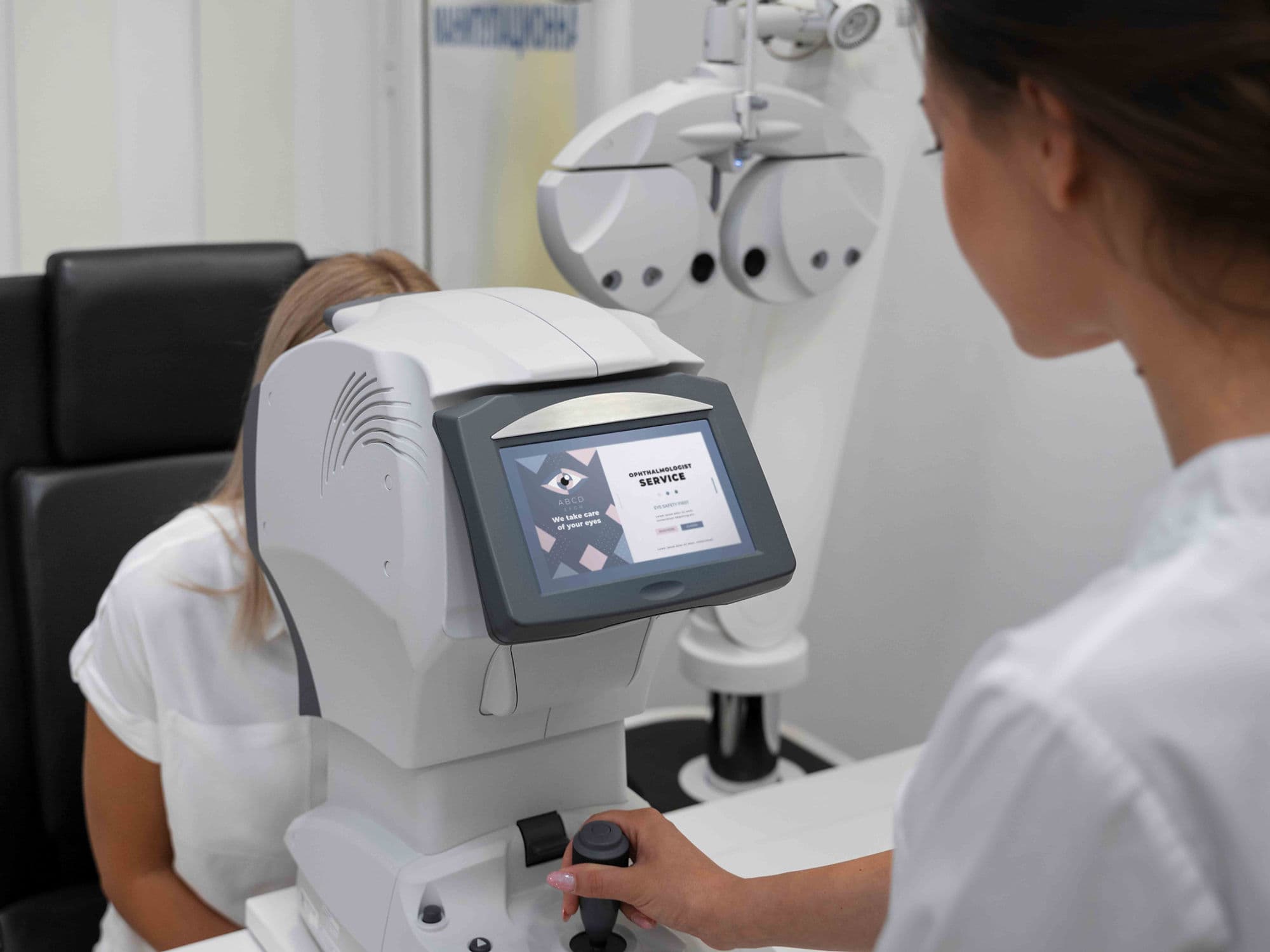
Tests Before Trabeculectomy
Visual Field Test: To document the current extent of peripheral vision loss and provide a baseline for post-operative monitoring.
Optical Coherence Tomography (OCT): To measure the thickness of the retinal nerve fiber layer around the optic nerve.
Gonioscopy: A specialized exam using a mirrored lens to view the drainage angle and plan the surgical entry point.
Pachymetry: Measuring corneal thickness, which can influence how eye pressure readings are interpreted.
Life After Trabeculectomy
Initial Vision: Vision is usually blurry for the first 2 to 4 weeks as the eye pressure stabilizes and the internal fluid levels adjust.
Frequent Check-ups: Close monitoring is required in the first few weeks to allow the surgeon to adjust stitches or manage the flow of fluid.
Medication Regimen: Patients must strictly use prescribed antibiotic and steroid eye drops for several weeks to prevent infection and control inflammation.
Activity Restrictions: Avoid heavy lifting, bending over, or strenuous exercise for about one month to prevent dangerous pressure spikes.
Long-Term Monitoring: Because there is a permanent "hole" in the eye, patients must watch for signs of Blebitis (infection of the bleb), such as sudden redness or pain.
Why Specialized Treatment Is Highly Effective
Significant Pressure Reduction: It is one of the most powerful tools available for achieving the very low pressures needed in advanced glaucoma.
Protects Remaining Sight: By reaching the target pressure, the procedure stops the ongoing "mechanical" damage to the optic nerve.
Outpatient Experience: Allows for major internal eye reconstruction with minimal downtime and a return to home on the same day.
Customizable Flow: The use of adjustable or "removable" sutures allows the surgeon to fine-tune the eye pressure in the office after the surgery.
Long-Lasting Results: When the bleb heals correctly and remains functional, it can provide years of stable pressure control without the need for additional drops.