Tracheostomy
Tracheostomy
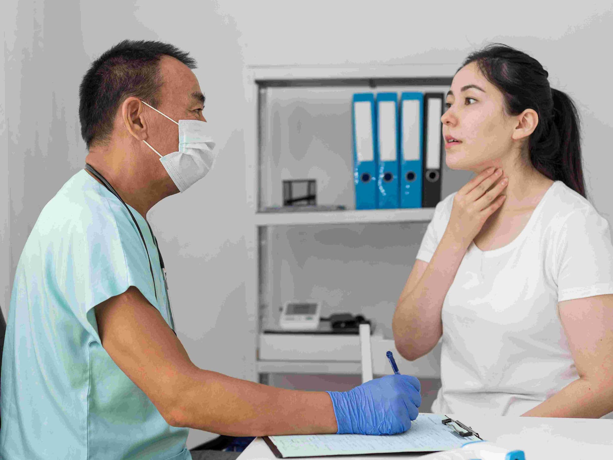
A tracheostomy is a specialized surgical procedure to create a functional opening (stoma) in the front of the neck directly into the trachea (windpipe). A tracheostomy tube is inserted into this opening to provide an alternative airway for breathing. This procedure is typically performed when the upper airway is obstructed or when a patient requires long-term mechanical ventilation to support lung function.
When You Should Consider a Tracheostomy
Acute Airway Obstruction: Due to tumors, severe facial trauma, vocal cord paralysis, or life-threatening swelling from allergic reactions.
Long-term Ventilation: For patients who cannot be "weaned" off a breathing machine (ventilator) through a standard tube in the mouth or nose.
Inability to Clear Secretions: To assist in suctioning thick mucus from the lungs in patients with weak cough reflexes, often seen in neuromuscular diseases.
Emergency Airway Access: When traditional intubation through the mouth is impossible due to physical blockages or extensive injury.
Bypassing Upper Airway Narrowing: To provide a stable airway for patients with chronic conditions like subglottic stenosis or laryngeal cancer.
How Is Performed
Anesthesia: The procedure is usually performed under general anesthesia in an operating room, though it can be done under local anesthesia at the bedside in emergency situations. It typically takes 20 to 45 minutes.
Incision: A precise horizontal or vertical cut is made in the lower neck, positioned between the thyroid cartilage and the sternum.
Access: The surgeon carefully moves aside the neck muscles and the thyroid gland to expose the underlying tracheal rings.
Tracheotomy: A small, controlled hole is created in the trachea.
Tube Insertion: A specialized tracheostomy tube is inserted into the opening. This tube often features an inflatable "cuff" that creates a seal to ensure air from a ventilator reaches the lungs.
Securing: The tube is held in place by a secure neck strap or temporary sutures to prevent accidental dislodgement.
Pre-Procedure Preparation
Neck Anatomy Assessment: Evaluating the physical structure of the neck to identify the best placement for the incision, especially in patients with a "short" neck or enlarged thyroid.
Coagulation Profile: A standard blood test to ensure the patient's blood clots correctly, minimizing the risk of internal bleeding.
Imaging (Optional): In complex cases, a CT scan or ultrasound of the neck may be used to map the blood vessels and thyroid position.
Consent and Communication: Discussing the temporary loss of voice and the specialized care required for the stoma after surgery.
Fasting: Adhering to strict "nothing by mouth" instructions if the procedure is a planned surgery under general anesthesia.
Tests Before Tracheostomy
Arterial Blood Gas (ABG): To measure oxygen and carbon dioxide levels in the blood, establishing a baseline for respiratory function.
Chest X-ray: To evaluate the current state of the lungs and the position of any existing breathing tubes.
Bronchoscopy: Using a thin camera to inspect the internal airway and confirm the level of obstruction.
Electrocardiogram (EKG): To ensure cardiac stability prior to the administration of anesthesia.
Life After Tracheostomy
Hospital Stay: Most patients remain in the hospital for several days to weeks to receive specialized nursing care and learn how to manage the stoma.
Communication: Initially, patients cannot speak because air exits through the tube instead of the vocal cords. As healing progresses, specialized speaking valves (such as a Passy-Muir valve) can be used.
Suctioning and Cleaning: The tube must be suctioned regularly to keep it clear of mucus. The "inner cannula" of the tube is also removed and cleaned daily.
Humidification: Because the nose (which naturally warms and moistens air) is bypassed, patients must breathe humidified air to prevent the windpipe from drying or becoming irritated.
Stoma Care: The skin around the tube must be kept clean and dry to prevent irritation and infection.
Emergency Awareness: Patients and caregivers are trained to recognize signs of a "mucus plug," which can block the tube and requires immediate clearing.
Why Specialized Treatment Is Highly Effective
Stable Long-term Airway: Provides a much more comfortable and secure breathing path than a tube through the mouth for patients requiring weeks or months of support.
Facilitates Oral Care: Unlike oral intubation, a tracheostomy allows for better mouth hygiene and, eventually, the possibility of eating by mouth.
Easier Weaning: A tracheostomy reduces the "work of breathing," making it easier for patients to gradually transition off a ventilator.
Improved Patient Comfort: Patients with a tracheostomy often require less sedation and are able to be more mobile and interactive during their recovery.
Direct Access for Suctioning: Allows for immediate removal of secretions from the lower airway, significantly reducing the risk of pneumonia in vulnerable patients.a