Medivisor India Treatment Logo
+91 83682 47758
info@medivisorhealth.com
  • Home
    • About Us
    • Our Services
    • Our Team
    • Our Safety Measures
    • Loading treatments...
    • Loading hospitals...
    • Loading doctors...
    • Treatment Cost
    • Treatment Process
    • Visa Process
    • Travel Guide
    • FAQs
    • Why Medivisor
    • Success Stories
    • Patient Activities
    • Social Activities
    • News Coverage
    • Blog
  • Become a Partner

Medivisor India Treatment is a trusted New Delhi–based medical travel company assisting international patients with advanced, affordable, and seamless treatment in India.

  • Medivisor House 359, Sector 1, Vaishali, Ghaziabad, (Delhi/NCR) India
  • +91 8368 247 758
  • info@medivisorhealth.com

About Us

  • About Us
  • Our Services
  • Our Team
  • Our Medical Advisors
  • Our Safety Measures
  • Join Our Network
  • Contact Us

India Treatment

  • Hospitals
  • Treatments
  • Treatment Cost
  • Treatment Process
  • Visa Process
  • Travel Guide
  • FAQs
  • Why Medivisor

Gallery

  • Patient Testimonials
  • Patient Activities
  • News Coverage
  • Blog

Get in Touch

© 2026 Medivisor India Treatment. All rights reserved.

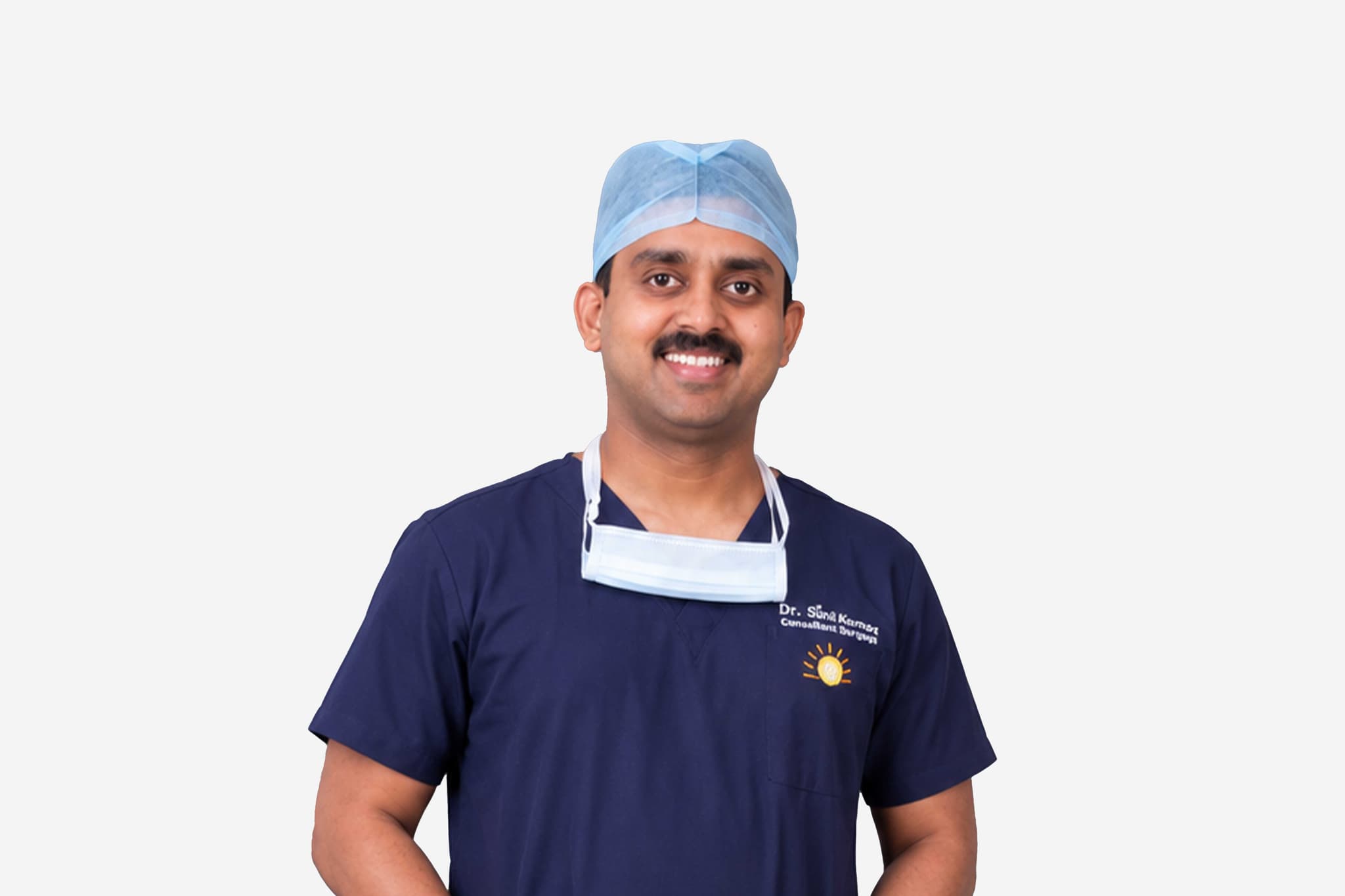
Dr Deepak Subramanian

Senior Consultant & Clinical Lead - General, Minimal Access (GI) & Bariatric Surgery

MGM Healthcare, Chennai

Bariatric SurgeonGeneral Surgeon

15+ years experience

Dr Deepak Subramanian, Senior Consultant & Clinical Lead - General, Minimal Access (GI) & Bariatric Surgery at MGM Healthcare, Chennai - MGM Healthcare, Chennai
  1. Home
  2. Doctors
  3. Dr Deepak Subramanian

About Dr Deepak Subramanian

Dr. Subramanian is a highly distinguished specialist in advanced gastrointestinal and abdominal wall surgery, recognized for his technical mastery in complex reconstructive procedures and minimally invasive interventions. He specializes in utilizing precision-guided laparoscopic and minimal access platforms to restore anatomical integrity and optimize patient outcomes.

Mastery in Advanced Laparoscopic Hernia Surgery

He specializes in the comprehensive management of various structural defects of the abdominal wall. His clinical practice focuses heavily on executing high-precision, minimally invasive repairs for inguinal, umbilical, and ventral hernias, employing modern tissue-reinforcement methodologies to ensure optimal repair strength and long-term durability.

Innovation in Complex Abdominal Wall Reconstruction

Dr. Subramanian possesses profound expertise in handling complex, recurrent, and large abdominal wall defects that require sophisticated reconstructive techniques. By utilizing advanced minimal access pathways, he performs intricate myofascial releases and component separations, minimizing tissue trauma and significantly reducing post-operative complications.

Precision Minimal Access Pathways and Accelerated Recovery

A primary focus of his surgical practice is the implementation of advanced laparoscopic techniques designed to minimize the surgical footprint. Patients benefit from sub-centimeter incisions, which dramatically reduce post-operative pain, lower the risk of surgical site infections, and facilitate accelerated recovery timelines and a faster return to independent mobility.

Clinical Governance and Evidence-Based Outcomes

Throughout his career, Dr. Subramanian has integrated modern surgical innovations with rigorous clinical protocols to ensure the highest standards of patient safety. His extensive procedural experience underpins his capability to deliver predictable, high-quality outcomes, establishing him as a trusted authority in advanced minimal access surgery.

Dr. Deepak Subramanian at a Glance

  • Specialist in Advanced Laparoscopic Surgery and Abdominal Wall Reconstruction.

  • Expert in minimally invasive repairs for inguinal, umbilical, and complex hernias.

  • Proficient in high-volume advanced laparoscopic interventions with a track record of safe outcomes.

  • Specialized in tissue-preserving techniques designed to accelerate post-operative recovery.

  • Focused on utilizing state-of-the-art surgical technology to minimize structural recurrence.

  • Dedicated to providing precision-guided, patient-centric clinical governance.

MBBS (Bachelor of Medicine and Bachelor of Surgery) 2002, Sri Ramachandra Medical College and Research Institute, Chennai, Tamil Nadu
MS (Master of Surgery) in General Surgery 2007, Sri Ramachandra Medical College and Research Institute, Chennai, Tamil Nadu
FIAGES (Fellowship in Minimal Access Surgery) 2008, Trichy, India
Diploma in Minimal Access Surgery 2008
Fellow in Minimal Access Surgery 2010, Madurai, India
MRCS (Royal College of Surgeons, Edinburgh 2010, Edinburgh, U.K
Fellow in Bariatric and Metabolic Surgery 2013, Mohak Institute of Bariatrics
MBA – International Healthcare Management 2015, Frankfurt School of Finance and Management
FICS- Fellow of International College of Surgeons 2015, International College of Surgeons
FALS- Fellow in Advanced Laparoscopic Surgery 2017, IAGES,India
FIBC- Fellow of International Bariatric Club 2017, International Bariatric Club
Board Certified in Bariatric Surgeon
“Best Laparoscopic & Bariatric Surgeon” – Awarded by Global Healthcare Excellence Award, 2018
Launched “ Happy Losers Club” – Weight loss Initiative Programme in October 2017
Authored the Chapter “Perioperative management of Medical Comorbidities after Bariatric Surgery” in the book – Bariatric Surgical Practice Guide
Observed multiple surgeries
maintained amicable rapports with eminent laparoscopic surgeons. Joel Leroy (France), Jacques Marescaux (France), Michel Gagner (Canada), Prof. C. Palanivelu (India)
Completed the Intercollegiate MRCS (Edinburgh) in 2010
Completed IELTS in Nov 2010 with an overall Band Score of
5
Life Member of IAGES (Indian Association of Gastrointestinal Endoscopic Surgeons)
Life member of IFSO
OSSI
Presented 2 international laparoscopic papers while representing India during a conference in laparoscopy conducted by the Society of Laparoendoscopic Surgeons (SLS) in Hawaii in Feb
Also a member of the organising committee.
Selected as an international delegate
Ambassador from India to represent SLS from 2010 onwards
Obtained the 2nd rank in the M.S. (General Surgery) examination conducted in April 2007

Affiliated Hospitals

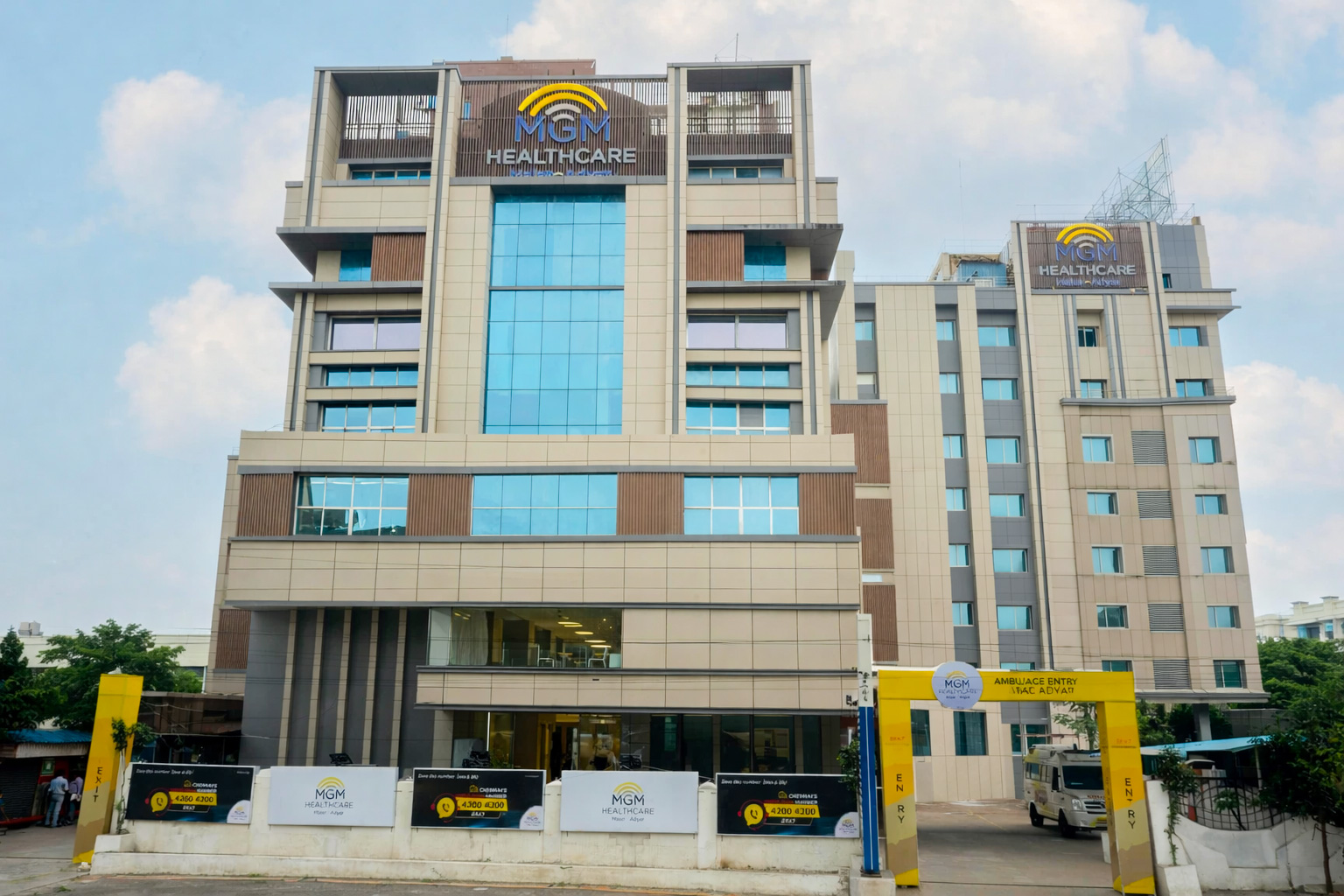
MGM Healthcare, Chennai

Multi Specialty

Chennai, Tamil Nadu

2019

Estd.

400+

Beds

250+

Doctors

May we help you?

Enter digits only (6-15 digits). We'll add your country code.

Related Treatments

Sleeve Gastrectomy
Sleeve Gastrectomy

Sleeve gastrectomy, often called the Gastric Sleeve, is a permanent weight-loss surgery that involves removing approximately 75–80% of the stomach. The remaining portion is joined together to form a narrow tube or "sleeve" about the size and shape of a banana. This procedure provides a powerful tool for long-term weight management by physically and hormonally changing how the body processes hunger and food intake.

How It Works

  • Physical Restriction: The significantly smaller stomach capacity limits the amount of food you can eat at one time, making you feel full much faster.

  • Hormonal Changes: By removing the upper portion of the stomach (the fundus), the surgery drastically reduces the production of Ghrelin, known as the "hunger hormone."

  • Appetite Suppression: The reduction in Ghrelin leads to a decreased appetite and significantly improved blood sugar control.

  • Natural Digestion: Unlike a gastric bypass, the intestines are not re-routed, meaning the natural digestion process remains largely intact.

When You Should Consider a Sleeve Gastrectomy

  • Body Mass Index (BMI): Typically recommended for individuals with a BMI of 40 or higher, or a BMI of 35 or higher with obesity-related health conditions.

  • Type 2 Diabetes: If you are struggling to manage blood sugar levels through diet and medication alone.

  • Hypertension: When high blood pressure persists despite medical intervention and lifestyle changes.

  • Obstructive Sleep Apnea: If excess weight is causing significant breathing interruptions during sleep.

  • Failed Weight Loss Efforts: For those who have been unable to achieve sustained weight loss through traditional diet and exercise programs.

How Is Performed

  • Anesthesia: The surgery is performed under general anesthesia and typically takes between 60 to 90 minutes.

  • Laparoscopic Approach: This is a minimally invasive "keyhole" surgery where the surgeon makes 4–5 small incisions in the abdomen to insert a camera and specialized instruments.

  • The Resection: The surgeon uses a surgical stapler to divide the stomach vertically, removing the outer curved portion of the organ.

  • Staple Line Reinforcement: The edges of the new stomach sleeve are securely stapled to ensure a watertight and airtight seal.

  • No Re-routing: The connection between the stomach and the small intestine is preserved, allowing for normal nutrient absorption.

Pre-Procedure Preparation

  • Nutritional Counseling: Working with a dietitian to understand the strict dietary phases required before and after surgery.

  • Psychological Evaluation: To ensure you have a strong support system and a clear understanding of the lifelong lifestyle changes required.

  • Medical Screening: A comprehensive review of heart and lung health to ensure you are a safe candidate for general anesthesia.

  • Pre-Op Liquid Diet: Most patients must follow a specific high-protein, low-calorie liquid diet for 1–2 weeks before surgery to shrink the liver.

  • Smoking Cessation: You must stop smoking at least 6 weeks before surgery to reduce the risk of staple line leaks and breathing complications.

Tests Before Sleeve Gastrectomy

  • Upper Endoscopy (EGD): To check the health of the stomach lining and rule out significant hiatal hernias or ulcers.

  • Abdominal Ultrasound: Specifically used to check for gallstones, which can be affected by rapid weight loss.

  • Blood Panels: Comprehensive testing of vitamin levels, blood sugar (HbA1c), and kidney function.

  • Sleep Study: To identify and treat undiagnosed sleep apnea prior to the administration of anesthesia.

Life After Sleeve Gastrectomy

  • Hospital Stay: Most patients remain in the hospital for 1 to 2 nights for monitoring and pain management.

  • Strict Dietary Phases: You must follow a multi-phase transition to allow the staple line to heal:
    Weeks 1–2: Clear liquids only.
    Weeks 3–4: Pureed and soft foods.
    Week 5+: Gradual introduction of solid, high-protein foods.

  • Lifelong Supplements: You must take daily vitamin and mineral supplements forever to prevent nutritional deficiencies.

  • Activity Restrictions: Most people return to work within 2 weeks but must avoid heavy lifting or core-straining exercise for a full 6 weeks.

  • Small Meals: You will transition to eating small, nutrient-dense meals throughout the day, as the stomach can no longer hold large volumes.

Why Specialized Treatment Is Highly Effective

  • Substantial Weight Loss: Most patients lose 60–70% of their excess body weight within the first 12–18 months.

  • Disease Resolution: The procedure can significantly improve or even resolve conditions like Type 2 diabetes, high blood pressure, and fatty liver disease.

  • Increased Longevity: By reducing the strain of obesity on the heart and joints, the surgery contributes to a longer, more active life.

  • Minimally Invasive: The laparoscopic technique results in less pain, smaller scars, and a much faster recovery than traditional open surgery.

  • Simpler Mechanism: Because the intestines are not re-routed, there is a lower risk of long-term malabsorption or "dumping syndrome" compared to other weight-loss surgeries.

Roux-en-Y Gastric Bypass

Roux-en-Y Gastric Bypass (RYGB) is considered the "gold standard" of weight-loss surgery. It is a complex, permanent procedure that involves creating a small stomach pouch and rerouting the small intestine to change how your body absorbs calories and nutrients.

How It Works

  • Restriction: The new stomach pouch is roughly the size of an egg (about 30ml), which drastically limits how much you can eat.

  • Malabsorption: By bypassing the first part of the small intestine, your body absorbs fewer calories and fats from the food you consume.

  • Hormonal Shifts: The surgery alters gut hormones (like GLP-1 and PYY) that suppress hunger, increase satiety, and rapidly improve blood sugar levels.

When You Should Consider a Gastric Bypass

  • Severe Obesity: Typically for individuals with a BMI of 40 or higher, or a BMI of 35 with serious weight-related health problems.

  • Type 2 Diabetes: When rapid and significant improvement in blood sugar control is a primary medical goal.

  • Chronic GERD: For patients with severe acid reflux that may be worsened by other types of weight-loss surgery.

  • Metabolic Syndrome: When a combination of high blood pressure, high blood sugar, and abnormal cholesterol levels requires a powerful intervention.

  • Failed Weight Loss: For those who have not achieved sufficient results through diet, exercise, or other non-surgical methods.

How Is Performed

  • Anesthesia: The surgery is performed under general anesthesia and typically takes 2 to 3 hours.

  • Laparoscopic Approach: Most procedures are performed using minimally invasive techniques, involving several small incisions in the abdomen for a camera and specialized tools.

  • Creating the Pouch: The surgeon staples the top section of the stomach to separate it from the rest, creating a small, functional pouch.

  • The "Roux" Limb: The small intestine is divided; the bottom end is lifted up and connected directly to the new small stomach pouch.

  • The Reconnection: The remaining part of the small intestine (still attached to the "old" stomach) is reconnected further down the "Roux" limb so that digestive juices can eventually mix with food.

Pre-Procedure Preparation

  • Nutritional Counseling: Extensive education on the strict dietary phases and lifelong eating habits required after surgery.

  • Psychological Clearance: To ensure you are emotionally prepared for the significant lifestyle changes and body image transitions.

  • Medical Screening: Comprehensive heart, lung, and gastrointestinal evaluations to minimize surgical risks.

  • Pre-Op Liquid Diet: A specialized diet for 1–2 weeks prior to surgery to reduce liver size and make the procedure safer.

  • Smoking Cessation: Patients must stop smoking at least 6 weeks before surgery to reduce the risk of ulcers and healing complications.

Tests Before Gastric Bypass

  • Upper Endoscopy (EGD): To examine the stomach lining and check for hiatal hernias or ulcers.

  • Blood Panels: Detailed testing for baseline vitamin and mineral levels, as well as kidney and liver function.

  • Sleep Study: To detect and treat obstructive sleep apnea, which is common in bariatric patients.

  • Cardiology Clearance: Including an EKG or stress test to ensure the heart can handle general anesthesia.

Life After Gastric Bypass

  • Hospital Stay: Usually involves 2 nights for observation and initial recovery.

  • Post-Op Diet Progression: A strict transition from clear liquids (weeks 1–2) to pureed foods (weeks 3–4) and finally soft solids.

  • Dumping Syndrome: A unique side effect where sugary or high-fat foods cause rapid heart rate, nausea, and sweating as food "dumps" too quickly into the small intestine.

  • Lifelong Supplements: Due to malabsorption, you must take daily multivitamins, calcium, B12, and iron for the rest of your life.

  • Activity Restrictions: Most patients return to work in 2 weeks but must avoid heavy lifting for 6 weeks to allow internal reconnections to heal.

Why Specialized Treatment Is Highly Effective

  • Significant Weight Loss: Patients typically lose 60% to 80% of their excess body weight within 12 to 18 months.

  • Diabetes Remission: Exceptionally effective at treating Type 2 diabetes, often leading to normal blood sugar levels within days of surgery.

  • Reflux Relief: Unlike some other procedures, a gastric bypass often cures chronic acid reflux (GERD).

  • Long-Term Health Benefits: Dramatically reduces the risk of stroke, heart disease, and several types of cancer.

  • Comprehensive Metabolic Control: Provides a multi-layered approach by combining physical restriction, malabsorption, and beneficial hormonal changes.

Mini Gastric Bypass
Mini Gastric Bypass

The Mini Gastric Bypass (MGB), also known as One-Anastomosis Gastric Bypass (OAGB), is a simplified, minimally invasive version of the traditional Roux-en-Y gastric bypass. It is considered both a restrictive and malabsorptive procedure, meaning it reduces the amount of food you can eat and limits the calories your body absorbs.

When You Should Consider a Mini Gastric Bypass

  • High BMI: Typically recommended for individuals with a Body Mass Index (BMI) of 40 or higher, or 35 with obesity-related health conditions.

  • Type 2 Diabetes: For patients seeking rapid and significant improvement in blood sugar control through metabolic surgery.

  • Hypertension: When high blood pressure persists despite medical intervention and significant weight gain.

  • Obstructive Sleep Apnea: If excess weight is causing breathing interruptions during sleep that impact daily energy and heart health.

  • Need for Shorter Surgery: For patients where a shorter operative time is preferred over the more complex traditional bypass.

How Is Performed

  • Anesthesia: The surgery is performed under general anesthesia and typically takes between 60 to 90 minutes.

  • Laparoscopic Approach: This is a "keyhole" surgery where the surgeon makes small incisions in the abdomen to insert a camera and specialized surgical instruments.

  • Creation of the Pouch: The surgeon staples the upper part of the stomach into a long, narrow tube (roughly the size of a banana) that holds about 30–50 ml of food.

  • Single Anastomosis (The Join): Unlike the two connections required in traditional bypass, the surgeon makes only one connection. A loop of the small intestine is brought up and attached directly to the new stomach pouch.

  • The Bypass: Approximately 150 to 200 cm of the small intestine is bypassed, redirecting food away from the area where most calories and fats are typically absorbed.

Pre-Procedure Preparation

  • Nutritional Counseling: Working with a specialist to understand the strict dietary phases and the importance of lifelong protein intake.

  • Psychological Evaluation: To ensure readiness for the significant lifestyle changes and dietary restrictions following surgery.

  • Medical Screening: A comprehensive review of cardiovascular and respiratory health to ensure safety during general anesthesia.

  • Pre-Op Liquid Diet: Most patients follow a high-protein, low-calorie liquid diet for 1–2 weeks before surgery to reduce liver size.

  • Smoking Cessation: You must stop smoking at least 6 weeks before surgery to minimize the risk of ulcers and complications at the surgical join.

Tests Before Mini Gastric Bypass

  • Upper Endoscopy (EGD): To examine the stomach lining and check for hiatal hernias, ulcers, or evidence of significant acid reflux.

  • Abdominal Ultrasound: Primarily used to check for gallstones, which can be affected by rapid weight loss.

  • Blood Panels: Comprehensive testing for baseline vitamin levels (B12, Iron, Vitamin D) and kidney/liver function.

  • Sleep Study: To identify and treat undiagnosed sleep apnea before the administration of anesthesia.

  • ECG: A standard heart check to ensure cardiac stability for the duration of the procedure.

Life After Mini Gastric Bypass

  • Hospital Stay: Most patients stay in the hospital for 1 to 2 nights for observation and pain management.

  • Staged Diet Progression: To allow the new joins to heal, patients follow a strict multi-phase plan:
    Days 1–3: Clear liquids only (water, broth).
    Week 2: Full liquids (protein shakes, thin soups).
    Weeks 3–4: Pureed foods (mashed vegetables, soft eggs).
    Week 5+: Gradual introduction of soft solids, moving toward regular healthy food.

  • Lifelong Supplements: Due to malabsorption, you must take daily multivitamins, calcium, B12, and iron for the rest of your life.

  • Activity Restrictions: Most patients return to work within 2 to 3 weeks but must avoid heavy lifting for 6 weeks to protect the internal staples.

Why Specialized Treatment Is Highly Effective

  • Significant Weight Loss: Patients often lose 60% to 75% of their excess body weight within the first year of surgery.

  • Metabolic Improvement: Highly effective at resolving or improving Type 2 diabetes, high blood pressure, and sleep apnea.

  • Technically Simpler: Having only one surgical join reduces operative time and lowers the risk of certain complications like internal hernias.

  • Potentially Reversible: Because no part of the stomach is removed from the body, the procedure is potentially reversible if ever medically necessary.

  • Durable Results: Combines the benefits of a smaller stomach with reduced calorie absorption for long-term weight maintenance and health.

Revisional Bariatric Surgery

Revisional bariatric surgery is a specialized procedure performed on patients who have already undergone a primary weight-loss surgery but require a second operation. This may be due to medical complications, insufficient weight loss, or significant weight regain. Because of existing scar tissue and altered anatomy, these procedures are more complex and carry higher risks than initial weight-loss surgeries.

When You Should Consider Revisional Surgery

  • Inadequate Weight Loss or Regain: If the initial stomach pouch has stretched over time or the body has adapted to the metabolic changes of the first surgery.

  • Severe Acid Reflux (GERD): Persistent or worsening reflux that does not respond to medication, frequently occurring after a gastric sleeve procedure.

  • Mechanical Complications: Issues such as a "slipped" gastric band, a staple line leak, or a narrowing (stricture) of a previous surgical connection.

  • Chronic Ulcers or "Dumping Syndrome": Persistent digestive issues following a bypass that cannot be managed through dietary changes alone.

  • Nutritional Malabsorption: Severe deficiencies in vitamins or protein that remain uncontrolled despite high-dose supplementation.

Common Revisional Procedures

  • Sleeve to Bypass Conversion: Converting a gastric sleeve into a Roux-en-Y gastric bypass. This is highly effective for treating severe reflux and triggering further weight loss.

  • Gastric Band Conversion: Removing a "Lap-Band" and converting the anatomy into either a sleeve or a bypass, often performed in a single or two-stage process.

  • Bypass Revision (Stoma Tightening): For a stretched gastric bypass, a surgeon may "re-tighten" the connection (stoma) to restore the feeling of fullness.

  • Intestinal Lengthening: Adjusting the "Roux" limb to increase the bypassed section of the intestine, thereby increasing calorie malabsorption.

  • Sleeve Re-Sleeving: Trimming a previously stretched gastric sleeve to restore its original narrow, restrictive shape.

How Is Performed

  • Anesthesia: The surgery is performed under general anesthesia and typically takes between 2 to 4 hours due to the precision required.

  • Surgical Approach: Almost always performed laparoscopically or robotically to navigate through previous scar tissue with high-definition visualization and accuracy.

  • Managing Adhesions: The surgeon must carefully separate internal scarring (adhesions) from previous operations before the new revision can begin.

  • Stapling and Joins: New surgical joins are created using specialized stapling instruments designed for thicker or scarred tissue.

  • Leak Testing: Extensive intra-operative testing is performed to ensure all new connections are watertight and airtight before finishing.

Pre-Procedure Preparation

  • Anatomy Mapping: Extensive imaging is required to visualize the current state of the stomach and intestines before entering the operating room.

  • Nutritional Optimization: Correcting any existing vitamin deficiencies through intensive supplementation prior to surgery.

  • Psychological Assessment: A thorough review to ensure the patient is prepared for the "reset" in dietary habits and lifestyle expectations.

  • Smoking Cessation: Patients must stop smoking at least 6 to 8 weeks before surgery, as nicotine significantly increases the risk of staple line leaks in scarred tissue.

  • Weight Management: Following a specific pre-operative diet to reduce liver size and improve surgical access.

Tests Before Revisional Surgery

  • Upper Endoscopy (EGD): Inserting a camera into the stomach to inspect the pouch and check for ulcers, inflammation, or stretching.

  • Barium Swallow (UGI Series): A specialized X-ray where the patient swallows contrast liquid to show the exact size and flow of the current anatomy.

  • Abdominal CT Scan: To provide a 3D view of the internal organs and identify any hidden hernias or mechanical issues.

  • Comprehensive Blood Panels: Detailed analysis of iron, B12, Vitamin D, and protein levels to establish a baseline.

Life After Revisional Surgery

  • Hospital Stay: Usually involves 2 to 3 nights of close monitoring to ensure the new surgical joins are healing correctly.

  • Dietary Reset: Patients must return to a "Phase 1" clear liquid diet and progress slowly through pureed and soft foods over a 4–6 week period.

  • Slower Weight Loss: It is important to note that weight loss after a revision is typically slower and less dramatic than after the first surgery.

  • Lifelong Monitoring: Regular follow-up appointments and blood tests are essential to monitor for malabsorption and nutritional health.

  • Activity Restrictions: Light walking is encouraged immediately, but heavy lifting and core-straining exercises are restricted for 6 weeks.

Why Specialized Treatment Is Highly Effective

  • Resolves Chronic Pain and Reflux: Provides immediate relief for patients suffering from the mechanical failures or side effects of a primary surgery.

  • Second Chance for Health: Allows patients who did not reach their goals to restart their weight-loss journey with a more powerful metabolic tool.

  • Precision Technology: The use of robotic platforms allows surgeons to operate safely in "hostile" abdomens filled with previous scar tissue.

  • Customized Anatomy: Every revision is tailored specifically to the patient’s existing anatomy, ensuring the most effective solution for their unique situation.

  • Prevents Long-Term Damage: Addressing issues like chronic ulcers or severe reflux prevents permanent damage to the esophagus and digestive tract.

Appendectomy (Open/Laparoscopic)

An Appendectomy is the surgical removal of the appendix, a small, finger-shaped pouch attached to the large intestine. It is the standard treatment for appendicitis, an inflammation of the organ that can lead to a life-threatening rupture if left untreated.

When You Should Consider Appendectomy

  • Pain that starts near the belly button and moves to the lower right abdomen.

  • "Rebound tenderness" or sharp pain in the lower right abdomen when pressure is released.

  • Fever and chills accompanying abdominal discomfort.

  • Nausea, vomiting, or loss of appetite.

  • High white blood cell counts detected during blood testing.

Methods of Appendectomy

  • Laparoscopic Appendectomy: The modern minimally invasive standard using three small incisions and a camera (laparoscope).

  • Open Appendectomy: A traditional approach using a single 2- to 4-inch incision, typically reserved for ruptured cases.

  • Emergency Appendectomy: Immediate surgical intervention performed to prevent or treat an appendix rupture.

  • Interval Appendectomy: A delayed surgery performed after a severe infection has been initially managed with antibiotics.

How Appendectomy Is Performed

  • Insufflation: In laparoscopic cases, carbon dioxide gas is used to inflate the abdomen for better visibility.

  • Appendix Identification: The surgeon locates the appendix and carefully ties off its base to separate it from the large intestine.

  • Irrigation: If the appendix has ruptured, the abdominal cavity is washed with saline to remove bacteria and pus.

  • Drainage: A small tube may be placed in the incision site for a few days to allow excess fluid to escape in complex cases.

  • Closure: Incisions are secured using absorbable sutures, surgical glue, or sterile strips.

Pre-Procedure Preparation

  • Maintaining NPO (nothing by mouth) status for several hours prior to the operation.

  • Starting Intravenous (IV) fluids to ensure proper hydration.

  • Administration of prophylactic antibiotics to reduce the risk of surgical site infection.

  • Undergoing general anesthesia to remain completely unconscious throughout the procedure.

Tests Before Appendectomy

  • Physical Examination: To check for localized pain and abdominal wall tension.

  • Ultrasound or CT Scan: Imaging used to confirm the inflammation or see if the appendix has burst.

  • Complete Blood Count (CBC): To identify signs of infection through elevated white blood cell levels.

  • Urine Test: Often performed to rule out a urinary tract infection or kidney stones which can mimic appendicitis symptoms.

Life After Appendectomy

  • For uncomplicated cases, hospital discharge usually occurs within 24 hours.

  • Recovery for laparoscopic surgery includes avoiding heavy lifting (over 5kg) for 1–2 weeks.

  • Recovery for open surgery requires avoiding strenuous exercise for 4–6 weeks to prevent hernia.

  • Diet begins with clear liquids and progresses as bowel function returns to normal.

  • Patients should monitor for signs of abscess, such as new fevers or persistent bloating.

Benefits of Appendectomy

  • Provides a permanent cure for appendicitis by removing the source of inflammation.

  • Prevents life-threatening complications associated with an appendix rupture or peritonitis.

  • Minimal scarring and faster recovery times when performed using laparoscopic techniques.

  • Rapidly alleviates severe abdominal pain and restores normal digestive health.

Cholecystectomy (Open/Laparoscopic)

A Cholecystectomy is the surgical removal of the gallbladder, a small organ located under the liver that stores bile. It is primarily performed to treat gallstones (cholelithiasis), gallbladder inflammation (cholecystitis), or biliary colic, which is intense pain caused by stones blocking the bile ducts.

When You Should Consider Cholecystectomy

  • Sudden and rapidly intensifying pain in the upper right portion of the abdomen.

  • Nausea or vomiting following high-fat meals.

  • Yellowing of the skin and the whites of the eyes (jaundice).

  • Fever or chills, which may indicate a gallbladder infection.

  • Confirmation of gallstones or a non-functional gallbladder through diagnostic testing.

Methods of Cholecystectomy

  • Laparoscopic Cholecystectomy: The modern "gold standard" minimally invasive approach used in over 90% of cases, involving four small incisions.

  • Open Cholecystectomy: A traditional surgical method using a larger incision, typically reserved for complex cases or severe scarring.

  • Emergency Cholecystectomy: Immediate removal performed when acute inflammation or infection poses a significant health risk.

  • Single-Incision Laparoscopic Surgery (SILS): A specialized technique where the gallbladder is removed through one small incision in the umbilicus.

How Cholecystectomy Is Performed

  • Pneumoperitoneum: During laparoscopic surgery, the abdomen is inflated with carbon dioxide gas to provide a clear workspace for the surgeon.

  • Duct and Artery Clipping: The surgeon identifies the cystic duct and cystic artery and secures them with permanent clips before separation.

  • Gallbladder Detachment: The organ is carefully separated from the liver bed using specialized surgical instruments.

  • Organ Extraction: The gallbladder is placed in a sterile bag and removed through one of the small abdominal incisions.

  • Drainage: In open or complex cases, a Jackson-Pratt (JP) drain may be placed temporarily to remove excess fluid or bile.

Pre-Procedure Preparation

  • Fasting (NPO) for at least 6 to 8 hours prior to the operation.

  • Discontinuing blood-thinning medications several days before surgery as directed by the surgical team.

  • Receiving general anesthesia and the placement of a breathing tube for the duration of the procedure.

  • Undergoing a pre-operative evaluation to check liver enzymes and white blood cell counts.

Tests Before Cholecystectomy

  • Abdominal Ultrasound: The "gold standard" imaging test used to identify gallstones and gallbladder wall thickening.

  • HIDA Scan: A specialized nuclear medicine test used to evaluate the functional emptying of the gallbladder.

  • Blood Panels: To check for elevated liver enzymes or signs of infection (leukocytosis).

  • MRCP or ERCP: Advanced imaging or endoscopic procedures used to check for stones that may have moved into the common bile duct.

Life After Cholecystectomy

  • Most laparoscopic patients return home the same day or after one overnight stay.

  • Recovery for open surgery involves a hospital stay of 3 to 5 days and restricted activity for 4 to 6 weeks.

  • A low-fat diet is recommended for the first few weeks as the body adjusts to bile flowing directly into the small intestine.

  • Patients may experience temporary shoulder pain or bloating due to the gas used during laparoscopic procedures.

  • Monitoring for Post-Cholecystectomy Syndrome (PCS), which affects 10–15% of patients and involves lingering indigestion or nausea.

Benefits of Cholecystectomy

  • Provides permanent relief from the recurring pain of biliary colic and gallstones.

  • Prevents serious complications such as gallbladder rupture, gangrene, or severe pancreatitis.

  • Minimally invasive techniques result in smaller scars, less post-operative pain, and a faster return to daily activities.

  • Eliminates the risk of future gallbladder infections and inflammation.

Hernia Repair (Inguinal/Umbilical)

Hernia repair is a surgical procedure to correct protrusions of tissue or organs through weakened areas of the abdominal wall. An inguinal hernia occurs in the groin area (the inguinal canal), while an umbilical hernia occurs at or near the navel (belly button). By reinforcing these weak points, surgery prevents the risk of "incarceration" or "strangulation," where the blood supply to the trapped tissue is cut off, making it an essential intervention for both comfort and long-term health.

When You Should Consider Hernia Repair

  • Persistent pain or a "dragging" sensation in the groin or abdomen, especially when lifting or coughing.

  • A visible bulge that may disappear when lying down but protrudes during physical activity.

  • Umbilical protrusions that become larger, more painful, or aesthetically concerning.

  • Inguinal hernias that cause discomfort during daily movements or exercise.

  • As a preventive measure to avoid the emergency complications of a trapped (strangulated) intestine.

  • When a hernia interferes with professional duties that require physical labor or heavy lifting.

Conditions That Require Specialized Care

  • Incarcerated Hernia: Tissue that is trapped in the abdominal wall and cannot be pushed back in.

  • Strangulated Hernia: A medical emergency where the blood supply to the trapped tissue is restricted.

  • Recurrent Hernia: A hernia that has returned after a previous surgical repair.

  • Bilateral Inguinal Hernias: When hernias are present on both the left and right sides of the groin simultaneously.

  • Large Incisional Hernias: Weakness occurring at the site of a previous surgical scar.

How Hernia Repair Is Performed

  • The procedure begins with Reduction, where the protruding fat or intestine is gently pushed back into the abdominal cavity.

  • Open Surgery involves a single incision made directly over the bulge to access the muscle defect.

  • Laparoscopic Surgery utilizes three to four "keyhole" incisions and a camera to repair the wall from the inside.

  • Robotic-Assisted Surgery provides the surgeon with high-definition 3D visualization and robotic precision for complex cases.

  • In most adult cases, a Synthetic Mesh is placed over the weak spot to act as a permanent reinforcement.

  • The muscle layers are sutured, and the skin is closed with dissolvable stitches, surgical tape, or skin glue.

Innovations in Hernia Surgery

  • Bio-Absorbable MeshesAdvanced mesh materials that provide a scaffold for the body’s own tissue to grow into before eventually dissolving, reducing long-term foreign body sensation.

  • Self-Gripping (ProGrip) MeshSpecialized mesh with micro-hooks that eliminate the need for traditional tacks or sutures, significantly reducing post-operative nerve pain.

  • 3D-Mapped Mesh ContouringThe use of meshes designed to anatomically fit the specific curvature of the groin or abdominal wall for a more natural feel.

  • Single-Port Robotic RepairThe ability to perform complex hernia reconstructions through just one tiny incision, further minimizing scarring.

  • Advanced Nerve MappingIntraoperative tools that help surgeons identify and avoid sensory nerves, lowering the risk of chronic post-surgical pain.

  • Glue-Based FixationUsing medical-grade adhesives instead of mechanical tacks to secure the mesh, leading to a smoother recovery.

Pre-Procedure Preparation

  • Physical examination and medical history to determine the most effective surgical approach (Open vs. Laparoscopic).

  • Guidance on smoking cessation, as nicotine can significantly weaken tissue healing and increase recurrence risk.

  • Review of medications, particularly anticoagulants (blood thinners) that may need to be paused.

  • Fasting (NPO) instructions starting the night before surgery to ensure a safe anesthetic experience.

  • Coordination for a "day-case" discharge, as most patients return home within hours of the procedure.

Pre-Procedure Tests

  • Ultrasound or CT Scan: To determine the exact size of the hernia and identify if any vital organs are involved.

  • Routine Blood Work (CBC/Electrolytes): To ensure general health and proper clotting function.

  • Electrocardiogram (ECG): For patients over 40 or those with cardiac history to ensure fitness for anesthesia.

  • Cough Test: A physical assessment to judge the stability of the hernia under abdominal pressure.

  • Urinalysis: To rule out any underlying infections prior to the placement of surgical mesh.

Why This Treatment Is Highly Effective

  • Prevents Emergencies: Repairing a hernia electively removes the life-threatening risk of bowel strangulation.

  • Minimal Recurrence: The use of modern mesh technology has reduced the risk of a hernia returning to less than 5%.

  • Rapid Return to Activity: Laparoscopic and robotic techniques allow most patients to return to desk work within a week.

  • Restored Core Strength: Reinforcing the abdominal wall improves overall stability and comfort during physical movement.

  • Cosmetic Improvement: Corrects the visible bulge, restoring the natural contour of the navel or groin.

Recovery and Monitoring

  • Patients are encouraged to walk within a few hours of surgery to promote blood flow and lung function.

  • Pain management typically transitions from clinical care to over-the-counter medications within 48 hours.

  • Heavy lifting (typically over 10–20 lbs) is restricted for 4 to 6 weeks to allow the mesh to integrate fully.

  • Most patients can resume driving once they can perform an emergency stop without abdominal discomfort.

  • Follow-up visits are scheduled to monitor the incision sites and ensure there is no fluid buildup (seroma).

Life After Hernia Repair

  • Freedom from the chronic aching or "heaviness" associated with the hernia.

  • Ability to return to high-impact sports and strenuous gym activities once full clearance is granted.

  • Permanent structural support of the abdominal wall through the integrated mesh.

  • Significant reduction in the psychological stress associated with a visible or painful bulge.

  • Long-term protection against the risk of intestinal complications.

Mesh Hernioplasty
Mesh Hernioplasty

Mesh Hernioplasty is the surgical repair of a hernia using a synthetic or biological flat sheet (mesh) to reinforce a weakened abdominal wall. Unlike a traditional herniorrhaphy, which simply stitches the muscle together, a hernioplasty is a tension-free repair. This approach is considered the global gold standard for significantly reducing the likelihood of a hernia returning.

When You Should Consider Mesh Hernioplasty

  • Presence of a visible bulge in the abdomen or groin that increases with physical strain.

  • Recurrent hernias where previous non-mesh (stitch-only) repairs have failed.

  • Large abdominal wall defects that cannot be closed securely with stitches alone.

  • Pain, pressure, or a pulling sensation at the site of a previous surgical scar or natural weakness.

  • Confirmation of a hernia defect through a physical exam or diagnostic imaging like a CT scan.

Methods of Mesh Hernioplasty

  • Open Hernioplasty: A traditional approach where a single incision is made directly over the hernia to place and secure the mesh.

  • Laparoscopic Hernioplasty: A minimally invasive "keyhole" technique using a camera and small ports to place mesh from the inside.

  • Robotic Hernioplasty: Utilizing robotic assistance for high-precision mesh placement and complex abdominal wall reconstruction.

  • Tension-Free Repair: A specialized technique where the mesh bridges the gap in the muscle without pulling the edges together, minimizing post-op pain.

How Mesh Hernioplasty Is Performed

  • Tissue Reduction: The bulging fat or intestine is gently returned to its proper position inside the abdominal cavity.

  • Mesh Customization: A mesh is selected and often trimmed to overlap the edges of the muscle defect by at least 3–5 cm.

  • Layered Placement: Depending on the technique, mesh is placed on top of the muscle (onlay), between muscle layers (sublay), or under the muscle (underlay).

  • Fixation: The mesh is secured using permanent sutures, specialized tacks, staples, or medical-grade surgical glue.

  • Integration: The surgeon ensures the mesh is flat and secure to allow natural fibrous tissue to grow into the mesh pores over time.

Pre-Procedure Preparation

  • Fasting (NPO) for 6–8 hours prior to the scheduled surgery.

  • Pausing blood-thinning medications to prevent the formation of hematomas around the mesh site.

  • Pre-operative evaluation to determine the most appropriate type of anesthesia (General, Spinal, or Local).

  • Measurement of the hernia defect via ultrasound or CT scan to ensure the correct mesh size is available.

Tests Before Mesh Hernioplasty

  • Physical Evaluation: To check the size of the bulge and determine if the tissue is "incarcerated" or trapped.

  • CT Scan or Ultrasound: Imaging used to measure the exact dimensions of the hole in the abdominal wall.

  • Blood Panels: Routine screens to ensure the patient is fit for the selected type of anesthesia.

  • Electrocardiogram (ECG): Often performed for older patients to assess heart health before the procedure.

Life After Mesh Hernioplasty

  • Most patients return home the same day, as the procedure is typically performed on a day-care basis.

  • Strict activity restrictions are required, including no heavy lifting (>5 kg) for at least 6 weeks.

  • Use of an abdominal binder or compression garment for approximately 4 weeks to support the repair.

  • Patients may feel "twinges" or minor soreness for a few months as the body integrates the mesh.

  • Return to light activities is encouraged early to promote circulation and healing.

Benefits of Mesh Hernioplasty

  • Significantly lowers the recurrence rate from up to 30% (with stitches alone) to less than 5%.

  • Provides a permanent, lifelong "scaffold" that reinforces the weakened abdominal wall.

  • The tension-free nature of the repair results in less post-operative pain compared to traditional methods.

  • High technical success rates and durability, as the mesh is designed to stay in the body indefinitely.

Incisional Hernia Repair

An Incisional Hernia Repair is a surgical procedure to fix a protrusion of tissue or an organ (usually the intestine) through a previously made surgical scar. This occurs when the abdominal wall muscles fail to heal completely or weaken over time following an earlier surgery.

When You Should Consider Incisional Hernia Repair

  • A visible bulge or lump at the site of a previous surgical scar.

  • A bulge that becomes more prominent or painful when coughing, straining, or lifting.

  • Persistent aching or pressure at the previous incision site.

  • Nausea or vomiting if the protruding tissue becomes trapped (incarcerated).

  • Confirmation of a muscle "defect" via CT scan or physical examination.

Methods of Incisional Hernia Repair

  • Open Repair: A traditional approach involving an incision over the old scar, often used for very large hernias or complex cases.

  • Laparoscopic Repair: A minimally invasive method using small "keyhole" incisions, CO2 gas, and a camera to repair the defect from the inside.

  • Robotic-Assisted Repair: A modern variation of laparoscopic surgery offering enhanced precision for medium to large hernias.

  • Tension-Free Mesh Repair: The use of synthetic or biologic mesh to reinforce the abdominal wall, significantly reducing the risk of recurrence.

How Incisional Hernia Repair Is Performed

  • Sac Reduction: The surgeon identifies the hernia sac and carefully pushes the protruding tissue or intestine back into the abdominal cavity.

  • Adhesiolysis: Freeing the intestine from old scar tissue or adhesions that may have formed since the previous surgery.

  • Mesh Placement: A flexible, permanent screen (mesh) is secured over or under the muscle layer to act as a scaffold for new tissue growth.

  • Fixation: The mesh is secured using sutures, tacks, or specialized surgical staples to ensure it remains in a "tension-free" position.

  • Abdominal Wall Reconstruction: In complex cases, the surgeon may rearrange muscle layers to achieve a more natural and strong closure.

Pre-Procedure Preparation

  • Quitting smoking for several weeks to improve blood flow and tissue healing.

  • Weight management and stabilization of blood sugar levels to minimize the risk of repair failure.

  • Fasting (NPO) as directed before undergoing general anesthesia.

  • Evaluation of the old surgical site to plan the best approach (open vs. minimally invasive).

Tests Before Incisional Hernia Repair

  • Physical Exam: To feel for the bulge and assess the strength of the surrounding abdominal wall.

  • CT Scan: The primary imaging tool used to measure the exact size of the muscle defect and identify the contents of the hernia.

  • Blood Panels: To check for signs of infection or uncontrolled diabetes that could affect recovery.

  • Pre-Surgical Optimization Review: Assessment of lifestyle factors (BMI, smoking status) that impact surgical success.

Life After Incisional Hernia Repair

  • Hospital stays range from same-day discharge for laparoscopic cases to 3–5 days for large open repairs.

  • Use of an abdominal binder (elastic wrap) for 4–6 weeks to support the new repair.

  • Strict activity restrictions, including no heavy lifting (>5 kg) for 6–8 weeks.

  • Immediate encouragement of gentle walking to prevent blood clots during the early recovery phase.

  • Monitoring for seromas (fluid collection) or signs of mesh infection at the incision site.

Benefits of Incisional Hernia Repair

  • Significantly reduces the risk of hernia recurrence from 30–50% (without mesh) to below 10% (with mesh).

  • Restores the structural integrity and strength of the abdominal wall.

  • Eliminates the risk of life-threatening complications such as bowel obstruction or strangulation.

  • Relieves chronic pain and discomfort associated with the protruding tissue.

Ventral Hernia Repair

A Ventral Hernia Repair is a surgical procedure to fix a hole or weakness in the abdominal wall muscles through which fat or organs (such as the intestine) bulge. A "ventral" hernia specifically refers to any hernia occurring on the front (anterior) of the abdomen, excluding the groin area.

When You Should Consider Ventral Hernia Repair

  • A visible bulge on the abdomen that may disappear when lying down or appear when straining.

  • Pain or discomfort at the hernia site, especially when lifting or coughing.

  • Nausea or vomiting, which may indicate that the bowel is trapped (incarcerated).

  • A bulge that becomes hard, red, or purple, signifying a potential medical emergency.

  • Significant epigastric or umbilical discomfort that interferes with daily physical activities.

Methods of Ventral Hernia Repair

  • Open Repair: A traditional method where an incision is made directly over the hernia to push the bulge back and close the muscle.

  • Laparoscopic Repair: A minimally invasive approach using "keyhole" incisions and CO2 gas to repair the defect from inside the abdominal cavity.

  • Robotic-Assisted Repair: Utilizing advanced technology to perform complex muscle rearrangements (like TAR) through small incisions.

  • Suture (Primary) Repair: Simply stitching the muscle together, typically reserved only for very small holes (less than 1–2 cm).

  • Mesh Reinforcement: Using a synthetic or biologic screen to provide a "tension-free" bridge across the weakened muscle wall.

How Ventral Hernia Repair Is Performed

  • Abdominal Contents Reduction: The surgeon carefully returns the protruding fat or organs to their proper place inside the abdomen.

  • Pneumoperitoneum: In laparoscopic or robotic cases, the abdomen is inflated with gas to provide a clear view and workspace.

  • Mesh Placement: A permanent synthetic mesh is secured on top of, between, or under the muscle layers to act as a scaffold.

  • TAR (Transversus Abdominis Release): A complex robotic or open technique used to close very large gaps by rearranging the abdominal muscle layers.

  • Closure: The incisions are closed with sutures or surgical glue, and the reinforced wall begins the process of growing into the mesh.

Pre-Procedure Preparation

  • Optimization of health factors, including quitting smoking and stabilizing blood sugar for diabetic patients.

  • Weight management, as a BMI over 35 can significantly increase the risk of the hernia returning.

  • Fasting (NPO) for a specified period before the administration of general anesthesia.

  • Discussion with the surgeon regarding the specific type of ventral hernia (epigastric, umbilical, or incisional).

Tests Before Ventral Hernia Repair

  • Physical Examination: To classify the hernia and check for "reducible" versus "incarcerated" tissue.

  • CT Scan: The primary diagnostic tool used to measure the exact size of the muscle defect and assess the involvement of bowel loops.

  • Blood Panels: To ensure the patient is fit for general anesthesia and to check for signs of inflammation.

  • Medical Clearance: Evaluation of heart and lung health, particularly for older patients or those undergoing complex robotic repairs.

Life After Ventral Hernia Repair

  • Small or simple repairs are often same-day procedures, while complex repairs may require a 3–5 day hospital stay.

  • Patients must wear an abdominal binder (compression belt) for 4–6 weeks to support the repair and reduce swelling.

  • Strict activity restrictions: no heavy lifting (>5 kg) for at least 6–8 weeks.

  • Avoidance of core-heavy exercises like planks or crunches until cleared by the surgical team.

  • Close monitoring for seromas (fluid pockets) or chronic pain at the mesh fixation site.

Benefits of Ventral Hernia Repair

  • Reduces the recurrence rate from approximately 30–40% (with stitches alone) down to 5–10% with mesh.

  • Prevents life-threatening emergencies such as bowel strangulation where blood supply is cut off.

  • Restores the strength and appearance of the abdominal wall.

  • Effectively relieves chronic pain and discomfort associated with the protrusion of abdominal contents.

Laparoscopic Hernia Repair

Laparoscopic Hernia Repair, also known as minimally invasive or keyhole surgery, uses specialized tools and a camera to repair a weakness in the abdominal wall from the inside out. It is most commonly used for inguinal (groin), umbilical (navel), and ventral hernias.

When You Should Consider Laparoscopic Hernia Repair

  • A visible bulge in the groin or abdomen that may become more prominent when standing or coughing.

  • Pain or pressure at the hernia site during physical activity or lifting.

  • Inguinal hernias that occur on both sides (bilateral) of the groin.

  • Recurrent hernias where a previous open surgical repair has failed.

  • Desire for a faster return to work and daily activities with minimal scarring.

Methods of Laparoscopic Hernia Repair

  • TAPP (Transabdominal Preperitoneal): The surgeon enters the peritoneal cavity where the organs are located to reach and repair the hernia.

  • TEP (Totally Extraperitoneal): The surgeon operates in the space between the muscle and the abdominal lining, avoiding the organ cavity entirely.

  • IPOM (Intraperitoneal Onlay Mesh): Primarily used for ventral hernias, where a specialized mesh is placed on the inside of the abdominal wall.

  • Robotic-Assisted Laparoscopy: Using a robotic interface for enhanced precision and 3D visualization during the repair.

How Laparoscopic Hernia Repair Is Performed

  • Insufflation: The abdominal cavity is inflated with Carbon Dioxide (CO2) gas to create a dome-shaped workspace.

  • Port Insertion: Three to four small incisions (0.5–1 cm) are made to allow the insertion of a laparoscope and long, thin instruments.

  • Reduction: Protruding tissue, such as fat or a loop of intestine, is carefully pulled back into the abdominal cavity from the inside.

  • Mesh Reinforcement: A lightweight, flexible synthetic mesh is unfurled over the defect to reinforce the weakened wall.

  • Fixation: The mesh is secured in place using surgical tacks, staples, or specialized surgical glue to prevent shifting.

Pre-Procedure Preparation

  • Fasting (NPO) for 6–8 hours prior to the surgery to ensure safety during anesthesia.

  • Pausing blood-thinning medications several days in advance as coordinated with the surgical team.

  • Medical clearance to ensure the patient can tolerate general anesthesia and abdominal CO2 inflation.

  • Discussing the specific approach (TAPP vs. TEP) based on the hernia's location and surgical history.

Tests Before Laparoscopic Hernia Repair

  • Physical Examination: The primary method to determine if the hernia is "reducible" or "incarcerated."

  • Ultrasound or CT Scan: Imaging used to confirm the diagnosis and map the size of the abdominal wall defect.

  • Electrocardiogram (ECG): Often required for patients over a certain age to ensure heart health for general anesthesia.

  • Blood Panels: To check for infection markers and ensure proper kidney and liver function.

Life After Laparoscopic Hernia Repair

  • Most patients return home the same day as the procedure (day-care surgery).

  • Walking is encouraged immediately following surgery to prevent blood clots.

  • Desk-based work can typically be resumed within 3 to 7 days.

  • Strict lifting restrictions—usually no more than 5–10 kg—must be followed for 4–6 weeks.

  • Monitoring for temporary shoulder pain, which is a common side effect of the CO2 gas used during surgery.

Benefits of Laparoscopic Hernia Repair

  • Results in significantly smaller scars and a lower risk of wound infection compared to open surgery.

  • Offers a faster recovery timeline and a quicker return to professional and athletic activities.

  • Provides a superior view for the surgeon to identify and repair multiple hernia defects through the same incisions.

  • Utilizes a "tension-free" mesh technique that lowers the risk of the hernia returning.

Realted Specialist

Dr Girish Bapat
Dr Girish Bapat
Bariatric Surgeon, General Surgeon
Jehangir Hospital
33+years experience
View Profile
Dr Hemanth Kumar
Dr Hemanth Kumar
Bariatric Surgeon, General Surgeon
Fortis Flt. Lt. Rajan Dhall Hospital, Vasant Kunj
20+years experience
View Profile
Dr Pradeep Chowbey
Dr Pradeep Chowbey
Bariatric Surgeon, General Surgeon
Max Super Specialty Hospital, Saket
50+years experience
View Profile
Dr Sumit Talwar
Dr Sumit Talwar
Bariatric Surgeon, General Surgeon
Manipal Hospital, Old Airport Road
30+years experience
View Profile
Dr Vivek Bindal
Dr Vivek Bindal
Bariatric Surgeon, General Surgeon
Max Super Specialty Hospital, Patparganj
17+years experience
View Profile
Dr A B Gopalamurugan
Electrophysiologist, Interventional Cardiologist
MGM Healthcare, Chennai
20+years experience
View Profile
Dr B M L Kapoor
Dr B M L Kapoor
General Surgeon
Indraprastha Apollo Hospital, Delhi
50+years experience
View Profile
Dr Deepraj Bhandarkar
General Surgeon
P D Hinduja Hospital, Mahim
35+years experience
View Profile
Dr G Moinoddin
Dr G Moinoddin
Bariatric Surgeon
Manipal Hospital, Hebbal, Bangalore
19+years experience
View Profile
Dr Govindaraj AB
Dr Govindaraj AB
Orthopedic Surgeon, Joint Replacement Surgeon
MGM Healthcare, Chennai
35+years experience
View Profile
Dr Jameel Akhter
Dr Jameel Akhter
General Surgeon
Apollo Hospital, Greams Road, Chennai
21+years experience
View Profile
Dr K R Balakrishnan
Dr K R Balakrishnan
Cardiothoracic & Vascular Surgeon (CTVS), Lung Transplant Surgeon
MGM Healthcare, Chennai
48+years experience
View Profile
Dr Kona Lakshmi Kumari
Gastroenterologist, Bariatric Surgeon
Yashoda Hospital, Somajiguda
27+years experience
View Profile
Dr Madva Jayashankar
General Surgeon
Manipal Hospital, Old Airport Road
38+years experience
View Profile
Dr Manoj Jain
Dr Manoj Jain
Bariatric Surgeon
Nanavati-Max Super Specialty Hospital, Mumbai
25+years experience
View Profile
Dr Muffazal Lakdawala
General Surgeon
Sir H. N. Reliance Foundation Hospital and Research Centre
19+years experience
View Profile
Dr Suresh Rao KG
Dr Suresh Rao KG
Cardiothoracic & Vascular Surgeon (CTVS)
MGM Healthcare, Chennai
38+years experience
View Profile
Dr Sushil Kumar Jain
General Surgeon
Indraprastha Apollo Hospital, Delhi
39+years experience
View Profile
Dr Usman Koya Shabeer Ahmed
Bariatric Surgeon
Fortis Hospital, BG Road, Bengaluru
36+years experience
View Profile
Dr Venkatasubramanian Rangarajan
General Surgeon
Apollo Hospital, Greams Road, Chennai
20+years experience
View Profile

May we help you?

Enter digits only (6-15 digits). We'll add your country code.