+91 83682 47758
info@medivisorhealth.com
Medivisor India Treatment Logo
  • Home
    • About Us
    • Our Services
    • Our Team
    • Our Safety Measures
    • Loading treatments...
    • Loading hospitals...
    • Loading doctors...
    • Loading tests & diagnoses...
    • Treatment Cost
    • Treatment Process
    • Visa Process
    • Travel Guide
    • FAQs
    • Why Medivisor
    • Success Stories
    • Patient Activities
    • Social Activities
    • News Coverage
    • Blog
  • Become a Partner

Medivisor India Treatment is a trusted New Delhi-based medical travel company assisting international patients with advanced, affordable, and seamless treatment in India.

  • Medivisor House 359, Sector 1, Vaishali, Ghaziabad, (Delhi/NCR) India
  • +91 8368 247 758
  • info@medivisorhealth.com

About Us

  • About Us
  • Our Services
  • Our Team
  • Our Safety Measures
  • Join Our Network
  • Contact Us

India Treatment

  • Hospitals
  • Treatments
  • Treatment Cost
  • Treatment Process
  • Visa Process
  • Travel Guide
  • FAQs
  • Why Medivisor

Gallery

  • Patient Testimonials
  • Patient Activities
  • News Coverage
  • Blog

Get in Touch

© 2026 Medivisor India Treatment. All rights reserved.

Dr Pradeep Chowbey

Group Chairman - Max Institute of Laparoscopic, Endoscopic, Bariatric & Gastrointestinal Surgery

Max Super Specialty Hospital, Saket

50+ years experience
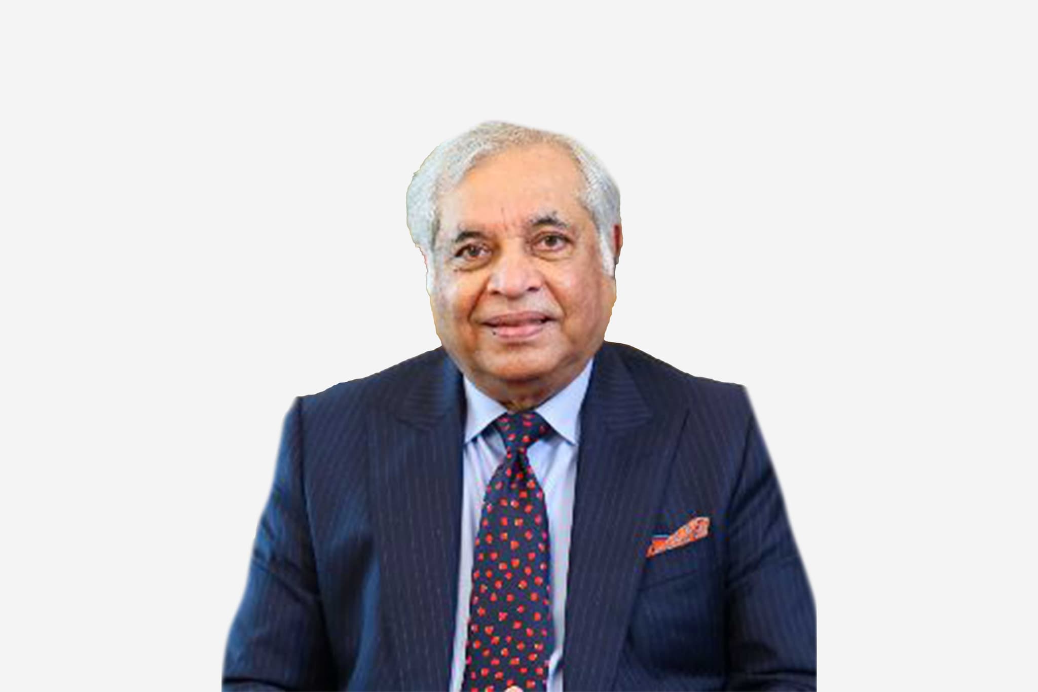
Dr Pradeep Chowbey, Group Chairman - Max Institute of Laparoscopic, Endoscopic, Bariatric & Gastrointestinal Surgery at Max Super Specialty Hospital, Saket - Max Healthcare
  1. Home
  2. Doctors
  3. Dr Pradeep Chowbey

About Dr Pradeep Chowbey

Dr. Pradeep Chowbey is a world-renowned pioneer in laparoscopic surgery and a foundational figure in the global evolution of minimal access surgery. With over 45 years of profound clinical experience and a record-breaking portfolio of nearly 98,300 surgical procedures, he currently serves as the Chairman of the Max Institute of Minimal Access, Metabolic & Bariatric Surgery at Max Super Speciality Hospital, Saket. His career is defined by his role as a "surgeon’s surgeon," having trained more than 20,000 surgeons from across the globe in the art of minimally invasive intervention.

Pioneering Breakthroughs in Laparoscopic and Endoscopic Innovation

Dr. Chowbey is credited with several landmark "firsts" that have redefined surgical standards in the Asian subcontinent. He was among the first to perform laparoscopic cholecystectomy (gallbladder removal) in North India and the first in the Asia-Pacific region to perform MAFT (Minimally Invasive Anal Fistula Treatment). A true innovator, he devised a unique method for Endoscopic Hernia Repair (TEP) and a novel endoscopic approach for neck surgeries, both of which have gained global clinical acceptance for their precision and safety.

National Leadership and Global Academic Excellence

In recognition of his monumental contributions to healthcare, the Government of India conferred the prestigious Padma Shri upon Dr. Chowbey. He has also served as the Honorary Surgeon to the President of India and the Dalai Lama. His institute at Max Healthcare was the first in India to receive "Center of Excellence" accreditation from the Surgical Review Corporation (USA) for Metabolic, Bariatric, and Hernia Surgery, solidifying its status as a premier global destination for specialized surgical care.

Advancing the Future of Metabolic and Bariatric Medicine

Dr. Chowbey has dedicated his career to the propagation of Metabolic and Bariatric surgery to treat obesity and type 2 diabetes. His research-driven approach is reflected in over 300 original scientific papers published in indexed international journals. As a prolific author, he has contributed to prestigious medical textbooks published by Springer, Elsevier, and Jaypee, ensuring that his surgical philosophies and technical innovations continue to guide the next generation of laparoscopic specialists.

Innovation and Clinical Expertise

A perennial fixture in the record books, Dr. Chowbey featured in the Guinness Book of World Records (1997) and the Limca Book of Records for 20 consecutive years (2000–2020) for performing the highest number of minimal access surgeries. His expertise spans a vast clinical repertoire, including complex hernia repairs, biliary tract surgeries, and advanced bariatric procedures. By combining high-volume surgical experience with a passion for technological advancement, he remains a central figure in the international surgical community.

Dr. Pradeep Chowbey at a Glance

  • Chairman of Minimal Access, Metabolic & Bariatric Surgery at Max Healthcare.

  • Recipient of the Padma Shri and Honorary Surgeon to the President of India.

  • Performed nearly 98,300 surgeries with over 45 years of clinical experience.

  • Global pioneer in MAFT technology and uniquely devised Endoscopic TEP Hernia Repair.

  • Trained over 20,000 international surgeons in minimally invasive techniques.

  • Guinness World Record holder for the highest number of minimal access surgeries.

MS (Jabalpur Medical College)
MBBS
MNAMS
FMAS
FALS
FIAGES
FACS
FICS
FAIS
FIMSA
FRCS (London)
Lifetime Achievement Award by IAGES in February,2015
Lifetime Achievement Award by Human Care Charitable Trust in August 2014
IMAAN INDIA SAMMAN award, October 2014
A Legend during the Lifetime Award by Balkan Ji Bari International, September 2013
Chairman’s Award by Max Healthcare, January 2012
Honorary Rotarian by Rotary Club of Delhi, May 2011
Doctor of Science (Honoris Causa Doctorate) by His Excellency Governor of MP
the Vice-Chancellor of the University of Jabalpur, October 2007
Dr. K.C Mahajan Award for Best Academician, January 2006
Gem of India Award for excellence in field, 2006
Arch of Excellence (Medicare) Award, 2006
Bharat Jyoti Award, 2006
Rashtriya Rattan Award & Gold Medal for Medical Excellence, December, 2005
Indira Gandhi Priyadarshini Award for Excellence in the laparoscopy field, November 2005
Dhanvantari Award for Excellence in Health Care, November, 2005
Awarded IMAGES Masters Virk Award – 2005 by Virk Hospital Centre for Human Reproduction
Awarded Padma Shri, the highest civilian honor, awarded by the Hon. President of India in January 2002
ACADIMA 2001, awarded in the field of Minimal Access Surgery
Aadharshila award for excellent work
contribution in the field of medicine
Placed in Limca Book of Records 2000 to 2009 consecutive years for most minimal access surgery
Ratna Award in Laparoscopic Surgery by Punjab Government in February 2000
Awarded by Guinness Book of Records for most minimal access surgery in 1997
Awarded Arya award for the most compassionate doctor in 1997
Vocational award 1995-96 by Rotary International
Awarded Gold Medal by Indian Medical Association as “Young Investigator of India 95”
Best performer National Board of Examination for MNAMS degree in 1979
Gold Medal
Best Performer, Govt. Medical College, Jabalpur (MP) at the MS (Gen. Surgery), 1977
Merit in all subjects, Govt. Medical College, Jabalpur (MP) at the MBBS examination ,1973
First Honorary Endoscopic & Laparoscopic Surgeon to the President of India, 2000
First Honorary Consultant in laparoscopic surgery to Armed Forces Medical Services of India(AFMC), 2000
The honour of conducting surgery on His Excellency Shri K R Narayanan for his gall bladder treatment (March 29, 2001)
May we help you?

Enter digits only (6-15 digits). We'll add your country code.

Related Treatments

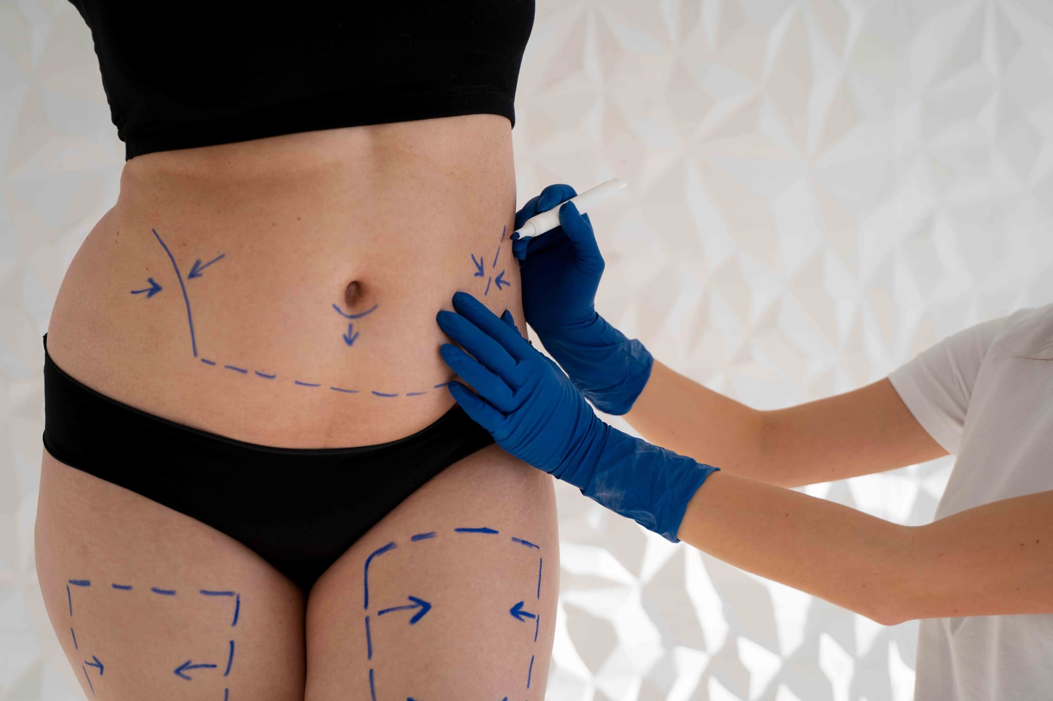
Body Contouring (Post Bariatric)

Body Contouring after bariatric surgery is a sequence of reconstructive procedures in 2026 designed to remove massive amounts of redundant skin and residual fat. It is the definitive final step for patients who have lost significant weight (50–100+ lbs), as excess skin can cause persistent hygiene issues, chronic infections, and physical discomfort that exercise alone cannot resolve.

When You Should Consider Body Contouring

This reconstructive journey is recommended for those who have completed their weight-loss phase and are seeking to improve their quality of life. Key indications include:

  • Redundant Skin Folds: Excessive skin on the abdomen, arms, or thighs that interferes with movement or clothing.

  • Chronic Skin Irritation: Recurring rashes or infections (intertrigo) within the skin folds.

  • Functional Limitations: Heavy skin that causes back pain, postural issues, or prevents a return to active fitness.

  • Stabilized Weight: Successful maintenance of a stable weight for at least 6 to 12 months.

  • Psychological Closure: A desire to have one's physical appearance reflect their health and weight-loss achievements.

Common Stages of Body Contouring

  • Phase 1 (The Core): Typically involves a Lower Body Lift or a Fleur-de-lis Abdominoplasty to stabilize the midsection.

  • Phase 2 (The Upper Body): Focuses on a Breast Lift (Mastopexy) and an Arm Lift (Brachioplasty).

  • Phase 3 (The Lower Extremities & Face): Includes a Thigh Lift or a Face and Neck Lift to address remaining laxity.

  • Auto-Augmentation: A 2026 standard where redundant tissue is used to rebuild volume in the breasts or buttocks, avoiding the need for synthetic implants.

  • Fleur-de-lis Technique: A specialized approach using both vertical and horizontal incisions to address massive skin redundancy in both directions.

How Body Contouring Is Performed

  • Staged Planning: Due to the extensive nature of these surgeries, procedures are spaced 4–6 months apart to allow the body to recover and ensure patient safety.

  • Incision & Removal: The surgeon makes precise incisions to remove wide sections of skin and fat, carefully repositioning the remaining tissue for a natural look.

  • Muscle & Tissue Repair: Weakened muscles (especially in the abdomen) are tightened to provide structural support for the new body shape.

  • Refined Contouring: Targeted liposuction is used to smooth the transitions between the areas where skin was removed.

  • Closure: Complex, multi-layered suturing is used to manage tension on the long incisions, and surgical drains are placed to prevent fluid buildup.

Pre-Procedure Preparation for Body Contouring

  • Nutritional Optimization: Surgeons require comprehensive blood panels to ensure protein, iron, and B12 levels are optimized, as post-bariatric patients often have deficiencies.

  • Weight Maintenance: Patients must demonstrate a stable "plateau" weight for at least 6 months prior to the first stage.

  • Mental Health Assessment: In 2026, professional support is often encouraged to help patients adjust to their changing body image.

  • Smoking/Vaping Cessation: Strict abstinence is mandatory for several weeks before and after to ensure tissue survival and wound healing.

Tests Before Body Contouring

  • Comprehensive Metabolic Panel: To check electrolyte balance, kidney function, and liver health.

  • Protein & Micronutrient Testing: To confirm the patient has the nutritional reserves (especially albumin) needed for massive tissue healing.

  • ECG & Cardiac Clearance: Essential due to the multiple, lengthy surgical sessions required.

  • Standard Blood Panel: To evaluate hemoglobin and clotting factors to minimize the risk of post-operative anemia.

Life After Body Contouring (Recovery)

  • High-Intensity Healing: The first week of each stage requires around-the-clock assistance and careful drain management.

  • Compression Support: Heavy-duty medical compression garments are worn for 6–8 weeks to support the tissues and minimize swelling.

  • Total Timeline: A full body transformation typically takes 12 to 18 months to complete all stages and see the final settled results.

  • Scar Management: While scars are extensive and permanent, 2026 protocols include advanced topical treatments and laser therapies to help them fade over time.

Benefits of Body Contouring

  • Elimination of Skin Issues: Directly resolves chronic rashes, infections, and hygiene struggles associated with skin folds.

  • Restored Mobility: Dramatically improves the ability to exercise, run, and move without the hindrance of heavy skin.

  • Permanent Tissue Correction: Once the redundant skin is surgically removed, it does not return if weight remains stable.

  • Complete Transformation: Provides the physical and emotional "finish line" to a significant weight-loss journey.

Starting Cost: $ 1500

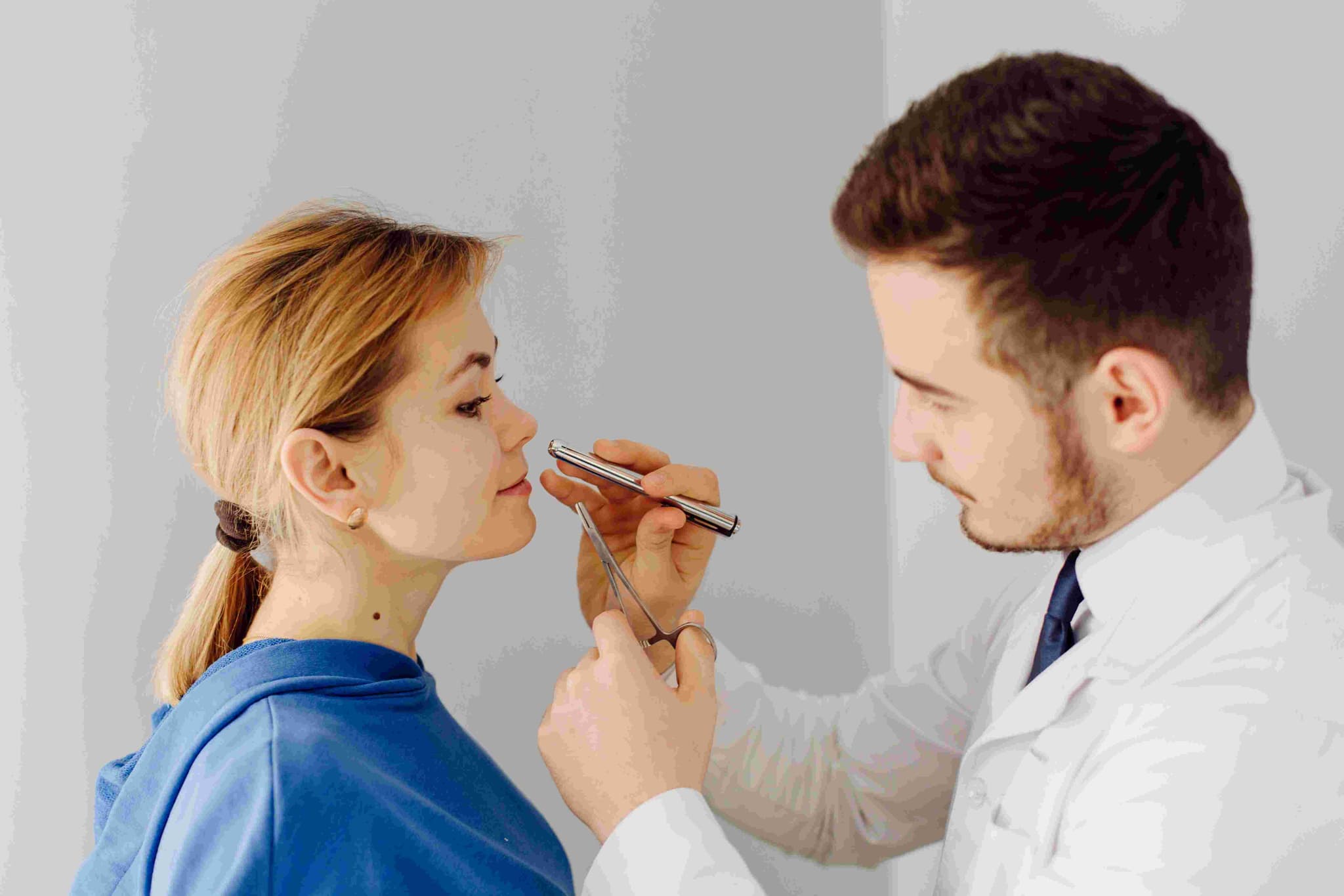
CSF Leak Repair (Endoscopic)

Endoscopic CSF (Cerebrospinal Fluid) leak repair is a specialized, minimally invasive surgery performed to seal a hole in the skull base—the critical bone barrier separating the brain from the nasal cavity. This procedure is essential for stopping the drainage of brain fluid and, more importantly, preventing bacteria from entering the central nervous system and causing life-threatening infections such as meningitis.

When You Should Consider CSF Leak Repair

  • Clear Nasal Drainage (Rhinorrhea): If you experience a persistent, watery discharge from one side of the nose that often increases when leaning forward or straining.

  • Salty or Metallic Taste: A distinct sensation in the back of the throat caused by spinal fluid draining down from the skull base.

  • Recurrent Meningitis: If you have suffered from unexplained or repeat bacterial infections of the brain lining.

  • Post-Traumatic Leaks: Following a head injury or facial fracture that has compromised the integrity of the skull base.

  • Iatrogenic Leaks: As a follow-up to previous sinus or neurosurgical procedures where a small opening in the dura was created.

  • Spontaneous Leaks: Often associated with high intracranial pressure, frequently seen in patients with idiopathic intracranial hypertension.

How CSF Leak Repair Is Performed

  • Anesthesia: The surgery is performed under general anesthesia and typically takes between 2 to 4 hours due to the microscopic precision required.

  • Endoscopic Approach: The surgeon operates entirely through the nostrils using a high-definition endoscope. There are no external incisions and no visible facial scarring.

  • Localization: Surgeons use the endoscope and sometimes a fluorescent dye (fluorescein) injected into the spinal fluid to pinpoint the exact site of the leak with total accuracy.

  • The Multi-Layer Repair: A "sandwich" technique is often used to ensure a watertight seal:
    Grafts: Small pieces of the patient’s own tissue (fat, muscle lining, or nasal mucosa) are placed over or into the defect.
    Vascularized Flap: For larger leaks, a Nasoseptal Flap—tissue with its own blood supply—is rotated from the septum to provide a robust, living cover for the repair.
    Sealants: Medical-grade "glue" or fibrin sealant is applied to bond the grafts to the bone.

  • Packing: The nose is packed with specialized sponge-like material (either dissolvable or removable) to maintain constant pressure on the repair site during the first stage of healing.

Pre-Procedure Preparation for CSF Leak Repair

  • High-Resolution Imaging: Combining CT scans (to see bone) and MRI (to see fluid) to create a precise surgical roadmap.

  • Beta-2 Transferrin Test: A laboratory confirmation that the fluid draining from the nose is indeed cerebrospinal fluid.

  • Pneumococcal Vaccination: Ensuring you are protected against the most common bacteria that cause meningitis before the skull base is manipulated.

  • Intracranial Pressure Assessment: Evaluating if you have high brain pressure that might have caused the leak and could potentially threaten the new repair.

  • Fasting: Following strict "nothing by mouth" instructions for the hours preceding your general anesthesia.

Tests Before CSF Leak Repair

  • Pledget Study: Placing small cotton sponges in the nose to absorb drainage, which are then tested for the presence of spinal fluid markers.

  • CT Cisternography: A specialized scan where contrast dye is injected into the spinal fluid to visualize the exact point of the "leakage."

  • Neurological Consultation: A full assessment of brain function and cranial nerves to establish a baseline before surgery.

  • Blood Clotting Panel: Ensuring your blood's ability to clot is optimal for a deep, vascularized repair.

Life After CSF Leak Repair

  • Hospital Monitoring: Patients typically remain in the hospital for 1 to 3 days. You may need to keep your head elevated at a 30–45 degree angle to reduce pressure on the repair.

  • Lumbar Drain: Occasionally, a small tube is placed in the lower back for 24–48 hours to divert spinal fluid and allow the nasal "patch" to bond without pressure.

  • The "No" Rules (4–6 Weeks): To protect the seal, you must strictly avoid:
    Blowing your nose: The most critical restriction to prevent air from being forced into the brain.
    Straining: Stool softeners are often prescribed to prevent "valsalva" maneuvers during bowel movements.
    Physical Exertion: No heavy lifting or intense exercise that could dislodge the graft.
    Closed-Mouth Sneezing: You must always sneeze with your mouth open to release internal pressure.

  • Sensation Changes: Headaches and a temporary decrease in the sense of smell (anosmia) are common as the nasal passages heal.

Why CSF Leak Repair Is Highly Effective

  • Prevents Life-Threatening Infection: By restoring the watertight barrier, the procedure eliminates the direct "highway" for bacteria to reach the brain.

  • High Success Rates: Modern endoscopic techniques have a success rate of 90% to 95% on the first attempt for most skull base defects.

  • Avoids Traditional Brain Surgery: This internal approach replaces the need for an "open" craniotomy, which involves removing a portion of the skull and retracting the brain.

  • Rapid Recovery: Because no external bone is cut and the brain is not manipulated, patients recover much faster and with fewer neurological side effects.

  • Preserves Nasal Function: Using localized grafts and flaps allows the surgeon to repair the defect while maintaining the health of the surrounding nasal lining.

Starting Cost: $ 2000

Endoscopic Endonasal Surgery

Endoscopic Endonasal Surgery (EES) is a minimally invasive surgical technique that uses the nose and sinuses as natural "corridors" to reach and treat conditions at the base of the brain or the top of the spine. Unlike traditional "open" brain surgery, it requires no external incisions on the face or scalp and avoids the need to remove large parts of the skull. This advanced approach allows surgeons to access deep-seated tumors and vascular issues with minimal disruption to healthy brain tissue.

When You Should Consider Endoscopic Endonasal Surgery

  • Pituitary Tumors: This is the primary and most common use for EES, particularly for adenomas affecting hormone levels or vision.

  • Skull Base Tumors: Including meningiomas, chordomas, and craniopharyngiomas located at the very bottom of the brain.

  • Cerebrospinal Fluid (CSF) Leaks: To repair physical holes or defects in the skull base where brain fluid is dripping through the nose.

  • Optic Nerve Compression: To decompress nerves that are being pinched by tumors, often leading to rapid vision improvement.

  • Sinus or Nasal Malignancies: For specialized removal of tumors that have invaded the bone separating the nose from the brain.

  • Top-of-Spine Disorders: To treat abnormalities where the skull meets the spinal column without traditional neck surgery.

How It Is Performed

  • Collaborative Team: The surgery is typically a joint effort between a neurosurgeon and an ENT (Otolaryngologist), performed under general anesthesia.

  • Access: A high-definition endoscope (a thin tube with a camera) is inserted through the nostrils. This provides a panoramic, high-magnification view of the surgical field.

  • Navigation: Surgeons use an image-guided system (neuronavigation), similar to a GPS for the brain, to map the exact location of the target in real-time using your pre-operative scans.

  • Removal: Specialized long-reach micro-instruments are passed through the nostrils to remove tumors or treat the affected area without any external skin cuts.

  • Reconstruction: To prevent brain fluid leaks, the surgeon may reconstruct the surgical site using a nasoseptal flap (the patient's own nasal tissue with its own blood supply) or specialized synthetic sealants.

  • Closing: Because there are no external incisions, no stitches are needed on the face or scalp; the internal nasal passages are simply cleaned and occasionally padded with temporary sponges.

Pre-Procedure Preparation

  • Skull Base MRI and CT: High-resolution scans to provide a 3D "roadmap" of your bone structure and major blood vessels.

  • Endocrine Evaluation: Comprehensive blood tests to check your current pituitary hormone levels.

  • Visual Field Testing: A detailed eye exam to establish a baseline for your vision and peripheral awareness.

  • Nasal Assessment: An ENT check-up to ensure your nasal anatomy (like the septum) is suitable for the endoscopic instruments.

  • Fasting: Following "nothing by mouth" instructions for 8 hours prior to your scheduled anesthesia.

Tests Before Endoscopic Endonasal Surgery

  • CT Navigation Scan: A specialized scan performed close to the surgery date to "calibrate" the surgical GPS system.

  • Hormone Panels: Specifically checking thyroid, adrenal, and growth hormones which may be impacted by the surgery.

  • Blood Panels: A routine check of your blood count, electrolytes, and clotting factors.

  • ECG: A standard heart check to confirm cardiovascular stability for the duration of the procedure.

Life After Endoscopic Endonasal Surgery

  • Hospital Stay: Patients typically stay in the hospital for 1 to 5 days, often spending the first night in a specialized Neuro-ICU.

  • Immediate Symptoms: It is normal to experience nasal congestion, mild headaches, and blood-tinged nasal drainage for 1 to 2 weeks.

  • Nasal Care: Regular saline nasal rinses are essential to keep the nasal passages clean, moist, and free of crusting.

  • The "No" Rules (4–6 Weeks): To protect the internal seal and prevent a brain fluid leak, you must strictly avoid:
    Blowing your nose: This can force air into the brain cavity.
    Straining: Stool softeners are often prescribed to prevent internal pressure.
    Heavy lifting: Nothing over 5–7 kg (11–15 lbs).
    Bending over at the waist: Keep your head above your heart level at all times.

  • Follow-up Debridement: You will visit your ENT specialist several times in the first month to have the nasal passages professionally cleaned (debrided).

Why Specialized Treatment Is Highly Effective

  • No External Scars: By utilizing natural pathways, there is no impact on your facial appearance or hairline.

  • Enhanced Visualization: The endoscope allows surgeons to see "around corners" and behind critical nerves that would be hidden in traditional surgery.

  • Faster Recovery: Avoiding a craniotomy (opening the skull) significantly reduces post-operative pain and shortens the hospital stay.

  • Direct Access: EES provides the shortest, most direct route to the pituitary gland and skull base, minimizing the "travel distance" through healthy brain tissue.

  • Superior Seal Techniques: The use of vascularized nasal flaps has revolutionized the safety of this procedure, dramatically reducing the risk of post-operative infections and leaks.

Starting Cost: $ 2500

Endoscopic Retrograde Cholangiopancreatography (ERCP)

Endoscopic Retrograde Cholangiopancreatography (ERCP) is a highly specialized procedure used primarily to treat disorders of the bile ducts and pancreatic ducts. It uniquely combines advanced endoscopy with real-time X-ray imaging (fluoroscopy). By 2026, ERCP has evolved from a diagnostic tool into a powerful therapeutic intervention, allowing physicians to clear obstructions, place stents, and repair leaks within the delicate ductal system of the liver and pancreas without the need for invasive abdominal surgery.

Why ERCP is Performed

  • Common Bile Duct Stones: Removing gallstones that have migrated out of the gallbladder and are blocking the main drainage duct.

  • Malignant Obstructions: Relieving blockages caused by tumors in the pancreas, bile ducts, or liver.

  • Biliary Strictures: Opening narrowed areas of the ducts caused by scar tissue or chronic inflammation.

  • Post-Surgical Bile Leaks: Sealing leaks that can occasionally occur following gallbladder or liver surgery.

  • Chronic Pancreatitis: Treating complications such as pancreatic stones or narrowings that cause severe, recurring pain.

  • Sphincter of Oddi Dysfunction: Managing issues with the muscular valve that controls the flow of digestive juices.

Conditions That Require Specialized Care

  • Massive Stone Burden: Cases with very large or numerous stones that may require advanced "fragmentation" techniques.

  • Anatomical Variations: Patients who have had previous gastric bypass or stomach surgery, requiring a modified "enteroscopy" approach.

  • Primary Sclerosing Cholangitis (PSC): A chronic condition requiring careful, repeated dilations of the bile ducts.

  • High-Risk Pancreatitis Patients: Individuals with a history of post-procedure inflammation who require preventative "stenting" of the pancreatic duct.

  • Pediatric ERCP: Rare cases in children involving congenital ductal abnormalities that require specialized pediatric endoscopes.

[Image showing the endoscope positioned in the duodenum with the catheter entering the bile duct]

How an ERCP Is Performed

  • Deep Sedation or Anesthesia: To ensure absolute stillness and comfort, the procedure is performed under deep sedation or general anesthesia.

  • Side-Viewing Endoscopy: A specialized duodenoscope is passed through the mouth and stomach to reach the "papilla"—the small opening where the ducts meet the intestine.

  • Cannulation: A tiny catheter is threaded into the papilla to gain access to the biliary or pancreatic tree.

  • Retrograde Contrast: A radio-opaque dye is injected "backward" into the ducts, highlighting the internal anatomy on a live X-ray monitor.

  • Sphincterotomy: Using a small electrified wire, the surgeon may make a tiny cut in the muscle valve (sphincter) to widen the opening for stone removal.

  • Mechanical Intervention: Tools such as baskets, balloons, or stents are passed through the scope to clear blockages or provide structural support.

Innovations in Duct Therapy

  • Disposable DuodenoscopesThe widespread adoption of single-use scopes to eliminate the risk of bacterial transmission and ensure the highest mechanical reliability for every patient.

  • SpyGlass™ CholangioscopyA "micro-scope" that is passed through the main endoscope, allowing the doctor to see directly inside the bile duct in high-definition to biopsy tumors or break up stones with lasers.

  • AI-Assisted FluoroscopyArtificial intelligence that enhances X-ray images in real-time, helping the doctor identify "shadows" or subtle strictures with greater clarity.

  • Biodegradable Biliary StentsNext-generation stents that provide support to a narrowed duct for several months and then dissolve, avoiding the need for a second procedure to remove them.

  • Laser Lithotripsy (EHL)Using targeted laser energy to "shatter" stubborn gallstones into tiny fragments that can be easily swept out of the duct.

  • Automated Risk-Prediction ModelsSoftware that analyzes a patient's health data before the procedure to suggest the exact dose of preventative medication needed to stop post-op inflammation.

Pre-Procedure Preparation

  • Fasting (NPO): A strictly empty stomach is required, usually meaning no food or drink for 8 to 12 hours prior to the procedure.

  • Antibiotic Prophylaxis: Certain patients may be given a dose of antibiotics before the start of the procedure to prevent ductal infection.

  • Blood Work: Checking coagulation (clotting) factors to ensure it is safe to perform a sphincterotomy or biopsy.

  • Medication Coordination: Careful management of blood thinners and diabetic medications is essential for a safe recovery.

  • Post-Op Plan: Because of the intensive nature of ERCP, patients must have a designated driver and a quiet place to recover for the first 24 hours.

Diagnostic and Safety Monitoring

  • Pancreatitis Prevention: Use of rectal medications or temporary "protective" pancreatic stents to lower the risk of post-ERCP inflammation.

  • Amylase/Lipase Checks: Blood tests performed if a patient experiences unusual pain after the procedure to check for pancreatic irritation.

  • Continuous Vital Monitoring: Specialized anesthesia teams monitor heart rhythm and oxygen levels throughout the entire intervention.

  • Radiation Safety: Using "low-dose" pulsed fluoroscopy to minimize X-ray exposure for both the patient and the medical team.

Why This Treatment Is Highly Effective

  • Avoids Major Surgery: ERCP provides a "keyhole" alternative to traditional open-belly surgery for clearing bile duct obstructions.

  • Immediate Obstruction Relief: Patients with jaundice (yellowing of the skin) often see a rapid improvement in symptoms once the duct is cleared or stented.

  • High Precision: The combination of live video and live X-ray ensures that the doctor knows exactly where the instruments are at all times.

  • Life-Saving for Sepsis: In cases of "cholangitis" (infected bile), ERCP provides urgent drainage that is critical for survival.

  • Dual Functionality: It allows for the immediate transition from identifying a problem to fixing it within the same 30-to-60-minute session.

Recovery and Aftercare

  • Immediate Observation: Most patients stay in a recovery unit for 1 to 2 hours to ensure the sedation has worn off and no immediate pain is present.

  • Sore Throat and Bloating: These are the most common minor side effects and typically resolve within 24 to 48 hours.

  • Dietary Progression: Patients usually start with clear liquids and move to a low-fat diet as tolerated by their digestive system.

  • Activity Limits: No driving or making major decisions for 24 hours. Most patients return to full activity within 2 to 3 days.

  • Follow-up Imaging: A simple ultrasound or blood test is often scheduled a few weeks later to confirm the ducts remain clear.

Life After ERCP

  • Rapid resolution of painful symptoms like biliary colic (gallstone pain) or chronic abdominal pressure.

  • Successful management of complex conditions like cancer-related blockages, allowing for a better quality of life.

  • Permanent removal of dangerous "trapped" stones that could otherwise lead to severe liver damage.

  • A return to a normal, healthy diet once the flow of digestive enzymes and bile has been restored.

  • Empowerment through the successful treatment of a condition that previously would have required a long, difficult surgical recovery.

Starting Cost: $ 300

Endoscopic Skull Base Surgery

Endoscopic Skull Base Surgery is a minimally invasive technique used to reach tumors and abnormalities at the very bottom of the brain and the top of the spine. Instead of utilizing large incisions or traditional "open" craniotomies, surgeons use the nose and sinuses as natural corridors to reach the target area. This advanced approach allows for the treatment of complex conditions located deep within the head without the need to retract or move the brain.

When You Should Consider Endoscopic Skull Base Surgery

  • Pituitary Adenomas: The most common use for this approach, particularly for tumors affecting hormone levels or vision.

  • Meningiomas & Chordomas: Tumors located at the base of the skull that would otherwise require highly invasive open surgery.

  • Craniopharyngiomas: Complex tumors located near the pituitary gland and optic nerves.

  • CSF Leaks: Repairing physical holes in the skull base where brain fluid is dripping from the nose.

  • Acoustic Neuromas: Certain tumors affecting the nerves responsible for balance and hearing.

  • Esthesioneuroblastoma: Rare tumors arising from the olfactory (smell) nerves at the roof of the nose.

How It Is Performed

  • Team Effort: The surgery is typically a collaborative effort between a Neurosurgeon and an ENT (Otolaryngologist), performed under general anesthesia over 3 to 7 hours.

  • Nasal Access: The ENT surgeon inserts a high-definition endoscope (a thin tube with a camera) through the nostrils. No external skin incisions are made on the face or scalp.

  • Navigation: Using a 3D "GPS" system mapped from your pre-operative MRI, the surgeons navigate through the sphenoid or ethmoid sinuses to reach the skull base bone.

  • Bone Opening: A small, precise window is created in the thin bone at the base of the skull to expose the protective lining of the brain (dura) or the tumor itself.

  • Tumor Removal: Using long, specialized micro-instruments through the other nostril, the tumor is removed in sections. The endoscope provides a close-up, panoramic view of critical structures like the optic nerves and carotid arteries.

  • Reconstruction: To prevent brain fluid leaks, the surgical opening is sealed using a nasoseptal flap (a flap of your own nasal tissue with its own blood supply), fat grafts, or specialized synthetic glues.

Pre-Procedure Preparation

  • High-Resolution Imaging: Dedicated skull base MRI and CT scans to map the bone anatomy and vascular structures.

  • Endocrine Testing: Comprehensive blood panels to check pituitary hormone function before the gland is approached.

  • Ophthalmology Review: Detailed visual field and acuity testing if the tumor is near the optic nerves.

  • Nasal Endoscopy: A quick office-based look at your nasal passages to ensure there are no obstructions like polyps or a severely deviated septum.

  • Fasting: Adhering to "nothing by mouth" instructions for 8 hours prior to your scheduled anesthesia.

Tests Before Endoscopic Skull Base Surgery

  • CT Navigation Scan: A specialized scan used to "sync" your anatomy with the surgical GPS system in the operating room.

  • Visual Field Test: To establish a baseline for your peripheral vision, which is often improved by the surgery.

  • Blood Panels: A routine check of your blood count, electrolytes, and clotting factors.

  • ECG: A standard heart check to confirm cardiovascular stability for a multi-hour procedure.

Life After Endoscopic Skull Base Surgery

  • Hospital Stay: Typically 2 to 4 days, with the first night often spent in the Neuro-ICU for intensive monitoring.

  • Immediate Symptoms: Nasal congestion, "crusting" inside the nose, and mild headaches are normal for 2 to 3 weeks as the sinuses heal.

  • Nasal Care: You will need to perform frequent saline nasal rinses to keep the surgical area clean and moist.

  • The "No" Rules (4–6 Weeks): To protect the internal seal and prevent a CSF leak, you must strictly avoid:
    Blowing your nose: This can force air into the brain cavity.
    Straining: Stool softeners are usually prescribed to prevent internal pressure.
    Heavy lifting: Nothing over 5 kg (11 lbs).
    Drinking through a straw: The suction pressure can disrupt the nasal flap.

  • Long-term Follow-up: Regular nasal debridement (cleaning) by your ENT and follow-up MRIs are required to monitor healing and ensure no tumor recurrence.

Why Specialized Treatment Is Highly Effective

  • No Visible Scars: By using the nose as a natural corridor, there are no incisions on the face, preserving your natural appearance.

  • Faster Recovery: Avoiding a traditional craniotomy means significantly less post-operative pain and a quicker return to daily activities.

  • Panoramic Visualization: The endoscope allows surgeons to see "around the corner" of critical nerves and arteries with better clarity than a traditional microscope.

  • Brain-Sparing Technique: Because the approach is from underneath the brain, there is no need for brain retraction, reducing the risk of post-operative swelling or cognitive changes.

  • Highly Precise Reconstruction: The use of vascularized nasoseptal flaps has revolutionized the success rate of sealing the skull base, making the procedure safer than ever before.

Starting Cost: $ 3000

Functional Endoscopic Sinus Surgery (FESS)

Functional Endoscopic Sinus Surgery (FESS) is a minimally invasive procedure used to treat chronic sinus conditions, such as persistent sinusitis and nasal polyps. By restoring natural drainage and airflow, FESS eliminates the root cause of chronic inflammation. The procedure is performed entirely through the nostrils using a specialized nasal endoscope (a thin tube with a magnified camera), meaning there are no external incisions or visible scars.

When You Should Consider FESS

  • Chronic Sinusitis: If you suffer from sinus inflammation lasting more than 12 weeks that hasn't responded to medical therapy.

  • Nasal Polyps: Benign growths in the nasal passages that physically obstruct your breathing and block natural fluid drainage.

  • Recurrent Acute Infections: When you experience four or more severe sinus infections per year, significantly impacting your quality of life.

  • Anatomical Obstructions: To correct issues such as a deviated septum or enlarged turbinates that contribute to chronic blockages.

  • Failed Medication: When antibiotics, steroid sprays, and saline rinses are no longer effective in managing your symptoms.

How Is Performed

  • Anesthesia: The surgery is usually performed as an outpatient (day-care) procedure under general anesthesia and typically lasts 2 to 3 hours.

  • Visualisation: The surgeon inserts the endoscope into the nostrils to obtain a high-definition, magnified 3D view of the internal sinus passages.

  • Tissue Removal: Using precise micro-instruments, the surgeon removes obstructions such as nasal polyps, diseased tissue, or small pieces of bone blocking the sinus openings.

  • Opening the Passageways: The natural drainage pathways (ostia) are carefully widened to improve long-term ventilation and allow future nasal rinses to reach deep into the sinus cavities.

  • Completion: To control minor bleeding, the surgeon may place dissolvable packing or temporary splints inside the nose, which do not require manual removal.

Pre-Procedure Preparation

  • CT Scan of the Sinuses: A mandatory high-resolution "map" used by the surgeon to navigate the complex anatomy near the eyes and brain.

  • Medication Review: You may be prescribed a short course of oral steroids or antibiotics before surgery to reduce inflammation and bleeding.

  • Smoking Cessation: Patients are strongly advised to stop smoking several weeks before surgery, as tobacco use significantly hinders the healing of sinus tissue.

  • Fasting: Following strict "nothing by mouth" instructions for the hours preceding your general anesthesia.

  • Transportation: Arranging for a ride home, as you will be drowsy and may have temporary nasal congestion affecting your comfort.

Tests Before FESS

  • Nasal Endoscopy: An in-office diagnostic check to visualize the extent of polyps or structural blockages.

  • Allergy Testing: To determine if underlying allergies need to be managed alongside surgery for the best long-term outcome.

  • Blood Tests: Standard pre-operative panels to ensure healthy clotting factors and general surgical readiness.

  • Olfactory Testing: Assessing your baseline sense of smell to monitor improvements after the airways are cleared.

Life After FESS

  • Immediate Symptoms: It is normal to experience stuffiness, mild facial pressure, and blood-stained drainage for the first 3 to 5 days.

  • Nasal Irrigation: Using a saline nasal wash (usually 4+ times daily) is essential to flush out debris, prevent crusting, and ensure the new openings stay clear.

  • Crucial Restrictions: You must strictly avoid blowing your nose for at least 7 to 14 days and avoid strenuous activity or heavy lifting for 2 to 3 weeks to prevent bleeding.

  • Debridement: Follow-up visits are vital for your surgeon to professionally clean out dried mucus and ensure the sinuses are healing without excessive scar tissue.

  • Healing Timeline: While most patients return to work or school within one week, full internal healing and the stabilization of the sinus lining can take several months.

Why Specialized Treatment Is Highly Effective

  • Targeted Precision: FESS focuses only on the diseased areas, preserving as much healthy nasal tissue and mucous membrane as possible.

  • No External Scarring: Because the surgery is entirely internal, there is no change to your physical appearance and no external stitches to remove.

  • High Safety Profile: With major complication rates of less than 0.5%, FESS is considered one of the safest and most effective ways to treat chronic sinus disease.

  • Long-Term Relief: By widening the natural ostia, the surgery provides a permanent structural improvement that allows for better drainage for years to come.

  • Improved Medication Delivery: Post-operative nasal sprays and rinses can finally reach the deep sinus cavities, making ongoing maintenance far more effective.

Starting Cost: $ 600

Pituitary Tumor Surgery (Endoscopic)

Endoscopic Pituitary Surgery, also known as Endoscopic Transsphenoidal Surgery, is a minimally invasive procedure that uses the nostrils as natural pathways to reach and remove tumors from the pituitary gland. Because it avoids large incisions and brain retraction, it typically offers a faster recovery and fewer side effects than traditional open surgery. This approach allows surgeons to access the "master gland" at the base of the brain with extreme precision.

When You Should Consider Endoscopic Pituitary Surgery

  • Hormone-Secreting Tumors: Such as those causing Cushing’s disease (excess cortisol), acromegaly (excess growth hormone), or prolactinomas.

  • Non-Functioning Macroadenomas: Large tumors that do not produce hormones but press on the optic nerves, causing vision loss, double vision, or chronic headaches.

  • Pituitary Apoplexy: An emergency condition where a tumor bleeds or outgrows its blood supply, requiring rapid decompression.

  • Failed Medical Management: When medications are unable to sufficiently control hormone levels or stop the growth of the tumor.

  • Rathke’s Cleft Cysts: Benign fluid-filled growths that can interfere with normal gland function or cause pressure symptoms.

How It Is Performed

  • Collaborative Team: The surgery is usually a joint effort between a neurosurgeon and an Ear, Nose, and Throat (ENT) surgeon, taking about 2 to 3 hours under general anesthesia.

  • Nasal Access: The ENT surgeon inserts a thin, lighted tube with a high-definition camera (endoscope) through one nostril to navigate to the very back of the nasal cavity.

  • Opening the Sphenoid Sinus: The surgeon opens the sphenoid sinus (an air-filled space behind the nose) to reach the sella turcica, the small bony compartment that houses the pituitary gland.

  • Tumor Removal: Using specialized long instruments through the other nostril, the neurosurgeon removes the tumor in small pieces. The endoscope provides a panoramic, high-magnification view of the area, including nearby carotid arteries and optic nerves.

  • Reconstruction: If needed, a small fat graft (often taken from the abdomen) or synthetic material is used to fill the space and seal the area to prevent cerebrospinal fluid (CSF) leaks.

Pre-Procedure Preparation

  • Endocrine Evaluation: Comprehensive blood and urine tests to establish your baseline hormone levels (growth hormone, ACTH, prolactin, etc.).

  • High-Resolution MRI: A dedicated "pituitary protocol" scan to map the tumor’s exact size and its relationship to the optic chiasm.

  • Ophthalmology Exam: A detailed visual field test to document any current vision loss before the surgery.

  • Nasal Assessment: An ENT evaluation to ensure your nasal passages are clear and suitable for the endoscopic approach.

  • Fasting: Following "nothing by mouth" instructions for 8 hours prior to your scheduled anesthesia.

Tests Before Endoscopic Pituitary Surgery

  • Visual Field Testing: To measure peripheral vision, which is often the first thing affected by pituitary tumors.

  • Dynamic Hormone Testing: Specialized "stimulation" or "suppression" tests to confirm the type of secreting tumor.

  • Carotid Imaging: Occasionally required if the tumor is very large and wrapping around the main arteries of the brain.

  • ECG: A standard heart check to confirm cardiovascular stability for the duration of the procedure.

Life After Endoscopic Pituitary Surgery

  • Hospital Stay: Typically 1 to 3 days, often starting with one night in the Intensive Care Unit (ICU) for close monitoring of your fluid balance and hormone levels.

  • Immediate Symptoms: It is normal to experience nasal congestion, mild headaches, and "watery" or blood-tinged nasal drainage for 1 to 2 weeks.

  • The "No" Rules: For 4 to 6 weeks, you must strictly avoid:
    Blowing your nose: To prevent pressure buildup that could cause a CSF leak.
    Lifting and Straining: No lifting objects over 5 lbs or heavy straining, which increases intracranial pressure.
    Drinking through straws: The suction can interfere with the healing of the nasal repairs.

  • Hormone Monitoring: You will work closely with an endocrinologist to check if your gland is producing the correct amount of hormones post-op.

  • Follow-up MRI: A baseline scan is usually performed 3 months after surgery to ensure the entire tumor was removed.

Why Specialized Treatment Is Highly Effective

  • No External Scars: By using the natural pathway of the nose, there are no visible incisions on the face or scalp.

  • Superior Visualization: The endoscope allows surgeons to "see around corners," identifying tumor tissue that might be missed with traditional microscopic surgery.

  • Rapid Vision Improvement: Decompressing the optic nerves often leads to a quick and significant improvement in peripheral vision and clarity.

  • Preserves Gland Function: The high-magnification view helps surgeons distinguish between the tumor and the healthy part of the pituitary gland.

  • Reduced Brain Trauma: Because the brain is not "moved" or retracted to reach the tumor, post-operative headaches and recovery times are greatly reduced.

Starting Cost: $ 2500

Revisional Bariatric Surgery

Revisional bariatric surgery is a specialized procedure performed on patients who have already undergone a primary weight-loss surgery but require a second operation. This may be due to medical complications, insufficient weight loss, or significant weight regain. Because of existing scar tissue and altered anatomy, these procedures are more complex and carry higher risks than initial weight-loss surgeries.

When You Should Consider Revisional Surgery

  • Inadequate Weight Loss or Regain: If the initial stomach pouch has stretched over time or the body has adapted to the metabolic changes of the first surgery.

  • Severe Acid Reflux (GERD): Persistent or worsening reflux that does not respond to medication, frequently occurring after a gastric sleeve procedure.

  • Mechanical Complications: Issues such as a "slipped" gastric band, a staple line leak, or a narrowing (stricture) of a previous surgical connection.

  • Chronic Ulcers or "Dumping Syndrome": Persistent digestive issues following a bypass that cannot be managed through dietary changes alone.

  • Nutritional Malabsorption: Severe deficiencies in vitamins or protein that remain uncontrolled despite high-dose supplementation.

Common Revisional Procedures

  • Sleeve to Bypass Conversion: Converting a gastric sleeve into a Roux-en-Y gastric bypass. This is highly effective for treating severe reflux and triggering further weight loss.

  • Gastric Band Conversion: Removing a "Lap-Band" and converting the anatomy into either a sleeve or a bypass, often performed in a single or two-stage process.

  • Bypass Revision (Stoma Tightening): For a stretched gastric bypass, a surgeon may "re-tighten" the connection (stoma) to restore the feeling of fullness.

  • Intestinal Lengthening: Adjusting the "Roux" limb to increase the bypassed section of the intestine, thereby increasing calorie malabsorption.

  • Sleeve Re-Sleeving: Trimming a previously stretched gastric sleeve to restore its original narrow, restrictive shape.

How Is Performed

  • Anesthesia: The surgery is performed under general anesthesia and typically takes between 2 to 4 hours due to the precision required.

  • Surgical Approach: Almost always performed laparoscopically or robotically to navigate through previous scar tissue with high-definition visualization and accuracy.

  • Managing Adhesions: The surgeon must carefully separate internal scarring (adhesions) from previous operations before the new revision can begin.

  • Stapling and Joins: New surgical joins are created using specialized stapling instruments designed for thicker or scarred tissue.

  • Leak Testing: Extensive intra-operative testing is performed to ensure all new connections are watertight and airtight before finishing.

Pre-Procedure Preparation

  • Anatomy Mapping: Extensive imaging is required to visualize the current state of the stomach and intestines before entering the operating room.

  • Nutritional Optimization: Correcting any existing vitamin deficiencies through intensive supplementation prior to surgery.

  • Psychological Assessment: A thorough review to ensure the patient is prepared for the "reset" in dietary habits and lifestyle expectations.

  • Smoking Cessation: Patients must stop smoking at least 6 to 8 weeks before surgery, as nicotine significantly increases the risk of staple line leaks in scarred tissue.

  • Weight Management: Following a specific pre-operative diet to reduce liver size and improve surgical access.

Tests Before Revisional Surgery

  • Upper Endoscopy (EGD): Inserting a camera into the stomach to inspect the pouch and check for ulcers, inflammation, or stretching.

  • Barium Swallow (UGI Series): A specialized X-ray where the patient swallows contrast liquid to show the exact size and flow of the current anatomy.

  • Abdominal CT Scan: To provide a 3D view of the internal organs and identify any hidden hernias or mechanical issues.

  • Comprehensive Blood Panels: Detailed analysis of iron, B12, Vitamin D, and protein levels to establish a baseline.

Life After Revisional Surgery

  • Hospital Stay: Usually involves 2 to 3 nights of close monitoring to ensure the new surgical joins are healing correctly.

  • Dietary Reset: Patients must return to a "Phase 1" clear liquid diet and progress slowly through pureed and soft foods over a 4–6 week period.

  • Slower Weight Loss: It is important to note that weight loss after a revision is typically slower and less dramatic than after the first surgery.

  • Lifelong Monitoring: Regular follow-up appointments and blood tests are essential to monitor for malabsorption and nutritional health.

  • Activity Restrictions: Light walking is encouraged immediately, but heavy lifting and core-straining exercises are restricted for 6 weeks.

Why Specialized Treatment Is Highly Effective

  • Resolves Chronic Pain and Reflux: Provides immediate relief for patients suffering from the mechanical failures or side effects of a primary surgery.

  • Second Chance for Health: Allows patients who did not reach their goals to restart their weight-loss journey with a more powerful metabolic tool.

  • Precision Technology: The use of robotic platforms allows surgeons to operate safely in "hostile" abdomens filled with previous scar tissue.

  • Customized Anatomy: Every revision is tailored specifically to the patient’s existing anatomy, ensuring the most effective solution for their unique situation.

  • Prevents Long-Term Damage: Addressing issues like chronic ulcers or severe reflux prevents permanent damage to the esophagus and digestive tract.

Starting Cost: $ 2000

Related Specialists

Dr Kumud Rai
Dr Kumud Rai
Principal Director - Vascular Surgery
Max Smart Super Specialty Hospital, Saket
42+years experience
View Profile
Dr Shaiwal Khandelwal
Director - Thoracic Surgery
Max Super Specialty Hospital, Saket
22+years experience
View Profile
Dr Balbir Singh
Dr Balbir Singh
Group Chairman - Cardiac Sciences
Max Super Specialty Hospital, Saket
35+years experience
View Profile
Dr Viveka Kumar
Dr Viveka Kumar
Chairman - Cardiology
Max Smart Super Specialty Hospital, Saket
27+years experience
View Profile
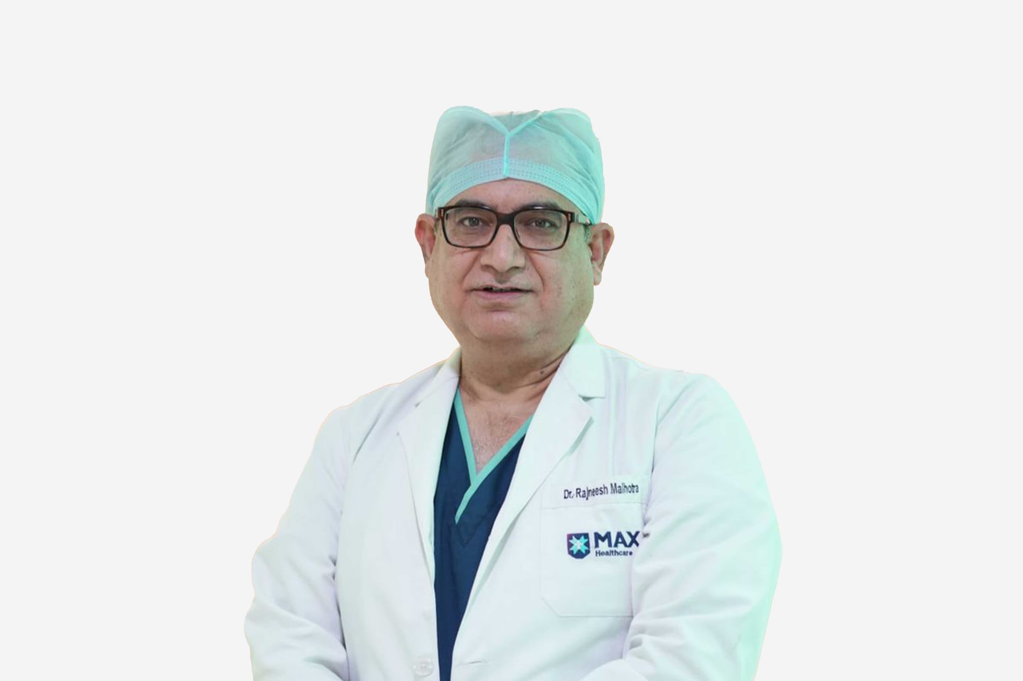
Dr Rajneesh Malhotra
Chairman & Head - CTVS
Max Super Specialty Hospital, Saket
34+years experience
View Profile
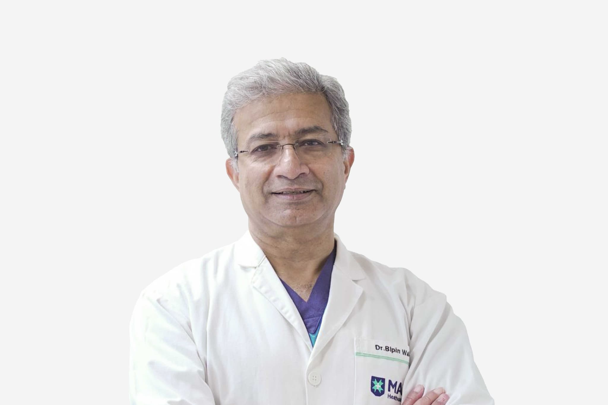
Dr Bipin Walia
Dr Bipin Walia
Chairman & Head – Neurosurgery & Neurospine
Max Smart Super Specialty Hospital, Saket
29+years experience
View Profile
Dr V P Singh
Dr V P Singh
Chairman - Neurosurgery
Max Smart Super Specialty Hospital, Saket
44+years experience
View Profile
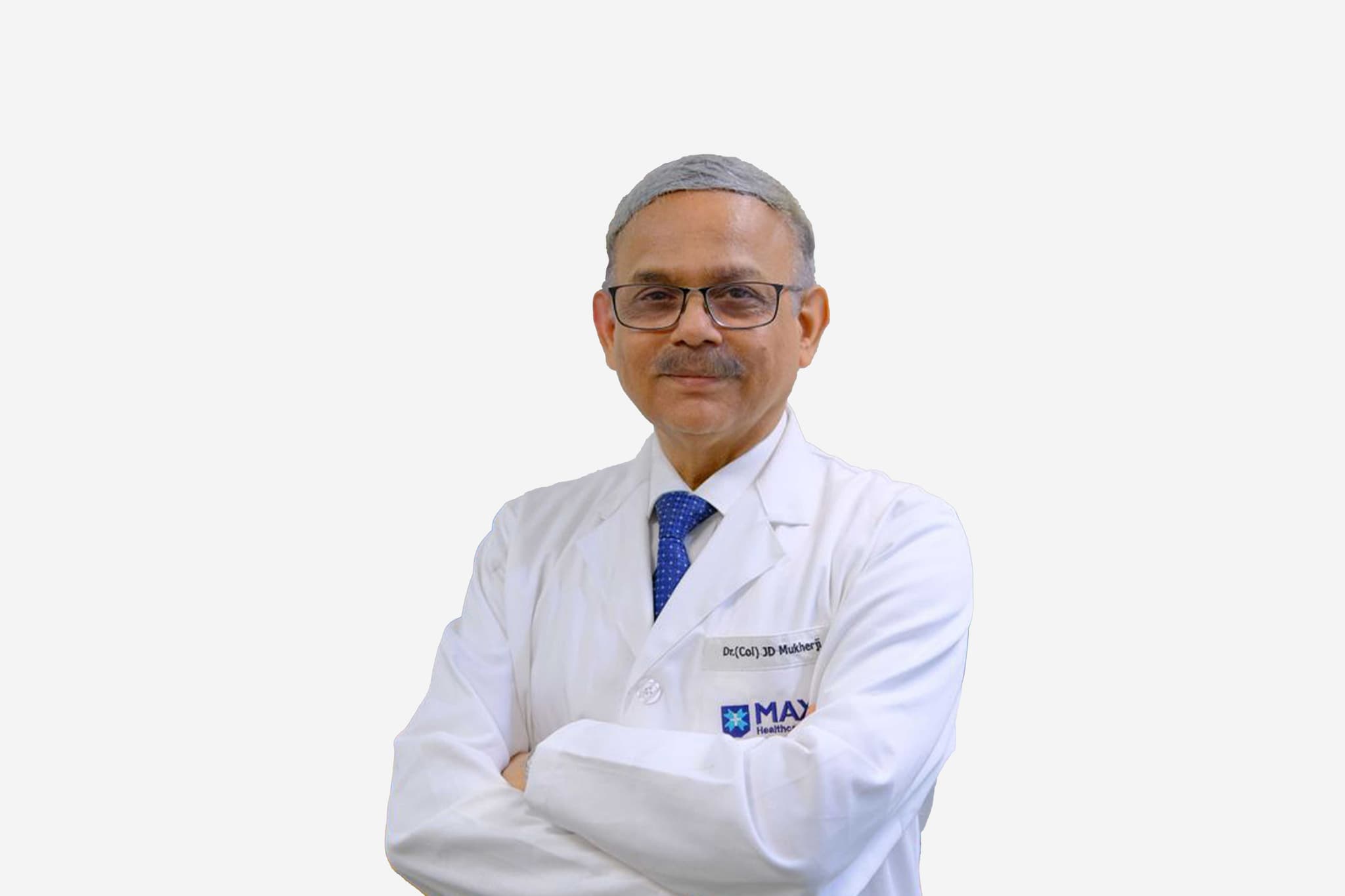
Dr Joy Dev Mukherji
Dr Joy Dev Mukherji
Chairman and Head - Neurology
Max Super Specialty Hospital, Saket
31+years experience
View Profile
Dr Anant Kumar
Dr Anant Kumar
Chairman - Urology Renal Transplant & Robotics
Max Super Specialty Hospital, Vaishali
39+years experience
View Profile
Dr Kamran Ali
Dr Kamran Ali
Associate Director - Thoracic Surgery
Max Smart Super Specialty Hospital, Saket
13+years experience
View Profile
Dr Anita Sethi
Dr Anita Sethi
Principal Director & HOD – Cataract, Lasik and Oculoplasty
Max Multi Specialty Centre, Panchsheel Park
36+years experience
View Profile
Dr Tarun Kapur
Dr Tarun Kapur
Senior Director & HOD - Ophthalmology
Max Smart Super Specialty Hospital, Saket
36+years experience
View Profile
Dr Sanjay Sachdeva
Dr Sanjay Sachdeva
Chairman - ENT
Max Super Specialty Hospital, Saket
40+years experience
View Profile
Dr Vineet Narula
Dr Vineet Narula
Associate Director - ENT (Ear Nose Throat)
Max Smart Super Specialty Hospital, Saket
15+years experience
View Profile
Dr Dinesh Khullar
Dr Dinesh Khullar
Group Chairman - Nephrology & Renal Transplant Medicine
Max Smart Super Specialty Hospital, Saket
33+years experience
View Profile
Dr Puneet Agarwal
Dr Puneet Agarwal
Vice Chairman - Neurology
Max Smart Super Specialty Hospital, Saket
26+years experience
View Profile
Dr Sameer Shrivastava
Chairman - Non Invasive Cardiology
BLK-Max Super Specialty Hospital, Delhi
31+years experience
View Profile
Dr Sandeep Budhiraja
Group Chairman – Internal Medicine & Group Medical Director – Max Healthcare
Max Multi Specialty Centre, Panchsheel Park
30+years experience
View Profile
Dr Vijay Kumar Chopra
Senior Director - Cardiology
Max Super Specialty Hospital, Saket
47+years experience
View Profile
Dr Subhash Gupta
Dr Subhash Gupta
Group Chairman - Centre for Liver & Biliary Sciences
Max Super Specialty Hospital, Vaishali
38+years experience
View Profile

May we help you?

Enter digits only (6-15 digits). We'll add your country code.