+91 83682 47758
info@medivisorhealth.com
Medivisor India Treatment Logo
  • Home
    • About Us
    • Our Services
    • Our Team
    • Our Safety Measures
    • Loading treatments...
    • Loading hospitals...
    • Loading doctors...
    • Treatment Cost
    • Treatment Process
    • Visa Process
    • Travel Guide
    • FAQs
    • Why Medivisor
    • Success Stories
    • Patient Activities
    • Social Activities
    • News Coverage
    • Blog
  • Become a Partner

Medivisor India Treatment is a trusted New Delhi-based medical travel company assisting international patients with advanced, affordable, and seamless treatment in India.

  • Medivisor House 359, Sector 1, Vaishali, Ghaziabad, (Delhi/NCR) India
  • +91 8368 247 758
  • info@medivisorhealth.com

About Us

  • About Us
  • Our Services
  • Our Team
  • Our Medical Advisors
  • Our Safety Measures
  • Join Our Network
  • Contact Us

India Treatment

  • Hospitals
  • Treatments
  • Treatment Cost
  • Treatment Process
  • Visa Process
  • Travel Guide
  • FAQs
  • Why Medivisor

Gallery

  • Patient Testimonials
  • Patient Activities
  • News Coverage
  • Blog

Get in Touch

© 2026 Medivisor India Treatment. All rights reserved.

Dr Kona Lakshmi Kumari

Consultant - Minimal Access & Robotic GI Surgery, Metabolic & Bariatric Surgery

Yashoda Hospital, Somajiguda

GastroenterologistBariatric Surgeon

27+ years experience
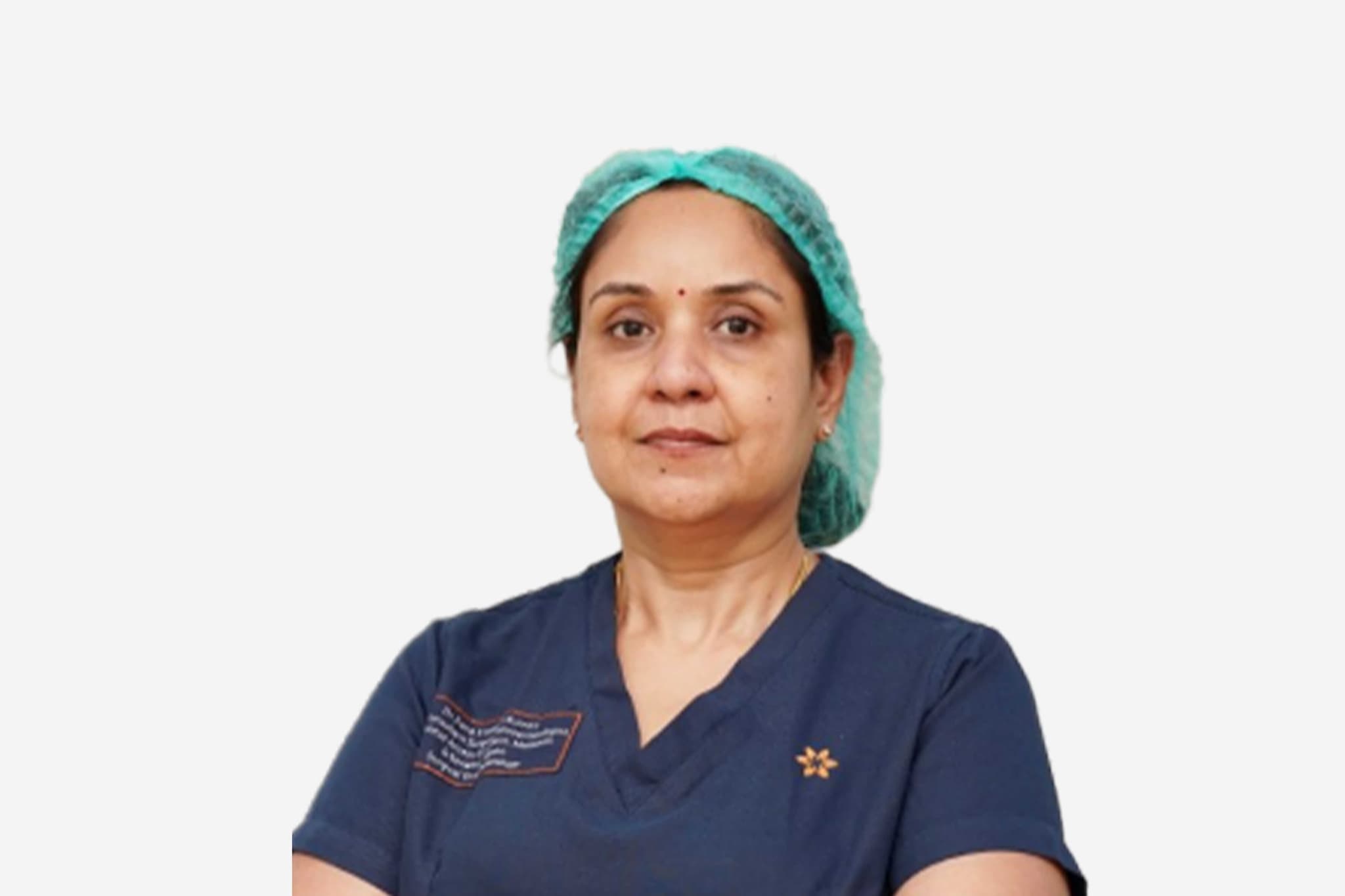
Dr Kona Lakshmi Kumari, Consultant - Minimal Access & Robotic GI Surgery, Metabolic & Bariatric Surgery at Yashoda Hospital, Somajiguda - Yashoda Hospitals
  1. Home
  2. Doctors
  3. Dr Kona Lakshmi Kumari

About Dr Kona Lakshmi Kumari

Dr. Kona Lakshmi Kumari is a highly distinguished specialist in surgical gastroenterology and bariatric surgery, recognized for her technical mastery in complex gastrointestinal interventions, advanced minimal-access procedures, and robotic-assisted surgeries. She specializes in utilizing precision-guided surgical platforms to manage deep visceral pathologies, perform metabolic revisions, and optimize long-term therapeutic outcomes.

Mastery in Advanced Surgical Gastroenterology and GI Surgery

She specializes in the advanced evaluation, diagnosis, and surgical management of a wide range of acute and chronic disorders affecting the digestive tract. His clinical practice leverages over 26 years of refined surgical acumen to execute high-precision abdominal and gastrointestinal procedures, ensuring optimal anatomical restoration, minimal tissue trauma, and predictable recovery timelines.

Innovation in Robotic and Minimal Access Pathways

Dr. Lakshmi Kumari possesses profound expertise in implementing state-of-the-art computer-assisted robotic platforms and advanced laparoscopic techniques within her operative framework. By avoiding traditional open access methodologies, she targets complex structural GI conditions with exceptional visualization and accuracy, significantly reducing post-operative discomfort and accelerating patient rehabilitation.

Specialized Metabolic and Bariatric Interventions

A primary focus of her surgical practice involves the delivery of highly specialized metabolic and bariatric interventions for severe obesity and metabolic disorders. Trained extensively at elite international centers, including the Cleveland Clinic in Ohio, USA, and the National University Hospital (NUH) in Singapore, she designs tailored operative pathways that promote safe, sustainable physiological revision and improved patient health.

Academic Leadership, Mentorship, and Institutional Governance

Throughout her extensive career, Dr. Lakshmi Kumari has combined modern surgical innovations with rigorous clinical governance and a strong commitment to medical education. Serving as a Consultant within the premier institutional framework at Yashoda Hospitals, Somajiguda, she actively mentors the next generation of minimally invasive surgeons and has held prominent leadership positions, including Vice President of IAGES and Organizing Secretary for elite national medical forums.

Dr. Kona Lakshmi Kumari at a Glance

  • Specialist in Surgical Gastroenterology, Minimal Access & Robotic GI Surgery, and Bariatric Interventions.

  • Extensive clinical experience with over 26 years of dedicated service in the medical and surgical field.

  • Elite international training in metabolic surgery from the Cleveland Clinic (USA) and NUH (Singapore).

  • Expert in utilizing state-of-the-art robotic systems to maximize precision during complex abdominal resections.

  • Prominent academic leader who has served in executive roles for major surgical bodies like IAGES.

  • Consultant delivering dedicated, evidence-based clinical governance at Yashoda Hospitals, Somajiguda.

Training in Bariatric Surgery, NUH
Training in Bariatric Surgery, Cleveland Clinic
Training in Minimal Access Bariatric Surgery, Gleneagles Global Hospitals
Fellowship, International College of Surgeons
2007: FIAGES, Indian Association of Gastrointestinal Endosurgeons (IAGES)
1997: MS (General Surgery), S.C.B Medical College and Hospital
1992: MBBS, S.C.B Medical College and Hospital
Board Certified in Gastroenterologist
1st Prize for Scientific Paper in ASICON
Best Paper Awards ASICON at Hyderabad, 2001
Vaidya Shiromani Award at Hyderabad, July 2012

Affiliated Hospitals

Yashoda Hospital, Somajiguda

Multi Specialty

Hyderabad, Telangana

1989

Estd.

620+

Beds

250+

Doctors

May we help you?

Enter digits only (6-15 digits). We'll add your country code.

Related Treatments

Endoscopic Retrograde Cholangiopancreatography (ERCP)

Endoscopic Retrograde Cholangiopancreatography (ERCP) is a highly specialized procedure used primarily to treat disorders of the bile ducts and pancreatic ducts. It uniquely combines advanced endoscopy with real-time X-ray imaging (fluoroscopy). By 2026, ERCP has evolved from a diagnostic tool into a powerful therapeutic intervention, allowing physicians to clear obstructions, place stents, and repair leaks within the delicate ductal system of the liver and pancreas without the need for invasive abdominal surgery.

Why ERCP is Performed

  • Common Bile Duct Stones: Removing gallstones that have migrated out of the gallbladder and are blocking the main drainage duct.

  • Malignant Obstructions: Relieving blockages caused by tumors in the pancreas, bile ducts, or liver.

  • Biliary Strictures: Opening narrowed areas of the ducts caused by scar tissue or chronic inflammation.

  • Post-Surgical Bile Leaks: Sealing leaks that can occasionally occur following gallbladder or liver surgery.

  • Chronic Pancreatitis: Treating complications such as pancreatic stones or narrowings that cause severe, recurring pain.

  • Sphincter of Oddi Dysfunction: Managing issues with the muscular valve that controls the flow of digestive juices.

Conditions That Require Specialized Care

  • Massive Stone Burden: Cases with very large or numerous stones that may require advanced "fragmentation" techniques.

  • Anatomical Variations: Patients who have had previous gastric bypass or stomach surgery, requiring a modified "enteroscopy" approach.

  • Primary Sclerosing Cholangitis (PSC): A chronic condition requiring careful, repeated dilations of the bile ducts.

  • High-Risk Pancreatitis Patients: Individuals with a history of post-procedure inflammation who require preventative "stenting" of the pancreatic duct.

  • Pediatric ERCP: Rare cases in children involving congenital ductal abnormalities that require specialized pediatric endoscopes.

[Image showing the endoscope positioned in the duodenum with the catheter entering the bile duct]

How an ERCP Is Performed

  • Deep Sedation or Anesthesia: To ensure absolute stillness and comfort, the procedure is performed under deep sedation or general anesthesia.

  • Side-Viewing Endoscopy: A specialized duodenoscope is passed through the mouth and stomach to reach the "papilla"—the small opening where the ducts meet the intestine.

  • Cannulation: A tiny catheter is threaded into the papilla to gain access to the biliary or pancreatic tree.

  • Retrograde Contrast: A radio-opaque dye is injected "backward" into the ducts, highlighting the internal anatomy on a live X-ray monitor.

  • Sphincterotomy: Using a small electrified wire, the surgeon may make a tiny cut in the muscle valve (sphincter) to widen the opening for stone removal.

  • Mechanical Intervention: Tools such as baskets, balloons, or stents are passed through the scope to clear blockages or provide structural support.

Innovations in Duct Therapy

  • Disposable DuodenoscopesThe widespread adoption of single-use scopes to eliminate the risk of bacterial transmission and ensure the highest mechanical reliability for every patient.

  • SpyGlass™ CholangioscopyA "micro-scope" that is passed through the main endoscope, allowing the doctor to see directly inside the bile duct in high-definition to biopsy tumors or break up stones with lasers.

  • AI-Assisted FluoroscopyArtificial intelligence that enhances X-ray images in real-time, helping the doctor identify "shadows" or subtle strictures with greater clarity.

  • Biodegradable Biliary StentsNext-generation stents that provide support to a narrowed duct for several months and then dissolve, avoiding the need for a second procedure to remove them.

  • Laser Lithotripsy (EHL)Using targeted laser energy to "shatter" stubborn gallstones into tiny fragments that can be easily swept out of the duct.

  • Automated Risk-Prediction ModelsSoftware that analyzes a patient's health data before the procedure to suggest the exact dose of preventative medication needed to stop post-op inflammation.

Pre-Procedure Preparation

  • Fasting (NPO): A strictly empty stomach is required, usually meaning no food or drink for 8 to 12 hours prior to the procedure.

  • Antibiotic Prophylaxis: Certain patients may be given a dose of antibiotics before the start of the procedure to prevent ductal infection.

  • Blood Work: Checking coagulation (clotting) factors to ensure it is safe to perform a sphincterotomy or biopsy.

  • Medication Coordination: Careful management of blood thinners and diabetic medications is essential for a safe recovery.

  • Post-Op Plan: Because of the intensive nature of ERCP, patients must have a designated driver and a quiet place to recover for the first 24 hours.

Diagnostic and Safety Monitoring

  • Pancreatitis Prevention: Use of rectal medications or temporary "protective" pancreatic stents to lower the risk of post-ERCP inflammation.

  • Amylase/Lipase Checks: Blood tests performed if a patient experiences unusual pain after the procedure to check for pancreatic irritation.

  • Continuous Vital Monitoring: Specialized anesthesia teams monitor heart rhythm and oxygen levels throughout the entire intervention.

  • Radiation Safety: Using "low-dose" pulsed fluoroscopy to minimize X-ray exposure for both the patient and the medical team.

Why This Treatment Is Highly Effective

  • Avoids Major Surgery: ERCP provides a "keyhole" alternative to traditional open-belly surgery for clearing bile duct obstructions.

  • Immediate Obstruction Relief: Patients with jaundice (yellowing of the skin) often see a rapid improvement in symptoms once the duct is cleared or stented.

  • High Precision: The combination of live video and live X-ray ensures that the doctor knows exactly where the instruments are at all times.

  • Life-Saving for Sepsis: In cases of "cholangitis" (infected bile), ERCP provides urgent drainage that is critical for survival.

  • Dual Functionality: It allows for the immediate transition from identifying a problem to fixing it within the same 30-to-60-minute session.

Recovery and Aftercare

  • Immediate Observation: Most patients stay in a recovery unit for 1 to 2 hours to ensure the sedation has worn off and no immediate pain is present.

  • Sore Throat and Bloating: These are the most common minor side effects and typically resolve within 24 to 48 hours.

  • Dietary Progression: Patients usually start with clear liquids and move to a low-fat diet as tolerated by their digestive system.

  • Activity Limits: No driving or making major decisions for 24 hours. Most patients return to full activity within 2 to 3 days.

  • Follow-up Imaging: A simple ultrasound or blood test is often scheduled a few weeks later to confirm the ducts remain clear.

Life After ERCP

  • Rapid resolution of painful symptoms like biliary colic (gallstone pain) or chronic abdominal pressure.

  • Successful management of complex conditions like cancer-related blockages, allowing for a better quality of life.

  • Permanent removal of dangerous "trapped" stones that could otherwise lead to severe liver damage.

  • A return to a normal, healthy diet once the flow of digestive enzymes and bile has been restored.

  • Empowerment through the successful treatment of a condition that previously would have required a long, difficult surgical recovery.

₹From From 300

Laparoscopic Polypectomy

Laparoscopic Polypectomy generally refers to the minimally invasive removal of polyps from the gallbladder or, in specific gynecological cases, subserosal uterine polyps. While most uterine polyps are removed via hysteroscopy (through the cervix), the laparoscopic approach is reserved for polyps or small growths located on the outer surface of an organ or within the abdominal cavity.

When You Should Consider Laparoscopic Polypectomy

  • Symptomatic Polyps: Growths causing localized pain or persistent digestive issues.

  • Suspect Features: Polyps showing irregular shapes or rapid growth during an ultrasound.

  • Size-Dependent Risks: Gallbladder polyps exceeding 1 cm in size, which is a standard surgical indication in 2026 guidelines.

  • Outer Organ Growth: When a polypoid growth is located on the exterior of the uterus or attached to pelvic ligaments.

  • Cancer Mitigation: To prevent the risk of malignancy in polyps that show signs of growth.

Methods of Removal and Techniques

  • Laparoscopic Cholecystectomy: Removal of the entire gallbladder for polyps larger than 10mm, as they cannot be safely scraped off the wall.

  • Laparoscopic Excision: The use of precision shears or lasers to excise a growth at its base from the exterior of an organ.

  • High-Definition Visualization: Utilizing zoomed-in views to identify a polyp’s blood supply and seal vessels before cutting.

  • Oncological Safety Protocols: The use of specimen bags during removal to ensure tissue does not contact the abdominal wall.

  • Hysteroscopy (Alternative): The standard approach for internal uterine polyps, distinct from the laparoscopic method used for external growths.

How Laparoscopic Polypectomy Is Performed

  • Anesthesia: The surgery is performed under general anesthesia to ensure the patient is comfortable and still.

  • Access Incisions: The surgeon makes 1 to 3 tiny incisions, ranging from 5–10 mm, in the abdominal wall.

  • Instrument Insertion: A laparoscope and specialized micro-instruments are inserted through the small incisions.

  • Vessel Sealing: Surgeons use high-definition cameras to seal blood vessels before removal, resulting in near-zero blood loss.

  • Abdominal Inflation: Carbon dioxide gas is used to create space, though this can lead to temporary post-operative bloating.

Pre-Procedure Preparation

  • Fasting Requirements: Patients must fast for 8-12 hours before the surgery or catheterization.

  • Medical Assessments: Blood tests, ECG, and chest X-rays are conducted to assess overall health.

  • Medication Adjustments: Current medications must be adjusted as directed by the medical team.

  • Allergy Discussion: Patients must discuss any allergies, particularly to materials used in surgical devices or dyes.

  • Post-Op Planning: Arranging for post-operative care and a support person for the recovery period.

Tests Before Laparoscopic Polypectomy

  • Ultrasound Imaging: Used to monitor the size and shape of the polyp and detect rapid growth.

  • Blood Tests: To assess overall health and check for markers of inflammation or infection.

  • ECG: To monitor the heart's electrical rhythm and check for conduction issues.

  • Pulse Oximetry: Used to evaluate oxygen saturation levels in the blood before anesthesia.

  • CT Scan: For detailed mapping of the abdominal cavity and complex growths.

Life After Laparoscopic Polypectomy

  • Hospital Stay: Usually a same-day discharge or a short 23-hour observation stay.

  • Immediate Recovery: Walking is encouraged immediately to aid digestion and prevent blood clots.

  • Pain Management: Discomfort is typically managed with over-the-counter anti-inflammatories.

  • Return to Work: Patients can generally return to work within 5 to 7 days post-procedure.

  • Activity Restrictions: Heavy lifting must be avoided for approximately 4 weeks to ensure proper healing.

Benefits of Laparoscopic Polypectomy

  • Minimal Tissue Disruption: Rapid recovery due to the use of tiny incisions rather than large open cuts.

  • Precise Visualization: High-definition cameras allow for the zoom-in on blood supplies for safer removal.

  • Near-Zero Blood Loss: Advanced sealing techniques minimize bleeding during the excision.

  • Strict Safety: The use of specialized bags prevents tissue fragments from remaining in the pelvic cavity.

  • Long-Term Cure: Provides a definitive treatment with high technical success rates for symptomatic growths.

₹From From 300

Liver Biopsy
Liver Biopsy

A liver biopsy is a medical procedure used to remove a small piece of liver tissue for laboratory examination. It is considered the definitive "gold standard" for identifying the precise cause of liver disease and determining the extent of organ damage. By 2026, advances in real-time imaging and needle technology have made the procedure faster and safer, allowing clinicians to distinguish between various types of inflammation, scarring, and tumors that blood tests or standard scans might miss.

When You Should Consider a Liver Biopsy

  • Unexplained Liver Enzymes: When blood tests show persistent elevations in ALT or AST that cannot be explained by standard diagnostic sets.

  • Staging Fibrosis: To determine the exact degree of scarring (cirrhosis) in patients with chronic conditions like Fatty Liver Disease (NASH/MASH).

  • Hepatitis Monitoring: Evaluating the severity of chronic Hepatitis B or C to decide on the most effective antiviral therapy.

  • Autoimmune Disorders: Diagnosing conditions where the body's immune system attacks the liver, such as Primary Biliary Cholangitis (PBC).

  • Transplant Monitoring: Checking a transplanted liver for early signs of organ rejection or medication toxicity.

  • Lumps or Masses: Investigating a suspicious nodule found on an ultrasound or MRI to rule out malignancy.

Types of Liver Biopsy

  • Percutaneous Biopsy: The standard approach. Under the guidance of ultrasound or CT, a specialized needle is inserted through the skin between the ribs directly into the liver to collect a tissue core.

  • Transjugular Biopsy: A specialized technique for patients with blood clotting issues or excess abdominal fluid (ascites). A catheter is threaded through the jugular vein in the neck and guided down to the liver's internal veins.

  • Laparoscopic Biopsy: Tissue is collected during a minimally invasive surgical procedure, allowing the doctor to visually inspect the liver surface and sample specific, hard-to-reach areas.

  • Fine-Needle Aspiration (FNA): Often used for specific liver tumors to collect cells for cytology, though it provides less structural information than a core biopsy.

How a Liver Biopsy Is Performed

  • Positioning: The patient typically lies on their back with the right arm raised above the head to widen the space between the ribs.

  • Imaging Guidance: A physician uses an ultrasound probe to identify the safest "window" for the needle, avoiding the gallbladder and major blood vessels.

  • Local Anesthesia: The skin and the lining of the chest wall are numbed; most patients feel a sensation of pressure rather than sharp pain.

  • The "Click": Many modern biopsy needles use a spring-loaded mechanism that captures the tissue sample in a fraction of a second.

  • Breath-Hold: The patient is asked to hold their breath for a few seconds during the needle insertion to keep the liver perfectly still.

  • Sample Verification: The tissue core is immediately inspected to ensure it is of sufficient length (usually 1.5 to 2 cm) for an accurate diagnosis.

Innovations in Liver Diagnosis

  • AI-Enhanced HistopathologyArtificial Intelligence algorithms that analyze biopsy slides to provide a more objective "score" for inflammation and fat content, reducing human variability.

  • Molecular ProfilingUsing biopsy tissue for genetic sequencing to identify the exact subtype of liver cancer, allowing for highly targeted chemotherapy.

  • Digital "Tension" MeasurementNeedles equipped with sensors that can measure liver stiffness (elasticity) at the exact moment the biopsy is taken.

  • Multiparametric MRI-Guided BiopsyA high-tech approach that overlays MRI data onto ultrasound monitors, helping surgeons target "hot spots" of metabolic activity.

  • Micro-RNA AnalysisAnalyzing the sample for specific RNA markers that can predict how fast a patient's liver disease might progress over the next decade.

  • Transjugular Pressure MonitoringMeasuring the pressure within the liver veins (HVPG) during a transjugular biopsy to provide critical data on portal hypertension.

Pre-Procedure Preparation

  • Coagulation Testing: Essential blood tests (INR and Platelets) are performed to ensure the blood clots normally before the procedure.

  • Medication Adjustment: Patients must stop taking aspirin, ibuprofen, and other blood thinners for 5–7 days prior to the biopsy.

  • NPO Guidelines: Standard fasting (no food or drink) for 6–8 hours before the appointment is typically required.

  • Observation Planning: Arranging for someone to drive you home, as you will need to remain under observation for several hours post-procedure.

  • Ultrasound Mapping: A pre-procedure scan to confirm the liver size and the absence of fluid that might interfere with the needle path.

Diagnostic and Safety Monitoring

  • Post-Biopsy Recovery Position: Patients must lie on their right side for 2–4 hours immediately after the procedure. This uses the body's own weight to apply pressure to the biopsy site, preventing bleeding.

  • Vital Sign Checks: Nurses monitor blood pressure and heart rate every 15 minutes for the first hour to detect any signs of internal bleeding.

  • Pain Assessment: Monitoring for "referred pain" in the right shoulder, which is a common but harmless sign of diaphragm irritation.

  • Follow-up Ultrasound: In rare cases of suspected bleeding, a quick bedside scan can confirm the stability of the liver.

Why This Treatment Is Highly Effective

  • Definitive Accuracy: While scans show the "shape" of the liver, a biopsy shows the "activity" of the cells, providing an answer that imaging cannot.

  • Personalized Care: Knowing the exact stage of disease allows doctors to prescribe the correct dosage of medication and avoid unnecessary treatments.

  • Cures Through Discovery: Often identifies rare, treatable conditions like Wilson’s disease or Hemochromatosis that might otherwise go undetected.

  • Proven Safety Record: With modern imaging guidance, the risk of serious complications has dropped to less than 1% in most clinical centers.

  • Minimal Recovery: Most patients return to their daily routine within 48 hours, making it a low-impact procedure with high-impact results.

Recovery and Aftercare

  • Activity Limits: Avoid heavy lifting, intense exercise, or "straining" the abdominal muscles for at least one week following the procedure.

  • Wound Care: The small puncture site usually only requires a simple bandage; it should be kept dry for 24 hours.

  • Pain Management: Over-the-counter acetaminophen is usually sufficient for any lingering site soreness; aspirin and NSAIDs should still be avoided for a few days.

  • Signs to Watch For: Patients are instructed to contact their doctor immediately if they experience severe abdominal pain, fainting, or fever.

  • Results Timeline: A preliminary report may be available in 2–3 days, while complex stains and genetic markers typically take 7–10 business days.

Life After a Liver Biopsy

  • A clear, evidence-based diagnosis that ends the "diagnostic mystery" of elevated liver enzymes.

  • A personalized roadmap for health, including specific dietary, lifestyle, and medication recommendations.

  • Peace of mind for transplant recipients knowing their new organ is being monitored with the highest level of detail.

  • Improved long-term outcomes through the early detection of fibrosis before it progresses to irreversible cirrhosis.

  • The opportunity to participate in clinical trials for new liver therapies based on the specific tissue characteristics found.

₹From From 200

Sleeve Gastrectomy
Sleeve Gastrectomy

Sleeve gastrectomy, often called the Gastric Sleeve, is a permanent weight-loss surgery that involves removing approximately 75–80% of the stomach. The remaining portion is joined together to form a narrow tube or "sleeve" about the size and shape of a banana. This procedure provides a powerful tool for long-term weight management by physically and hormonally changing how the body processes hunger and food intake.

How It Works

  • Physical Restriction: The significantly smaller stomach capacity limits the amount of food you can eat at one time, making you feel full much faster.

  • Hormonal Changes: By removing the upper portion of the stomach (the fundus), the surgery drastically reduces the production of Ghrelin, known as the "hunger hormone."

  • Appetite Suppression: The reduction in Ghrelin leads to a decreased appetite and significantly improved blood sugar control.

  • Natural Digestion: Unlike a gastric bypass, the intestines are not re-routed, meaning the natural digestion process remains largely intact.

When You Should Consider a Sleeve Gastrectomy

  • Body Mass Index (BMI): Typically recommended for individuals with a BMI of 40 or higher, or a BMI of 35 or higher with obesity-related health conditions.

  • Type 2 Diabetes: If you are struggling to manage blood sugar levels through diet and medication alone.

  • Hypertension: When high blood pressure persists despite medical intervention and lifestyle changes.

  • Obstructive Sleep Apnea: If excess weight is causing significant breathing interruptions during sleep.

  • Failed Weight Loss Efforts: For those who have been unable to achieve sustained weight loss through traditional diet and exercise programs.

How Is Performed

  • Anesthesia: The surgery is performed under general anesthesia and typically takes between 60 to 90 minutes.

  • Laparoscopic Approach: This is a minimally invasive "keyhole" surgery where the surgeon makes 4–5 small incisions in the abdomen to insert a camera and specialized instruments.

  • The Resection: The surgeon uses a surgical stapler to divide the stomach vertically, removing the outer curved portion of the organ.

  • Staple Line Reinforcement: The edges of the new stomach sleeve are securely stapled to ensure a watertight and airtight seal.

  • No Re-routing: The connection between the stomach and the small intestine is preserved, allowing for normal nutrient absorption.

Pre-Procedure Preparation

  • Nutritional Counseling: Working with a dietitian to understand the strict dietary phases required before and after surgery.

  • Psychological Evaluation: To ensure you have a strong support system and a clear understanding of the lifelong lifestyle changes required.

  • Medical Screening: A comprehensive review of heart and lung health to ensure you are a safe candidate for general anesthesia.

  • Pre-Op Liquid Diet: Most patients must follow a specific high-protein, low-calorie liquid diet for 1–2 weeks before surgery to shrink the liver.

  • Smoking Cessation: You must stop smoking at least 6 weeks before surgery to reduce the risk of staple line leaks and breathing complications.

Tests Before Sleeve Gastrectomy

  • Upper Endoscopy (EGD): To check the health of the stomach lining and rule out significant hiatal hernias or ulcers.

  • Abdominal Ultrasound: Specifically used to check for gallstones, which can be affected by rapid weight loss.

  • Blood Panels: Comprehensive testing of vitamin levels, blood sugar (HbA1c), and kidney function.

  • Sleep Study: To identify and treat undiagnosed sleep apnea prior to the administration of anesthesia.

Life After Sleeve Gastrectomy

  • Hospital Stay: Most patients remain in the hospital for 1 to 2 nights for monitoring and pain management.

  • Strict Dietary Phases: You must follow a multi-phase transition to allow the staple line to heal:
    Weeks 1–2: Clear liquids only.
    Weeks 3–4: Pureed and soft foods.
    Week 5+: Gradual introduction of solid, high-protein foods.

  • Lifelong Supplements: You must take daily vitamin and mineral supplements forever to prevent nutritional deficiencies.

  • Activity Restrictions: Most people return to work within 2 weeks but must avoid heavy lifting or core-straining exercise for a full 6 weeks.

  • Small Meals: You will transition to eating small, nutrient-dense meals throughout the day, as the stomach can no longer hold large volumes.

Why Specialized Treatment Is Highly Effective

  • Substantial Weight Loss: Most patients lose 60–70% of their excess body weight within the first 12–18 months.

  • Disease Resolution: The procedure can significantly improve or even resolve conditions like Type 2 diabetes, high blood pressure, and fatty liver disease.

  • Increased Longevity: By reducing the strain of obesity on the heart and joints, the surgery contributes to a longer, more active life.

  • Minimally Invasive: The laparoscopic technique results in less pain, smaller scars, and a much faster recovery than traditional open surgery.

  • Simpler Mechanism: Because the intestines are not re-routed, there is a lower risk of long-term malabsorption or "dumping syndrome" compared to other weight-loss surgeries.

₹From From 2000

Roux-en-Y Gastric Bypass

Roux-en-Y Gastric Bypass (RYGB) is considered the "gold standard" of weight-loss surgery. It is a complex, permanent procedure that involves creating a small stomach pouch and rerouting the small intestine to change how your body absorbs calories and nutrients.

How It Works

  • Restriction: The new stomach pouch is roughly the size of an egg (about 30ml), which drastically limits how much you can eat.

  • Malabsorption: By bypassing the first part of the small intestine, your body absorbs fewer calories and fats from the food you consume.

  • Hormonal Shifts: The surgery alters gut hormones (like GLP-1 and PYY) that suppress hunger, increase satiety, and rapidly improve blood sugar levels.

When You Should Consider a Gastric Bypass

  • Severe Obesity: Typically for individuals with a BMI of 40 or higher, or a BMI of 35 with serious weight-related health problems.

  • Type 2 Diabetes: When rapid and significant improvement in blood sugar control is a primary medical goal.

  • Chronic GERD: For patients with severe acid reflux that may be worsened by other types of weight-loss surgery.

  • Metabolic Syndrome: When a combination of high blood pressure, high blood sugar, and abnormal cholesterol levels requires a powerful intervention.

  • Failed Weight Loss: For those who have not achieved sufficient results through diet, exercise, or other non-surgical methods.

How Is Performed

  • Anesthesia: The surgery is performed under general anesthesia and typically takes 2 to 3 hours.

  • Laparoscopic Approach: Most procedures are performed using minimally invasive techniques, involving several small incisions in the abdomen for a camera and specialized tools.

  • Creating the Pouch: The surgeon staples the top section of the stomach to separate it from the rest, creating a small, functional pouch.

  • The "Roux" Limb: The small intestine is divided; the bottom end is lifted up and connected directly to the new small stomach pouch.

  • The Reconnection: The remaining part of the small intestine (still attached to the "old" stomach) is reconnected further down the "Roux" limb so that digestive juices can eventually mix with food.

Pre-Procedure Preparation

  • Nutritional Counseling: Extensive education on the strict dietary phases and lifelong eating habits required after surgery.

  • Psychological Clearance: To ensure you are emotionally prepared for the significant lifestyle changes and body image transitions.

  • Medical Screening: Comprehensive heart, lung, and gastrointestinal evaluations to minimize surgical risks.

  • Pre-Op Liquid Diet: A specialized diet for 1–2 weeks prior to surgery to reduce liver size and make the procedure safer.

  • Smoking Cessation: Patients must stop smoking at least 6 weeks before surgery to reduce the risk of ulcers and healing complications.

Tests Before Gastric Bypass

  • Upper Endoscopy (EGD): To examine the stomach lining and check for hiatal hernias or ulcers.

  • Blood Panels: Detailed testing for baseline vitamin and mineral levels, as well as kidney and liver function.

  • Sleep Study: To detect and treat obstructive sleep apnea, which is common in bariatric patients.

  • Cardiology Clearance: Including an EKG or stress test to ensure the heart can handle general anesthesia.

Life After Gastric Bypass

  • Hospital Stay: Usually involves 2 nights for observation and initial recovery.

  • Post-Op Diet Progression: A strict transition from clear liquids (weeks 1–2) to pureed foods (weeks 3–4) and finally soft solids.

  • Dumping Syndrome: A unique side effect where sugary or high-fat foods cause rapid heart rate, nausea, and sweating as food "dumps" too quickly into the small intestine.

  • Lifelong Supplements: Due to malabsorption, you must take daily multivitamins, calcium, B12, and iron for the rest of your life.

  • Activity Restrictions: Most patients return to work in 2 weeks but must avoid heavy lifting for 6 weeks to allow internal reconnections to heal.

Why Specialized Treatment Is Highly Effective

  • Significant Weight Loss: Patients typically lose 60% to 80% of their excess body weight within 12 to 18 months.

  • Diabetes Remission: Exceptionally effective at treating Type 2 diabetes, often leading to normal blood sugar levels within days of surgery.

  • Reflux Relief: Unlike some other procedures, a gastric bypass often cures chronic acid reflux (GERD).

  • Long-Term Health Benefits: Dramatically reduces the risk of stroke, heart disease, and several types of cancer.

  • Comprehensive Metabolic Control: Provides a multi-layered approach by combining physical restriction, malabsorption, and beneficial hormonal changes.

₹From From 2000

Mini Gastric Bypass
Mini Gastric Bypass

The Mini Gastric Bypass (MGB), also known as One-Anastomosis Gastric Bypass (OAGB), is a simplified, minimally invasive version of the traditional Roux-en-Y gastric bypass. It is considered both a restrictive and malabsorptive procedure, meaning it reduces the amount of food you can eat and limits the calories your body absorbs.

When You Should Consider a Mini Gastric Bypass

  • High BMI: Typically recommended for individuals with a Body Mass Index (BMI) of 40 or higher, or 35 with obesity-related health conditions.

  • Type 2 Diabetes: For patients seeking rapid and significant improvement in blood sugar control through metabolic surgery.

  • Hypertension: When high blood pressure persists despite medical intervention and significant weight gain.

  • Obstructive Sleep Apnea: If excess weight is causing breathing interruptions during sleep that impact daily energy and heart health.

  • Need for Shorter Surgery: For patients where a shorter operative time is preferred over the more complex traditional bypass.

How Is Performed

  • Anesthesia: The surgery is performed under general anesthesia and typically takes between 60 to 90 minutes.

  • Laparoscopic Approach: This is a "keyhole" surgery where the surgeon makes small incisions in the abdomen to insert a camera and specialized surgical instruments.

  • Creation of the Pouch: The surgeon staples the upper part of the stomach into a long, narrow tube (roughly the size of a banana) that holds about 30–50 ml of food.

  • Single Anastomosis (The Join): Unlike the two connections required in traditional bypass, the surgeon makes only one connection. A loop of the small intestine is brought up and attached directly to the new stomach pouch.

  • The Bypass: Approximately 150 to 200 cm of the small intestine is bypassed, redirecting food away from the area where most calories and fats are typically absorbed.

Pre-Procedure Preparation

  • Nutritional Counseling: Working with a specialist to understand the strict dietary phases and the importance of lifelong protein intake.

  • Psychological Evaluation: To ensure readiness for the significant lifestyle changes and dietary restrictions following surgery.

  • Medical Screening: A comprehensive review of cardiovascular and respiratory health to ensure safety during general anesthesia.

  • Pre-Op Liquid Diet: Most patients follow a high-protein, low-calorie liquid diet for 1–2 weeks before surgery to reduce liver size.

  • Smoking Cessation: You must stop smoking at least 6 weeks before surgery to minimize the risk of ulcers and complications at the surgical join.

Tests Before Mini Gastric Bypass

  • Upper Endoscopy (EGD): To examine the stomach lining and check for hiatal hernias, ulcers, or evidence of significant acid reflux.

  • Abdominal Ultrasound: Primarily used to check for gallstones, which can be affected by rapid weight loss.

  • Blood Panels: Comprehensive testing for baseline vitamin levels (B12, Iron, Vitamin D) and kidney/liver function.

  • Sleep Study: To identify and treat undiagnosed sleep apnea before the administration of anesthesia.

  • ECG: A standard heart check to ensure cardiac stability for the duration of the procedure.

Life After Mini Gastric Bypass

  • Hospital Stay: Most patients stay in the hospital for 1 to 2 nights for observation and pain management.

  • Staged Diet Progression: To allow the new joins to heal, patients follow a strict multi-phase plan:
    Days 1–3: Clear liquids only (water, broth).
    Week 2: Full liquids (protein shakes, thin soups).
    Weeks 3–4: Pureed foods (mashed vegetables, soft eggs).
    Week 5+: Gradual introduction of soft solids, moving toward regular healthy food.

  • Lifelong Supplements: Due to malabsorption, you must take daily multivitamins, calcium, B12, and iron for the rest of your life.

  • Activity Restrictions: Most patients return to work within 2 to 3 weeks but must avoid heavy lifting for 6 weeks to protect the internal staples.

Why Specialized Treatment Is Highly Effective

  • Significant Weight Loss: Patients often lose 60% to 75% of their excess body weight within the first year of surgery.

  • Metabolic Improvement: Highly effective at resolving or improving Type 2 diabetes, high blood pressure, and sleep apnea.

  • Technically Simpler: Having only one surgical join reduces operative time and lowers the risk of certain complications like internal hernias.

  • Potentially Reversible: Because no part of the stomach is removed from the body, the procedure is potentially reversible if ever medically necessary.

  • Durable Results: Combines the benefits of a smaller stomach with reduced calorie absorption for long-term weight maintenance and health.

₹From From 2000

Revisional Bariatric Surgery

Revisional bariatric surgery is a specialized procedure performed on patients who have already undergone a primary weight-loss surgery but require a second operation. This may be due to medical complications, insufficient weight loss, or significant weight regain. Because of existing scar tissue and altered anatomy, these procedures are more complex and carry higher risks than initial weight-loss surgeries.

When You Should Consider Revisional Surgery

  • Inadequate Weight Loss or Regain: If the initial stomach pouch has stretched over time or the body has adapted to the metabolic changes of the first surgery.

  • Severe Acid Reflux (GERD): Persistent or worsening reflux that does not respond to medication, frequently occurring after a gastric sleeve procedure.

  • Mechanical Complications: Issues such as a "slipped" gastric band, a staple line leak, or a narrowing (stricture) of a previous surgical connection.

  • Chronic Ulcers or "Dumping Syndrome": Persistent digestive issues following a bypass that cannot be managed through dietary changes alone.

  • Nutritional Malabsorption: Severe deficiencies in vitamins or protein that remain uncontrolled despite high-dose supplementation.

Common Revisional Procedures

  • Sleeve to Bypass Conversion: Converting a gastric sleeve into a Roux-en-Y gastric bypass. This is highly effective for treating severe reflux and triggering further weight loss.

  • Gastric Band Conversion: Removing a "Lap-Band" and converting the anatomy into either a sleeve or a bypass, often performed in a single or two-stage process.

  • Bypass Revision (Stoma Tightening): For a stretched gastric bypass, a surgeon may "re-tighten" the connection (stoma) to restore the feeling of fullness.

  • Intestinal Lengthening: Adjusting the "Roux" limb to increase the bypassed section of the intestine, thereby increasing calorie malabsorption.

  • Sleeve Re-Sleeving: Trimming a previously stretched gastric sleeve to restore its original narrow, restrictive shape.

How Is Performed

  • Anesthesia: The surgery is performed under general anesthesia and typically takes between 2 to 4 hours due to the precision required.

  • Surgical Approach: Almost always performed laparoscopically or robotically to navigate through previous scar tissue with high-definition visualization and accuracy.

  • Managing Adhesions: The surgeon must carefully separate internal scarring (adhesions) from previous operations before the new revision can begin.

  • Stapling and Joins: New surgical joins are created using specialized stapling instruments designed for thicker or scarred tissue.

  • Leak Testing: Extensive intra-operative testing is performed to ensure all new connections are watertight and airtight before finishing.

Pre-Procedure Preparation

  • Anatomy Mapping: Extensive imaging is required to visualize the current state of the stomach and intestines before entering the operating room.

  • Nutritional Optimization: Correcting any existing vitamin deficiencies through intensive supplementation prior to surgery.

  • Psychological Assessment: A thorough review to ensure the patient is prepared for the "reset" in dietary habits and lifestyle expectations.

  • Smoking Cessation: Patients must stop smoking at least 6 to 8 weeks before surgery, as nicotine significantly increases the risk of staple line leaks in scarred tissue.

  • Weight Management: Following a specific pre-operative diet to reduce liver size and improve surgical access.

Tests Before Revisional Surgery

  • Upper Endoscopy (EGD): Inserting a camera into the stomach to inspect the pouch and check for ulcers, inflammation, or stretching.

  • Barium Swallow (UGI Series): A specialized X-ray where the patient swallows contrast liquid to show the exact size and flow of the current anatomy.

  • Abdominal CT Scan: To provide a 3D view of the internal organs and identify any hidden hernias or mechanical issues.

  • Comprehensive Blood Panels: Detailed analysis of iron, B12, Vitamin D, and protein levels to establish a baseline.

Life After Revisional Surgery

  • Hospital Stay: Usually involves 2 to 3 nights of close monitoring to ensure the new surgical joins are healing correctly.

  • Dietary Reset: Patients must return to a "Phase 1" clear liquid diet and progress slowly through pureed and soft foods over a 4–6 week period.

  • Slower Weight Loss: It is important to note that weight loss after a revision is typically slower and less dramatic than after the first surgery.

  • Lifelong Monitoring: Regular follow-up appointments and blood tests are essential to monitor for malabsorption and nutritional health.

  • Activity Restrictions: Light walking is encouraged immediately, but heavy lifting and core-straining exercises are restricted for 6 weeks.

Why Specialized Treatment Is Highly Effective

  • Resolves Chronic Pain and Reflux: Provides immediate relief for patients suffering from the mechanical failures or side effects of a primary surgery.

  • Second Chance for Health: Allows patients who did not reach their goals to restart their weight-loss journey with a more powerful metabolic tool.

  • Precision Technology: The use of robotic platforms allows surgeons to operate safely in "hostile" abdomens filled with previous scar tissue.

  • Customized Anatomy: Every revision is tailored specifically to the patient’s existing anatomy, ensuring the most effective solution for their unique situation.

  • Prevents Long-Term Damage: Addressing issues like chronic ulcers or severe reflux prevents permanent damage to the esophagus and digestive tract.

₹From From 2000

Laparoscopic Gastric Bypass Surgery

Gastric Sleeve and Gastric Bypass are the primary surgical treatments for severe obesity and its associated metabolic conditions, such as Type 2 diabetes and hypertension. While both procedures aim to facilitate significant weight loss and metabolic health, they utilize different surgical mechanisms—one focusing on reducing the stomach's size and the other involving the rerouting of the digestive tract. By 2026, the use of robotic-assisted platforms has become the standard for these procedures, providing surgeons with the precision needed to ensure optimal outcomes and faster patient recovery.

When You Should Consider Bariatric Surgery

  • Class II or III Obesity: Generally recommended for individuals with a BMI over 35, or over 30 when accompanied by serious health conditions.

  • Uncontrolled Type 2 Diabetes: When blood sugar remains high despite intensive medical management.

  • Severe Obstructive Sleep Apnea: When weight-related airway obstruction impacts cardiovascular health and daily energy.

  • Non-Alcoholic Fatty Liver Disease (NAFLD): To reduce liver fat and prevent the progression toward cirrhosis.

  • Chronic Joint Pain: For patients whose weight has led to severe debilitating osteoarthritis in the knees or hips.

  • Failed Conventional Weight Loss: After multiple documented attempts at diet and exercise have failed to produce sustainable results.

Conditions That Require Specialized Care

  • Severe Acid Reflux (GERD): Patients with significant reflux are often steered toward Gastric Bypass, as the Gastric Sleeve can sometimes exacerbate the condition.

  • Sweet Eaters: Those with a high intake of processed sugars may benefit from the "Dumping Syndrome" feedback of Gastric Bypass to help modify behavior.

  • Previous Abdominal Surgeries: Presence of extensive scar tissue that may require a robotic-assisted approach for safe navigation.

  • Polycystic Ovary Syndrome (PCOS): Requiring careful hormonal monitoring as rapid weight loss begins to restore fertility and metabolic balance.

  • Extreme BMI (>60): Sometimes managed with a "staged" approach—performing a sleeve first and converting to a bypass later if needed.

How These Procedures Are Performed

  • Robotic-Assisted Access: Both surgeries are performed through several tiny "keyhole" incisions, utilizing a 3D high-definition camera and robotic wrists for extreme dexterity.

  • Sleeve Gastrectomy (Gastric Sleeve): Approximately 75–80% of the stomach is permanently removed along the "greater curvature," leaving a narrow, sleeve-like pouch.

  • Gastric Bypass (RYGB): A small stomach pouch (roughly the size of an egg) is created and connected directly to the middle part of the small intestine, bypassing the rest of the stomach.

  • Hormonal Modulation: Both procedures involve the removal or bypassing of the area where "Ghrelin" (the hunger hormone) is produced, significantly reducing appetite.

  • Staple Line Reinforcement: Surgeons use advanced stapling technology, often reinforced with specialized sutures or "buttress" material, to ensure a leak-proof seal.

  • Leak Testing: Before finishing the surgery, a specialized dye or air test is performed to confirm the absolute integrity of all new connections.

Innovations in Bariatric Surgery

  • Robotic Precision PlatformsAdvanced systems that provide surgeons with tremor-free movement and 3D visualization, making the complex suturing of a bypass significantly safer.

  • Digital Health IntegrationModern care involves wearable tech and apps that sync with the bariatric team, tracking protein intake and hydration in real-time.

  • Smart StaplersDevices that automatically sense tissue thickness and adjust the firing pressure to create more consistent and secure staple lines.

  • Endoscopic Revision (TORE)The ability to "tighten" a previous bypass or sleeve using a camera through the mouth, avoiding the need for a second major surgery if weight is regained.

  • Fluorescence-Guided SurgeryUsing injectable dyes that glow under specialized light to ensure a perfect blood supply to the newly created stomach pouch.

  • Vagal Nerve BlockingExperimental neuro-modulators that can be adjusted after surgery to help control hunger signals and improve long-term success.

Pre-Procedure Preparation

  • Psychological Evaluation: To ensure the patient has the mental health support and coping strategies needed for a major lifestyle transition.

  • Nutritional Counseling: Multiple sessions with a bariatric dietitian to learn the "protein-first" rule and the importance of lifelong vitamins.

  • Pre-Op Liquid Diet: A 1-to-2-week specialized diet intended to shrink the liver, making the surgery safer and easier for the surgeon.

  • Cardiac and Sleep Clearance: Ensuring the heart and lungs are strong enough for general anesthesia and the post-operative healing phase.

  • Smoking Cessation: A mandatory requirement for many programs, as nicotine significantly increases the risk of stomach ulcers and poor healing.

Diagnostic and Safety Monitoring

  • Upper GI Series (Swallow Test): An X-ray performed shortly after surgery to verify that liquids are passing correctly through the new anatomy.

  • Vitamin Level Tracking: Regular blood tests to monitor levels of Iron, B12, Vitamin D, and Calcium to prevent long-term deficiencies.

  • Body Composition Analysis: Using advanced scales to ensure that weight loss is coming from fat mass rather than muscle tissue.

  • Gallbladder Ultrasound: Monitoring for gallstones, which can occasionally form during the rapid weight loss phase following surgery.

  • Hemoglobin A1c Monitoring: Tracking the rapid (often near-instant) improvement in blood sugar levels in diabetic patients.

Why This Treatment Is Highly Effective

  • Metabolic Reset: These surgeries do more than "shrink the stomach"; they fundamentally change the body's hormonal response to food and insulin.

  • Sustainable Results: Unlike traditional dieting, bariatric surgery alters the body's "set point," making it easier to maintain weight loss long-term.

  • Resolution of Co-morbidities: Many patients see a complete remission of Type 2 diabetes, sleep apnea, and high blood pressure within months.

  • Increased Longevity: Studies consistently show that bariatric surgery reduces the long-term risk of heart disease, stroke, and certain cancers.

  • Enhanced Quality of Life: Patients often report a dramatic increase in mobility, energy levels, and social confidence.

Recovery and Aftercare

  • Early Mobilization: Walking is required within 4 hours of surgery to promote circulation and help clear the surgical gas from the body.

  • Phased Diet: A strict transition from clear liquids to purees, then soft foods, and finally solid proteins over a 6-to-8-week period.

  • Lifelong Supplementation: Daily bariatric-specific multivitamins are required to compensate for the changes in nutrient absorption.

  • Support Groups: Regular attendance at peer support groups is highly correlated with long-term weight loss success.

  • Follow-up Schedule: Frequent visits with the surgical team during the first year (1, 3, 6, and 12 months) to monitor progress and nutrition.

Life After Bariatric Surgery

  • A transformed relationship with food, where "fullness" is felt quickly and hunger signals are significantly muted.

  • The ability to engage in physical activities that were previously impossible, such as running, hiking, or playing with children.

  • Significant improvement in self-image and mental well-being as health goals are met and surpassed.

  • A requirement for ongoing mindful eating—surgery is a powerful "tool," but long-term success depends on a healthy partnership with that tool.

  • A renewed lease on life, often described by patients as a "second chance" at a healthy, active future.

₹From From 1500

Related Specialists

Dr BSV Raju
Dr BSV Raju
Spine Surgeon, Neurosurgeon
Yashoda Hospital, Somajiguda
29+years experience
View Profile
Dr C Raghu
Dr C Raghu
Cardiologist, Interventional Cardiologist
Yashoda Hospital, Somajiguda
28+years experience
View Profile
Dr Deepak Subramanian
Bariatric Surgeon, General Surgeon
MGM Healthcare, Chennai
15+years experience
View Profile
Dr G Moinoddin
Dr G Moinoddin
Bariatric Surgeon
Manipal Hospital, Hebbal, Bangalore
19+years experience
View Profile
Dr Girish Bapat
Dr Girish Bapat
Bariatric Surgeon, General Surgeon
Jehangir Hospital
33+years experience
View Profile
Dr Hemanth Kumar
Dr Hemanth Kumar
Bariatric Surgeon, General Surgeon
Fortis Flt. Lt. Rajan Dhall Hospital, Vasant Kunj
20+years experience
View Profile
Dr Koushik Amancharla
Urologist
Yashoda Hospital, Somajiguda
14+years experience
View Profile
Dr Manoj Jain
Dr Manoj Jain
Bariatric Surgeon
Nanavati-Max Super Specialty Hospital, Mumbai
25+years experience
View Profile
Dr Parag Patel
Dr Parag Patel
Bariatric Surgeon
Shalby Multispecialty Hospital, Ahmedabad
20+years experience
View Profile
Dr Pradeep Chowbey
Dr Pradeep Chowbey
Bariatric Surgeon, General Surgeon
Max Super Specialty Hospital, Saket
50+years experience
View Profile
Dr Pradeep Kumar Jain
Bariatric Surgeon
Fortis Hospital, Shalimar Bagh, New Delhi
35+years experience
View Profile
Dr Raj Palaniappan
Dr Raj Palaniappan
Bariatric Surgeon
Apollo Hospital, Greams Road, Chennai
22+years experience
View Profile
Dr Rajnesh Chander Reddy
Gastroenterologist, General Surgeon
Apollo Health City, Jubilee Hills
30+years experience
View Profile
Dr Ravi Suman Reddy
Dr Ravi Suman Reddy
Spine Surgeon, Neurosurgeon
Yashoda Hospital, Somajiguda
21+years experience
View Profile
Dr Sarada Vani N
Dr Sarada Vani N
Gynecologist, Obstetrician
Yashoda Hospital, Somajiguda
23+years experience
View Profile
Dr Siddhartha Bhattacharya
Bariatric Surgeon
Manipal Hospital, Salt Lake
21+years experience
View Profile
Dr Rajasekara Chakravarthi Madarasu
Nephrologist
Yashoda Hospital, Hitec City
30+years experience
View Profile
Dr S Srikanth Raju
Dr S Srikanth Raju
Vascular Surgeon
Yashoda Hospital, Hitec City
16+years experience
View Profile
Dr V Rajasekhar
Dr V Rajasekhar
Cardiologist, Electrophysiologist, Interventional Cardiologist
Yashoda Hospital, Hitec City
30+years experience
View Profile
Dr Vishal Khante
Dr Vishal Khante
Cardiothoracic & Vascular Surgeon, Heart Transplant Surgeon, Interventional Cardiologist
Yashoda Hospital, Secunderabad, Hyderabad
11+years experience
View Profile

May we help you?

Enter digits only (6-15 digits). We'll add your country code.