Dr Pratik Ranjan Sen
Retina Specialist
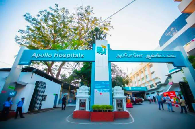
Apollo Hospital, Greams Road, Chennai
Retina Specialist
20+ years experience

About Dr Pratik Ranjan Sen
Dr. Sen is a highly distinguished specialist in ophthalmology, recognized for his technical mastery in advanced microsurgical interventions and comprehensive ocular care. He specializes in utilizing precision-guided diagnostic and therapeutic technologies to restore visual acuity and preserve long-term structural eye health.
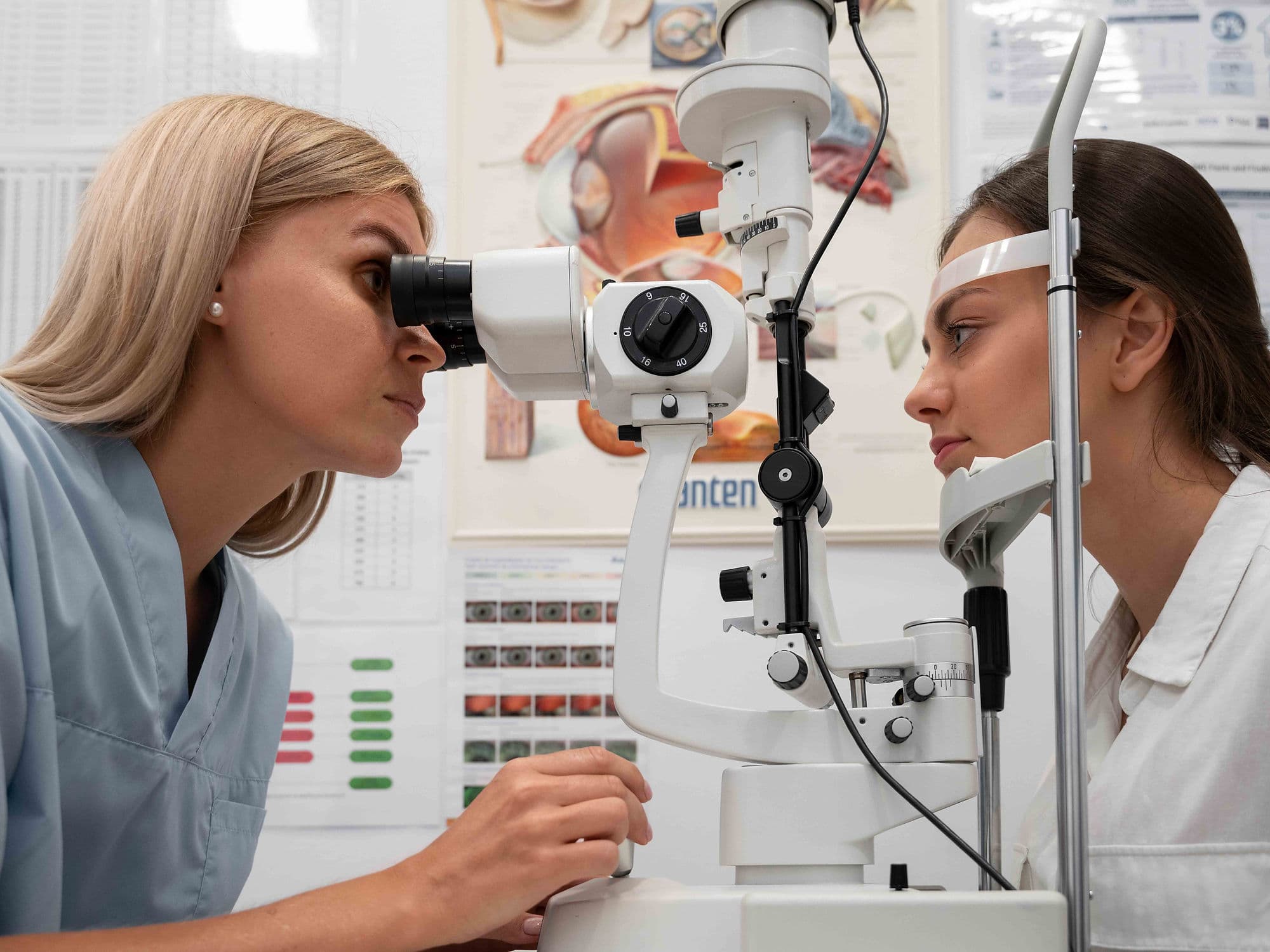
Mastery in Ophthalmic Microsurgery and Vision Restoration
He specializes in high-precision microsurgical procedures designed to treat complex intraocular pathologies. His clinical practice focuses on employing state-of-the-art operative methodologies to deliver safe, effective surgical solutions, ensuring anatomical alignment and optimized visual recovery for his patients.
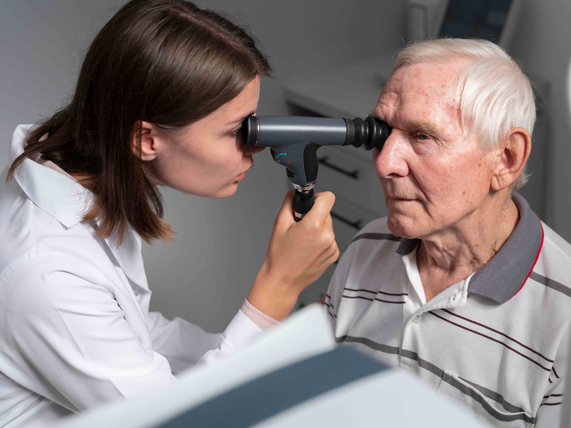
Advanced Care for Complex Ocular Disorders
Dr. Sen possesses profound expertise in diagnosing and managing a wide spectrum of complex eye conditions and degenerative ocular diseases. By integrating detailed clinical evaluations with targeted treatment pathways, he targets the underlying mechanisms of visual impairment to prevent progressive vision loss.
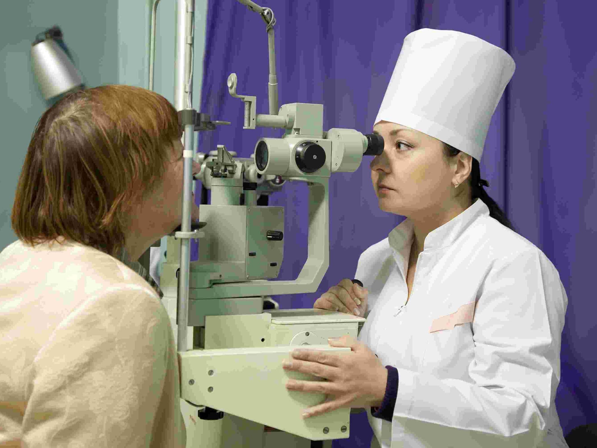
Precision Diagnostic Frameworks and Clinical Governance
A primary focus of his practice is the implementation of advanced ophthalmic imaging and diagnostic mapping tools. His meticulous approach to patient care ensures that every therapeutic or surgical strategy is precisely tailored to the specific anatomical profile of the patient, significantly reducing procedural risks.
Evidence-Based Pathways and Comprehensive Eye Health
Throughout his extensive career, Dr. Sen has combined modern ophthalmic innovations with rigorous clinical protocols to maintain the highest standards of safety. His evidence-based methodology is structured to deliver predictable, high-quality outcomes, establishing him as a trusted name in advanced vision care.
Dr. Pratik Ranjan Sen at a Glance
Specialist in Advanced Ophthalmology, Intraocular Microsurgery, and Vision Care.
Extensive experience delivering dedicated specialist services within the field of eye health.
Expert in precision-guided diagnostics and customized ophthalmic treatment pathways.
Focused on utilizing state-of-the-art technology to optimize visual acuity outcomes.
Specialized in tissue-preserving surgical interventions and long-term ocular stability.
Dedicated to providing comprehensive, evidence-based clinical governance.
MBBS, MS, DO
Board Certified in Retina Specialist
No awards & achievements available