Ankle Arthroscopy
Ankle Arthroscopy
Ankle Arthroscopy (keyhole surgery) is a minimally invasive procedure used to diagnose and treat issues inside the ankle joint using a tiny camera and specialized tools. It is widely preferred over open surgery because it allows for high-precision joint repair with faster recovery times and a lower risk of wound-related complications.
When You Should Consider Ankle Arthroscopy
Persistent Impingement: Pain or limited motion caused by inflamed soft tissue or bone spurs (osteophytes) that have not responded to physical therapy.
Mechanical Symptoms: Feeling the joint "lock," "catch," or "click," often due to free-floating fragments of bone or cartilage.
Cartilage Damage: Treatment of Osteochondral Lesions of the Talus (OLT) where the smooth surface of the joint has been chipped or worn down.
Chronic Synovitis: Recurring inflammation of the joint lining caused by trauma, overuse, or rheumatoid arthritis.
Joint Instability: Evaluation and repair of torn ligaments (such as the ATFL) when the ankle feels "loose" or gives way frequently.
Methods of Ankle Arthroscopy
Debridement: Trimming away inflamed tissue or smoothing out frayed cartilage to reduce pain and friction.
Microfracture: Making tiny holes in the bone to stimulate a healing response and the growth of new fibrocartilage.
Synovectomy: Removing the diseased or inflamed lining of the joint.
Ligament Stabilization: Using arthroscopic techniques to tighten or reattach torn ligaments to restore joint stability.
Bone Spur Removal: Shaving down bony growths that pinch the joint during movement (anterior or posterior impingement).
How Ankle Arthroscopy Is Performed
Portal Creation: The surgeon makes 2–3 tiny incisions (less than 1 cm each), typically at the front of the ankle (anteromedial and anterolateral portals).
Joint Distention: The ankle is expanded with sterile saline or a mechanical distraction device to create a clear workspace for the camera.
Visualization: A miniature high-definition camera (arthroscope) is inserted to project live images of the joint internal structures onto a monitor.
Surgical Intervention: Miniature shavers, burrs, and graspers are inserted through the other portals to repair or remove damaged tissue.
Closure: Once the repair is complete, the fluid is drained and the tiny portals are closed with a single stitch or sterile tape.
Pre-Procedure Preparation
Diagnostic confirmation through physical exams and imaging to identify the specific source of joint pain.
Fasting (NPO) as directed by the surgical team prior to the procedure.
Evaluation for regional anesthesia, which numbs the leg and provides several hours of pain relief after the surgery.
Arrangement for a post-operative walking boot or splint, depending on the anticipated type of repair.
Tests Before Ankle Arthroscopy
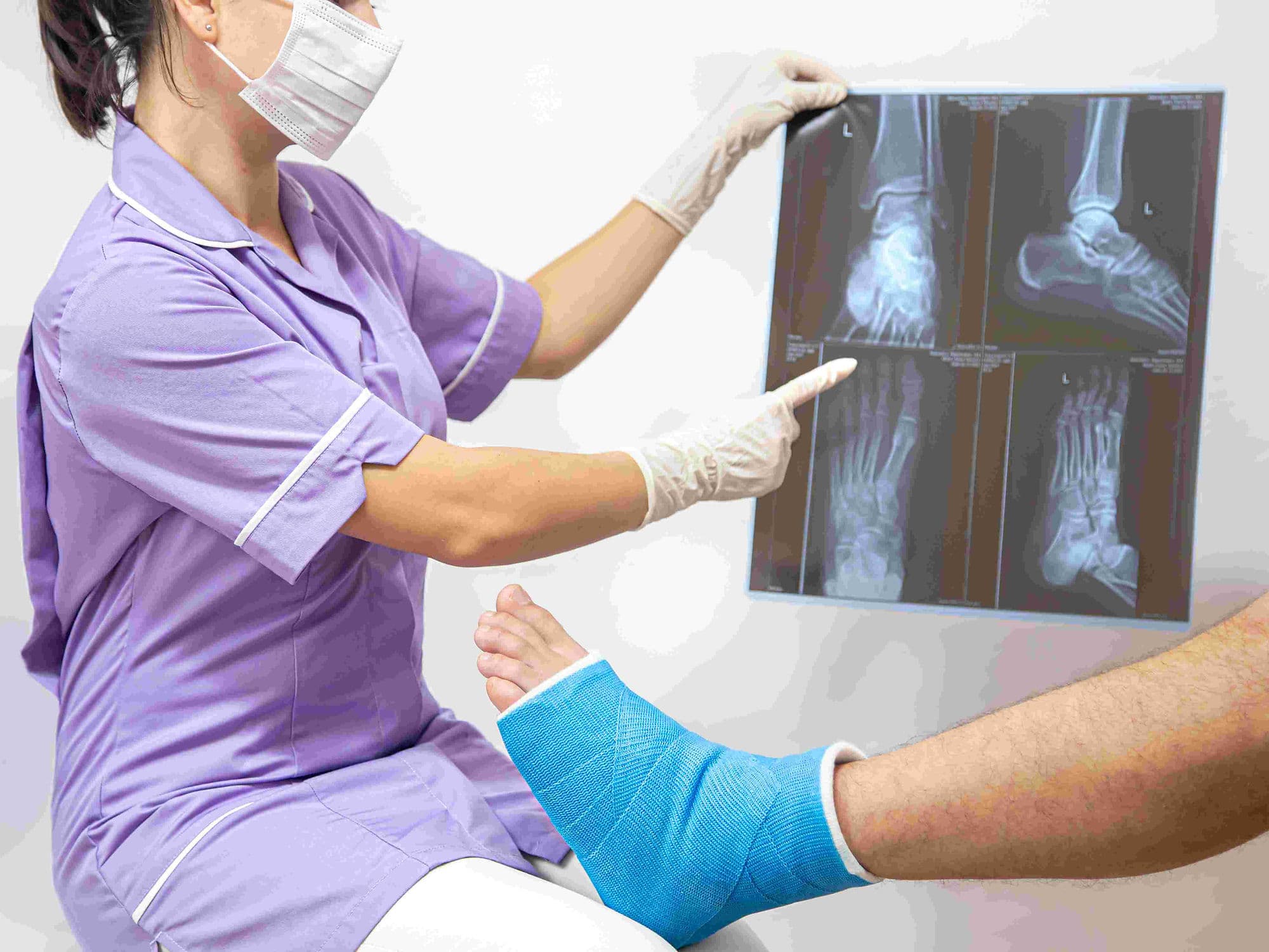
Ankle MRI: The primary tool for assessing soft tissue damage, ligament tears, and cartilage lesions.
X-rays: Used to identify bone spurs or loose bone fragments that may be contributing to mechanical joint issues.
CT Scan: Occasionally used to provide a detailed view of complex bone anatomy or "bony" impingement.
Blood Panels: Routine testing to ensure the patient is fit for general anesthesia and outpatient surgery.
Life After Ankle Arthroscopy
Almost always an outpatient (daycare) procedure, with patients returning home the same day.
Strict elevation of the ankle above the heart for the first 2–3 days is critical to minimize swelling and pain.
Weight-bearing status varies: simple trimming allows immediate walking, while cartilage repairs may require crutches for 4–8 weeks.
Physical therapy typically begins around 2 weeks post-op to regain range of motion and ankle strength.
Return to desk work is often possible in 1–2 weeks, while high-impact sports usually take 3 to 6 months.
Benefits of Ankle Arthroscopy
Minimally invasive nature results in significantly less post-operative pain and swelling than traditional open incisions.
Tiny incisions lead to a lower risk of infection and faster overall healing of the surgical site.
Provides a dynamic and comprehensive view of the joint, allowing the surgeon to address multiple issues in one session.
High success rates for resolving mechanical symptoms like locking or catching in the ankle.