+91 83682 47758
info@medivisorhealth.com
Medivisor India Treatment Logo
  • Home
    • About Us
    • Our Services
    • Our Team
    • Our Safety Measures
    • Loading treatments...
    • Loading hospitals...
    • Loading doctors...
    • Loading tests & diagnoses...
    • Treatment Cost
    • Treatment Process
    • Visa Process
    • Travel Guide
    • FAQs
    • Why Medivisor
    • Success Stories
    • Patient Activities
    • Social Activities
    • News Coverage
    • Blog
  • Become a Partner

Medivisor India Treatment is a trusted New Delhi-based medical travel company assisting international patients with advanced, affordable, and seamless treatment in India.

  • Medivisor House 359, Sector 1, Vaishali, Ghaziabad, (Delhi/NCR) India
  • +91 8368 247 758
  • info@medivisorhealth.com

About Us

  • About Us
  • Our Services
  • Our Team
  • Our Safety Measures
  • Join Our Network
  • Contact Us

India Treatment

  • Hospitals
  • Treatments
  • Treatment Cost
  • Treatment Process
  • Visa Process
  • Travel Guide
  • FAQs
  • Why Medivisor

Gallery

  • Patient Testimonials
  • Patient Activities
  • News Coverage
  • Blog

Get in Touch

© 2026 Medivisor India Treatment. All rights reserved.

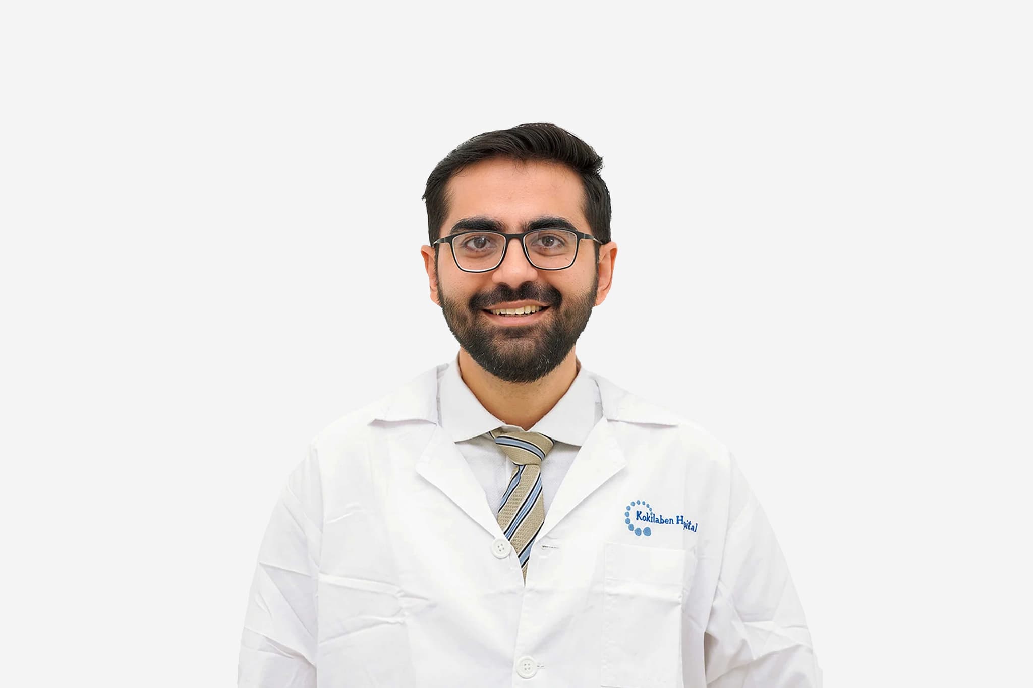
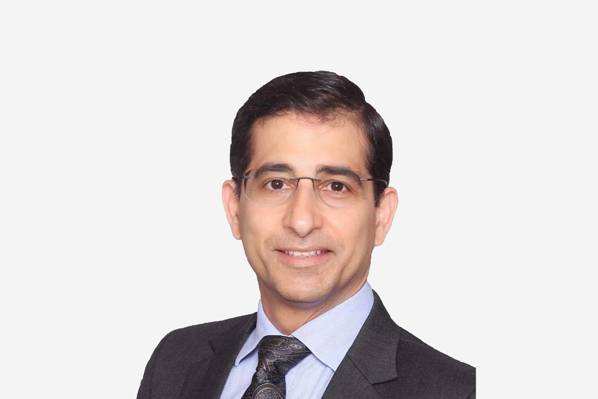
Dr Shreyash Gajjar

Consultant, Unit Lead - Sports Orthopedics, Arthroscopy & Trauma

Kokilaben Dhirubhai Ambani Hospital

27+ years experience
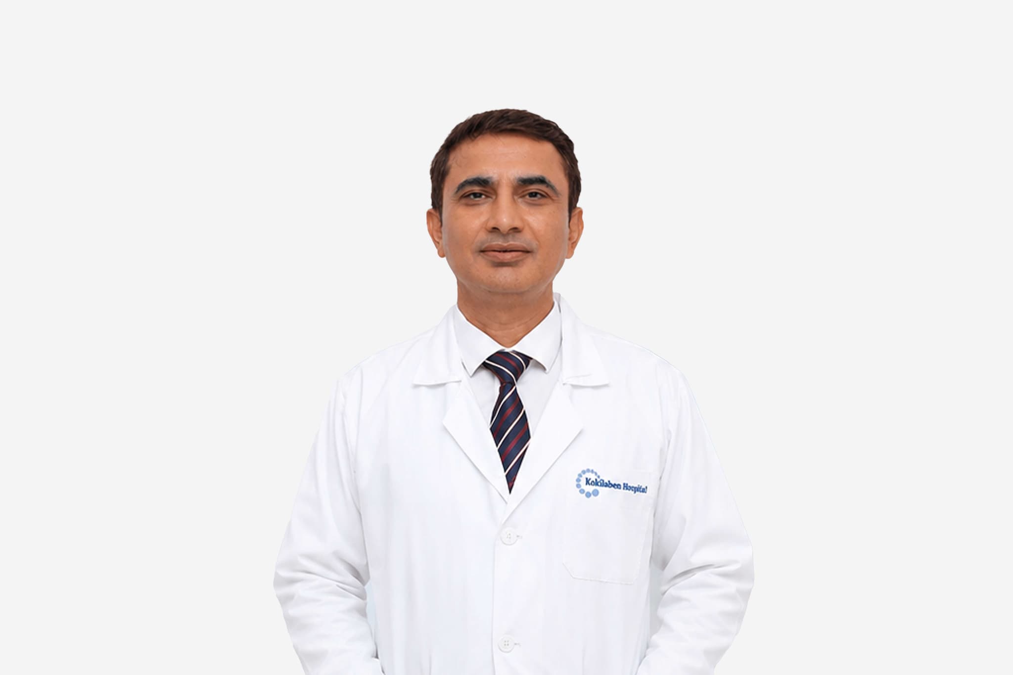
Dr Shreyash Gajjar, Consultant, Unit Lead - Sports Orthopedics, Arthroscopy & Trauma at Kokilaben Dhirubhai Ambani Hospital - Kokilaben Dhirubhai Ambani Hospital
  1. Home
  2. Doctors
  3. Dr Shreyash Gajjar

About Dr Shreyash Gajjar

Dr. Shreyash Gajjar is a highly specialized Orthopedic Surgeon with a dedicated focus on Sports Medicine and Arthroscopic Surgery. Currently practicing at Kokilaben Dhirubhai Ambani Hospital, Mumbai, he is recognized for his commitment to evidence-based healthcare and his passion for restoring mobility and function to patients of all activity levels.

Journey into Sports Orthopedics

Driven by a lifelong aspiration to practice medicine, Dr. Gajjar discovered his calling in Orthopedic surgery during his foundational medical training. His personal interest and active participation in sports naturally led him to pursue advanced sub-specialization in the fields of Sports Medicine and Sports Orthopedic Surgery. This specialized path allows him to combine his surgical expertise with an athlete's understanding of physical performance and recovery.

Clinical Philosophy and Patient Care

Dr. Gajjar’s practice is built on a "patient-first" philosophy. He strongly believes that modern medical advancements should be accessible to everyone, regardless of age or gender. His approach involves:

  • Comprehensive Counseling: Ensuring patients fully understand their condition and the available therapeutic pathways.

  • Functional Restoration: Tailoring treatments to help individuals regain their pre-injury level of activity, whether they are professional athletes or recreational fitness enthusiasts.

  • Evidence-Based Practice: Regularly updating his technical skills and knowledge to ensure his patients benefit from the latest global standards in sports medicine.

Specialized Areas of Expertise

Dr. Gajjar is proficient in managing a wide range of sports-related injuries and degenerative conditions through minimally invasive techniques:

  • Arthroscopic Surgery: Utilizing keyhole techniques for the knee, shoulder, and ankle to ensure smaller incisions and faster rehabilitation.

  • Ligament Reconstruction: Expertise in ACL, PCL, and multi-ligament knee repairs.

  • Shoulder Interventions: Management of recurrent dislocations, rotator cuff tears, and labral injuries.

  • Sports Traumatology: Specialized care for acute fractures and soft tissue injuries sustained during athletic activities.

Dr. Shreyash Gajjar at a Glance

  • Consultant in Sports Medicine and Sports Orthopedic Surgery.

  • Specialist in Arthroscopic (Keyhole) Surgery for the knee and shoulder.

  • Academic foundation in Orthopedics with elite sub-specialization in Sports Medicine.

  • Dedicated to evidence-based treatment and functional recovery.

  • Active contributor to the field through regular skill updates and clinical research.

  • Renowned for a compassionate, counseling-based approach to patient management.

MS - Orthopedics
DNB - Orthopedics
D - Orthopedics
FCPS - Orthopedics (CCT (UK)
FRCS Ed(Tr. & Orth)
FRCS Ed(Surg)
Dr. Shreyash M. Gajjar is an invited Reviewer for the prestigious international journals - The American Journal of Sports Medicine (AJSM)
Journal of Arthroscopic & Related Surgery (USA). He is also the Ex-Editor of the Indian Arthroscopy Society.
He is member in good-standing of Indian Arthroscopy Society (IAS), Shoulder & Elbow Society of India (SESI), International Society of Arthroscopy, Knee Surgery
Orthopaedic Sports Medicine (ISAKOS), Indian Orthopaedic Association (IOA)
Bombay Orthopaedic Society (BOS).
He has been faculty on Arthroscopy courses/meetings
contributed in Numerous International
National Presentations & Publications.
He has been awarded Fellowships in General Surgery (FRCSEd)
Trauma & Orthopaedic Surgery (FRCSEd - Tr. & Orth.) by the Royal College of Surgeons of Edinburgh, UK.
ISAKOS Scientific Committee Member 2023-27
ISAKOS Shoulder Committee Member 2025-27
Editorial Board (Arthrosocopy & Sports section)- Indian Journal of Orthopaedics
Elite Reviewer JISAKOS, AJSM, OJSM
Past Editor-Indian Arthroscopy Society (IAS)
May we help you?

Enter digits only (6-15 digits). We'll add your country code.

Related Treatments

Ankle Arthroscopy
Ankle Arthroscopy

Ankle Arthroscopy (keyhole surgery) is a minimally invasive procedure used to diagnose and treat issues inside the ankle joint using a tiny camera and specialized tools. It is widely preferred over open surgery because it allows for high-precision joint repair with faster recovery times and a lower risk of wound-related complications.

When You Should Consider Ankle Arthroscopy

  • Persistent Impingement: Pain or limited motion caused by inflamed soft tissue or bone spurs (osteophytes) that have not responded to physical therapy.

  • Mechanical Symptoms: Feeling the joint "lock," "catch," or "click," often due to free-floating fragments of bone or cartilage.

  • Cartilage Damage: Treatment of Osteochondral Lesions of the Talus (OLT) where the smooth surface of the joint has been chipped or worn down.

  • Chronic Synovitis: Recurring inflammation of the joint lining caused by trauma, overuse, or rheumatoid arthritis.

  • Joint Instability: Evaluation and repair of torn ligaments (such as the ATFL) when the ankle feels "loose" or gives way frequently.

Methods of Ankle Arthroscopy

  • Debridement: Trimming away inflamed tissue or smoothing out frayed cartilage to reduce pain and friction.

  • Microfracture: Making tiny holes in the bone to stimulate a healing response and the growth of new fibrocartilage.

  • Synovectomy: Removing the diseased or inflamed lining of the joint.

  • Ligament Stabilization: Using arthroscopic techniques to tighten or reattach torn ligaments to restore joint stability.

  • Bone Spur Removal: Shaving down bony growths that pinch the joint during movement (anterior or posterior impingement).

How Ankle Arthroscopy Is Performed

  • Portal Creation: The surgeon makes 2–3 tiny incisions (less than 1 cm each), typically at the front of the ankle (anteromedial and anterolateral portals).

  • Joint Distention: The ankle is expanded with sterile saline or a mechanical distraction device to create a clear workspace for the camera.

  • Visualization: A miniature high-definition camera (arthroscope) is inserted to project live images of the joint internal structures onto a monitor.

  • Surgical Intervention: Miniature shavers, burrs, and graspers are inserted through the other portals to repair or remove damaged tissue.

  • Closure: Once the repair is complete, the fluid is drained and the tiny portals are closed with a single stitch or sterile tape.

Pre-Procedure Preparation of Ankle Arthroscopy

  • Diagnostic confirmation through physical exams and imaging to identify the specific source of joint pain.

  • Fasting (NPO) as directed by the surgical team prior to the procedure.

  • Evaluation for regional anesthesia, which numbs the leg and provides several hours of pain relief after the surgery.

  • Arrangement for a post-operative walking boot or splint, depending on the anticipated type of repair.

Tests Before Ankle Arthroscopy

  • Ankle MRI: The primary tool for assessing soft tissue damage, ligament tears, and cartilage lesions.

  • X-rays: Used to identify bone spurs or loose bone fragments that may be contributing to mechanical joint issues.

  • CT Scan: Occasionally used to provide a detailed view of complex bone anatomy or "bony" impingement.

  • Blood Panels: Routine testing to ensure the patient is fit for general anesthesia and outpatient surgery.

Life After Ankle Arthroscopy

  • Almost always an outpatient (daycare) procedure, with patients returning home the same day.

  • Strict elevation of the ankle above the heart for the first 2–3 days is critical to minimize swelling and pain.

  • Weight-bearing status varies: simple trimming allows immediate walking, while cartilage repairs may require crutches for 4–8 weeks.

  • Physical therapy typically begins around 2 weeks post-op to regain range of motion and ankle strength.

  • Return to desk work is often possible in 1–2 weeks, while high-impact sports usually take 3 to 6 months.

Benefits of Ankle Arthroscopy

  • Minimally invasive nature results in significantly less post-operative pain and swelling than traditional open incisions.

  • Tiny incisions lead to a lower risk of infection and faster overall healing of the surgical site.

  • Provides a dynamic and comprehensive view of the joint, allowing the surgeon to address multiple issues in one session.

  • High success rates for resolving mechanical symptoms like locking or catching in the ankle.

Starting Cost: $ 1000

Knee Arthroscopy Surgery

Knee Arthroscopy is a minimally invasive surgical procedure used to diagnose and treat problems in the knee joint through tiny incisions using a camera. This specialized "keyhole" approach allows surgeons to view and repair joint structures without the need for a large open incision.

When You Should Consider Knee Arthroscopy

  • Persistent knee pain that has not responded to rest, physical therapy, or injections.

  • Mechanical symptoms such as the knee "locking," "catching," or "giving way" during movement.

  • Swelling and stiffness caused by loose fragments of bone or cartilage within the joint.

  • Confirmed soft tissue injuries, such as a meniscus tear or a ligament injury (ACL/PCL).

  • Chronic inflammation of the joint lining (synovium) that requires surgical removal.

Methods of Knee Arthroscopy

  • Diagnostic Arthroscopy: A procedure focused on exploring the joint to identify the source of unexplained pain or instability.

  • Meniscectomy: Trimming away the damaged or frayed edges of a torn meniscus.

  • Meniscal Repair: Suturing a torn meniscus back together to preserve the natural cushioning of the knee.

  • Chondroplasty: Smoothing out damaged or irregular articular cartilage to improve joint glide.

  • Loose Body Removal: Extracting floating bits of bone or cartilage that cause painful mechanical interference.

How Knee Arthroscopy Is Performed

  • Portal Creation: The surgeon makes two or three tiny incisions, or portals (about 0.5 cm each), around the kneecap.

  • Saline Insufflation: The joint is expanded with sterile saline to stretch the capsule, providing a clear view and space for the tools.

  • Visualization: A high-definition fiber-optic camera (arthroscope) is inserted through one portal to project live images onto a monitor.

  • Surgical Intervention: Miniature specialized tools, such as shavers or probes, are used to repair, trim, or remove damaged tissue.

  • Closure: After the saline is drained, the tiny portals are closed with a single stitch or sterile tape (Steri-Strips).

Pre-Procedure Preparation

  • Fasting (NPO) for 6–8 hours prior to the surgery to ensure safety during anesthesia.

  • Evaluation of current medications; blood thinners are typically only stopped if significant bleeding is anticipated.

  • Choosing the most appropriate anesthesia option, which may include general, spinal, or a regional nerve block.

  • Diagnostic confirmation through physical stability tests and imaging to map out the internal damage.

Tests Before Knee Arthroscopy

  • MRI Scan: The primary imaging tool used to visualize soft tissues like ligaments, tendons, and cartilage.

  • X-rays: Used to rule out significant bone-on-bone arthritis or fractures that may not be treatable via arthroscopy.

  • Physical Stability Exams: Specialized maneuvers (like the Lachman or McMurray tests) to assess ligament and meniscus integrity.

  • Blood Panels: Routine screens to ensure general health and fitness for an outpatient surgical procedure.

Life After Knee Arthroscopy

  • Almost always an outpatient procedure, with most patients returning home 2–3 hours after waking up.

  • Adherence to the R.I.C.E. protocol (Rest, Ice, Compression, Elevation) is critical for the first 48–72 hours.

  • Weight-bearing status depends on the procedure: simple trimming allows walking in days, while repairs may require crutches for 4–6 weeks.

  • Driving is typically restricted for 1–2 weeks, particularly if the surgery was on the right leg or the patient drives a manual vehicle.

  • Physical therapy focuses on restoring range of motion and strengthening the muscles surrounding the knee joint.

Benefits of Knee Arthroscopy

  • Minimally invasive nature results in significantly less post-operative pain and swelling than open surgery.

  • Tiny incisions lead to faster wound healing and minimal surgical scarring.

  • High success rates for resolving mechanical issues like joint locking or catching.

  • Reduced risk of infection and lower blood loss compared to traditional joint procedures.

Starting Cost: $ 800

Shoulder Arthroscopy

Shoulder Arthroscopy is a minimally invasive surgical procedure used to diagnose and treat various joint problems, such as rotator cuff tears and labral injuries. Using a tiny camera called an arthroscope, surgeons can see inside the joint and perform high-precision repairs through small, buttonhole-sized incisions.

When You Should Consider Shoulder Arthroscopy

  • Persistent shoulder pain that has not improved with physical therapy, rest, or injections.

  • Loss of range of motion or weakness, often associated with a torn rotator cuff.

  • Recurrent shoulder dislocations or a feeling of "looseness" in the joint (instability).

  • Mechanical symptoms such as painful catching, clicking, or "locking" of the shoulder.

  • Presence of bone spurs that cause "impingement," where tendons are pinched during overhead movement.

Methods of Shoulder Arthroscopy

  • Rotator Cuff Repair: Reattaching torn tendons to the humerus (arm bone) using specialized suture anchors.

  • Labral Repair (Bankart or SLAP Repair): Stitching the cartilage ring (labrum) back to the socket to restore stability.

  • Subacromial Decompression: Shaving down bone spurs and inflamed tissue to create more space for the tendons to move.

  • Biceps Tenodesis: Relocating a damaged biceps tendon to a new attachment point to relieve pain.

  • Capsular Release: Surgically stretching or cutting tight tissue to treat severe "frozen shoulder."

How Shoulder Arthroscopy Is Performed

  • Fluid Insufflation: The surgeon injects sterile saline into the shoulder to inflate the joint capsule, providing a clear workspace and view.

  • Portal Placement: Two to three small incisions (0.5–1 cm) are made around the shoulder to serve as entry points for the camera and tools.

  • Joint Inspection: The arthroscope is inserted to project high-definition images of the tendons, ligaments, and cartilage onto a monitor.

  • Specialized Repair: Miniature tools, such as shavers or suture passers, are used to trim damaged tissue or anchor tendons back to the bone.

  • Portal Closure: Once the repair is complete, the saline is drained and the tiny incisions are closed with a single stitch or sterile tape.

Pre-Procedure Preparation

  • Diagnostic confirmation through MRI or X-rays to map the internal damage and plan the surgical approach.

  • Fasting (NPO) for 6–12 hours prior to the procedure to ensure safety during anesthesia.

  • Coordination of a regional nerve block, which numbs the entire arm for up to 24 hours to assist with immediate pain control.

  • Pausing certain medications, such as blood thinners or anti-inflammatories, as directed by the surgical team.

Tests Before Shoulder Arthroscopy

  • Shoulder MRI: The gold standard for visualizing soft tissue injuries like rotator cuff tears and labral damage.

  • MRI Arthrogram: An MRI where dye is injected into the joint to highlight small tears that might be missed on a standard scan.

  • X-rays: Used to identify bone spurs, arthritis, or fractures that may contribute to shoulder pain.

  • Physical Strength Testing: Assessing the deltoid and rotator cuff muscles to determine the functional impact of the injury.

Life After Shoulder Arthroscopy

  • This is typically an outpatient procedure, allowing most patients to return home the same day.

  • A shoulder sling is mandatory for 1 to 6 weeks, depending on the complexity of the repair (e.g., longer for a rotator cuff repair).

  • Pendulum exercises, involving gently swinging the arm, are often started within days to prevent "frozen shoulder" stiffness.

  • Driving is generally restricted for at least 1 week, or until the patient has stopped taking narcotic pain medications.

  • Heavy lifting and overhead sports are avoided for 3 to 6 months while the repaired tendons fully bond to the bone.

Benefits of Shoulder Arthroscopy

  • Significantly less post-operative pain and swelling compared to traditional "open" shoulder surgery.

  • Smaller incisions result in minimal scarring and a lower risk of wound-related complications.

  • High success rate for restoring shoulder stability and relieving chronic pain.

  • Allows for a more precise diagnosis by giving the surgeon a dynamic, 360-degree view of the internal joint structures.

Starting Cost: $ 1000

Related Specialists

Dr Manish Hinduja
Dr Manish Hinduja
Consultant - Adult Cardiac Surgery & Cardiothoracic & Vascular Surgery
Kokilaben Dhirubhai Ambani Hospital
15+years experience
View Profile
Dr Jamshed Dalal
Dr Jamshed Dalal
Director - Cardiac Sciences
Kokilaben Dhirubhai Ambani Hospital
45+years experience
View Profile
Dr Rajesh Mistry
Dr Rajesh Mistry
Senior Director (Group), Oncology
Kokilaben Dhirubhai Ambani Hospital
39+years experience
View Profile
Dr Dinshaw Pardiwala
Director - Arthroscopy, Sports Orthopedics & Shoulder Service
Kokilaben Dhirubhai Ambani Hospital
27+years experience
View Profile
Dr Abhaya Kumar
Dr Abhaya Kumar
Head - Neurosurgery & Consultant - Minimally Invasive Spine Surgery
Kokilaben Dhirubhai Ambani Hospital
20+years experience
View Profile
Dr Mohit Bhatt
Dr Mohit Bhatt
Director - Neurosciences, Consultant - Neurology, Director - Medical Research
Kokilaben Dhirubhai Ambani Hospital
35+years experience
View Profile
Dr Raghuram Sekhar
Dr Raghuram Sekhar
Head - Vascular Surgery
Kokilaben Dhirubhai Ambani Hospital
35+years experience
View Profile
Dr Prashant Bobhate
Dr Prashant Bobhate
Consultant - Children's Heart Centre
Kokilaben Dhirubhai Ambani Hospital
16+years experience
View Profile
Dr Sanjay Pandey
Dr Sanjay Pandey
Director, Urology; Head, Andrology & Reconstructive Urology; Consultant, Robotic Urology & Gender Reassignment Surgery
Kokilaben Dhirubhai Ambani Hospital
25+years experience
View Profile

May we help you?

Enter digits only (6-15 digits). We'll add your country code.