Clubfoot Correction
Clubfoot Surgery
Clubfoot correction via surgery is typically reserved for severe cases or when non-surgical methods, such as the Ponseti method (casting), fail. The surgery aims to realign the foot by releasing or lengthening tight tissues to allow for a functional, pain-free position. While the procedure is highly effective, the affected foot and calf may remain slightly smaller than the unaffected side throughout the child's life.
When You Should Consider Surgery
Severe Deformity: For cases where the foot is rigidly fixed in an abnormal position.
Failed Casting: When traditional serial casting (Ponseti method) does not achieve the necessary correction.
Relapsed Clubfoot: If the deformity returns after initial successful non-surgical treatment.
Late Diagnosis: In older children where the bones and tissues are less flexible and require structural realignment.
How Is Performed
Anesthesia: Most clubfoot surgeries are performed under general anesthesia to ensure the child is comfortable.
Incision & Release: The surgeon makes one or two incisions, usually on the back and inside of the foot, to access tight structures.
Tissue Lengthening: Surgeons meticulously lengthen tight tendons, such as the Achilles, and release tight ligaments around the joints.
Stabilization: In complex cases, small metal pins, screws, or plates may be inserted to hold bones in their new, correct positions during healing.
Duration: The surgical procedure typically takes between 2 and 3 hours to complete.
Pre-Procedure Preparation
Medical Evaluation: The healthcare provider performs a physical exam, reviews medical history, and orders X-rays of the foot.
Blood Tests: Standard tests, including a complete blood count (CBC) and checks for clotting factors, are required.
Medication Audit: Parents are typically instructed to stop giving the child blood-thinning medications, such as ibuprofen, roughly 10 days before the operation.
Fasting (NPO): The child must not eat or drink anything for 4 to 6 hours before surgery to minimize anesthesia risks.
Hygiene: Bathe the child with antiseptic soap the night before or the morning of the surgery to reduce infection risks.
Tests Before Clubfoot Surgery
Foot X-rays: To visualize the alignment of the tarsal bones and plan the surgical correction.
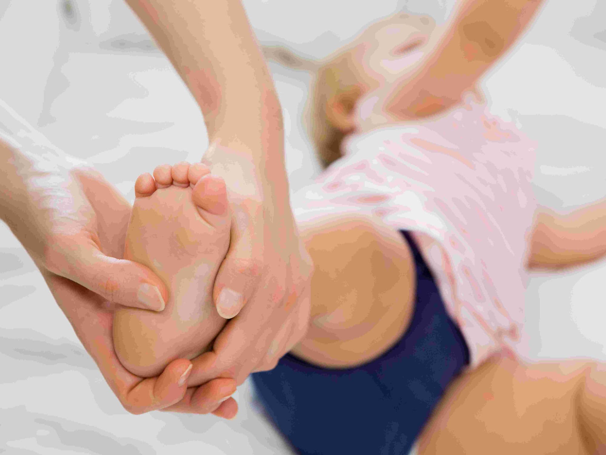
Physical Assessment: To document the range of motion and the flexibility of the foot structures.
Complete Blood Count (CBC): To ensure there are no underlying infections or issues with blood cell levels.
Clotting Profile: To confirm the blood can clot properly during and after the surgical incisions.
Life After Clubfoot Surgery
Hospital Stay: Depending on complexity, the child may stay in the hospital for 1 to 3 days for monitoring.
Casting Phase: A long-leg cast is applied initially; these are changed every few weeks for a total of 6 to 12 weeks.
Pin Removal: If metal pins were used for stabilization, they are typically removed in the office 4 to 6 weeks after surgery.
Bracing Phase: Once the final cast is removed, a brace (orthosis) is required to prevent the foot from returning to the clubfoot position.
Physical Therapy: A therapist guides the family through exercises to strengthen the repaired foot and improve its range of motion.
Why Specialized Treatment Is Highly Effective
Structural Realignment: Directly addresses the tight ligaments and tendons that prevent the foot from sitting flat.
Long-Term Function: Most children achieve a functional foot and can lead active, athletic lives.
Customized Bracing: Post-operative bracing plans are tailored to the child's growth to maintain the correction.
Comprehensive Care: Involves a multidisciplinary team of surgeons and therapists to manage healing and strength