Osteotomy
Osteotomy
An osteotomy is a surgical procedure where a bone is precisely cut, reshaped, or removed to change its alignment or length. In the context of limb lengthening, it is the foundational step that allows for new bone growth. This specialized intervention triggers the body's natural healing mechanisms to bridge gaps or correct structural deformities.
Types of Osteotomy
Closing Wedge: A wedge of bone is removed to straighten a tilted bone, a technique commonly used in "knock-knee" corrections.
Opening Wedge: A cut is made and the bone is pulled open to create a gap, which is then filled with a bone graft or allowed to grow new bone.
Rotational: The bone is cut and turned to correct a twist or "torsion" within the limb.
Corticotomy: A specific type used in lengthening where only the hard outer shell (cortex) is cut, preserving the inner marrow and blood supply to speed up healing.
How Is Performed
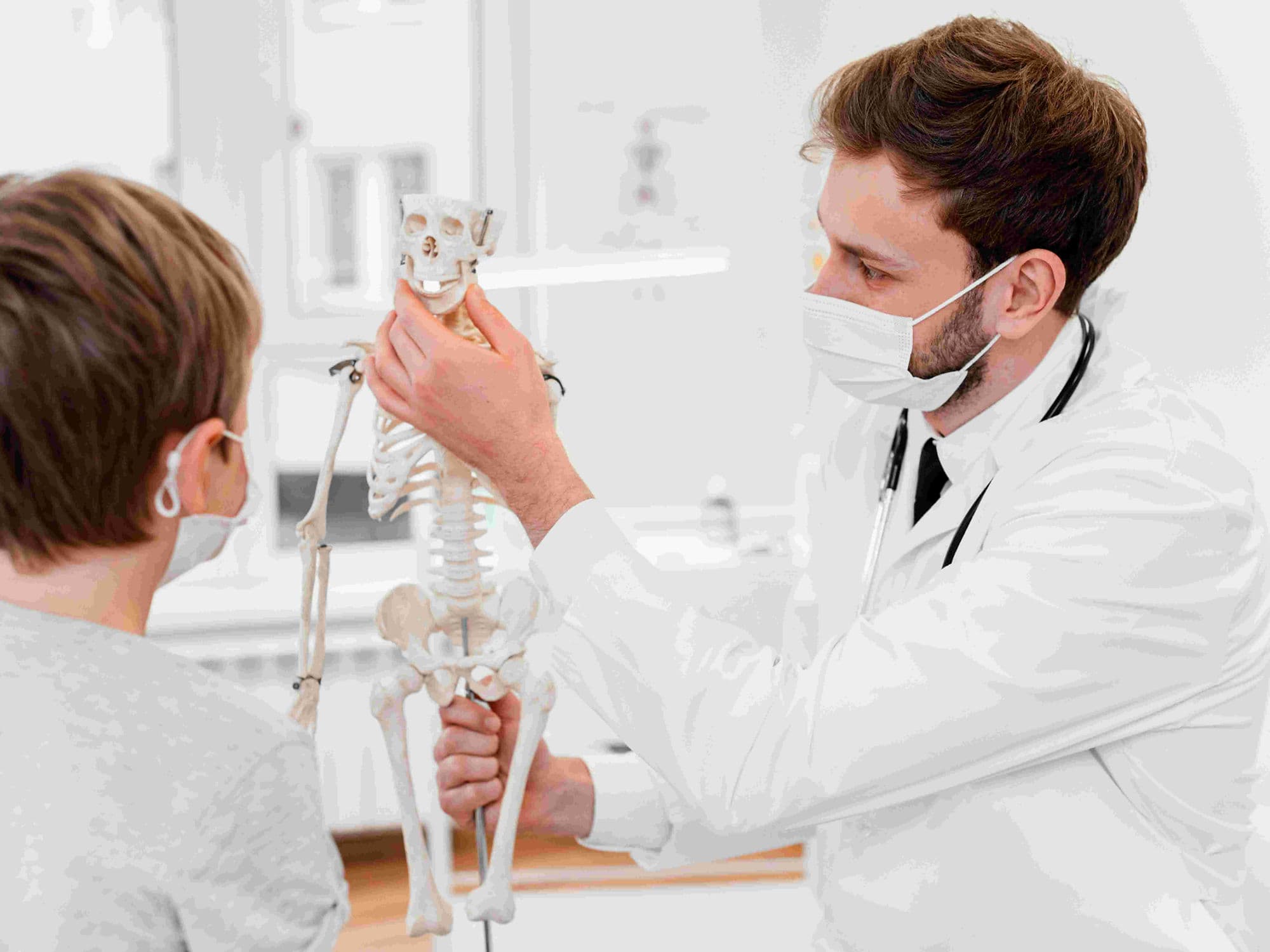
Incision: The surgeon makes a small skin incision to access the target bone, usually the femur or tibia.
Protection: Surrounding nerves, blood vessels, and muscles are retracted and shielded using specialized tools during the procedure.
The Cut: Using a surgical saw, drill, or osteotome (a chisel-like tool), the surgeon performs a "low-energy" cut to minimize heat damage to the bone cells.
Hardware Fixation: Once the bone is cut, an internal rod (intramedullary nail) or an external fixator (pins and frames) is attached to hold the segments in the new position.
Biological Healing (The "Glow")
The primary goal of an osteotomy in lengthening is to trigger Distraction Osteogenesis:
Hematoma Formation: Immediately after the cut, blood fills the gap, creating a "scaffold" for the healing process.
Callus Formation: Within days, the body sends "osteoblasts" (bone-building cells) to create a soft, cartilage-like bridge called a callus.
Tension-Stress Effect: By slowly pulling the two cut pieces apart (distraction), the body is "tricked" into continuously creating more callus, which eventually hardens into solid bone.
Pre-Procedure Preparation
Imaging Workup: Detailed X-rays or CT scans are required to plan the exact angle and location of the bone cut.
Vascular Assessment: Ensuring healthy blood flow to the limb is critical, as the bone depends on this supply to grow new tissue.
Medication Audit: Patients must pause anti-inflammatory drugs (NSAIDs) or blood thinners that could interfere with hematoma formation and bone healing.
Smoking Cessation: Nicotine must be avoided entirely, as it constricts blood vessels and significantly increases the risk of the bone failing to knit back together.
Tests Before Osteotomy
Weight-Bearing X-rays: To assess the overall mechanical axis of the leg and determine the degree of correction needed.
Blood Panels: Checking calcium, Vitamin D, and alkaline phosphatase levels to ensure the body has the mineral resources for bone growth.
CT Scan (3D Reconstruction): Provides a precise anatomical map for complex rotational or multi-planar corrections.
Nerve Conduction Study: May be performed if there is a pre-existing nerve issue to establish a baseline before the bone is realigned.
Life After Osteotomy
Nerve/Vessel Monitoring: Surgeons monitor the limb post-op for "compartment syndrome" or nerve compression because the bone has been physically severed.
Pain Management: The first 48–72 hours involve the most acute pain as the bone ends and surrounding tissue settle.
Weight-Bearing Restrictions: Weight-bearing is strictly limited until X-rays show "bridging" (new bone crossing the gap) to prevent hardware failure or bone shifting.
Physical Therapy: Early motion of the joints above and below the osteotomy is encouraged to prevent stiffness while the bone heals.
Why Specialized Treatment Is Highly Effective
Permanent Realignment: Corrects the root cause of joint pain and uneven wear by shifting the load to healthy areas of the bone.
Bone Preservation: Modern "low-energy" techniques preserve the biological vitality of the bone, leading to faster consolidation.
Customized Hardware: 2026-standard internal nails and external frames allow for microscopic adjustments to ensure a perfect final alignment.
Prevents Arthritis: By correcting a tilted or twisted bone early, an osteotomy can often delay or eliminate the need for a joint replacement later in life.