Total Knee Replacement (TKR)
Total Knee Replacement (TKR)
Total Knee Replacement (TKR), also known as Total Knee Arthroplasty, is a major surgical procedure to resurface a damaged, arthritic, or diseased knee joint with artificial components (prostheses). It is most commonly performed for end-stage osteoarthritis where conservative treatments have failed.
When You Should Consider Total Knee Replacement
Severe knee pain or stiffness that limits everyday activities, such as walking or climbing stairs.
Moderate or severe knee pain while resting, either day or night.
Chronic knee inflammation and swelling that does not improve with rest or medications.
Knee deformity, such as a bowing in or out of the knee (knock-knees or bowlegs).
Failure to substantially improve with other treatments such as anti-inflammatory medications, cortisone injections, or physical therapy.
Methods of Total Knee Replacement
Standard TKR: The traditional surgical approach involving an 8- to 10-inch incision to resurface the entire joint.
Robotic-Assisted TKR: Utilizing advanced systems for ultra-precise bone cuts and ligament balancing to achieve a more "natural" joint feel.
Cemented Fixation: Using specialized bone cement (polymethylmethacrylate) to secure the metal components to the bone.
Cementless (Press-fit) Fixation: Relying on new bone growing into the surface of the implant, typically preferred for younger or more active patients.
Patellar Resurfacing: A specific technique where the undersurface of the kneecap is replaced with a plastic button.
How Total Knee Replacement Is Performed
Bone Preparation: Damaged cartilage and a small amount of underlying bone are removed from the ends of the femur and tibia.
Implant Positioning: A metal femoral shell and a metal tibial plate are precisely fixed to the prepared bone surfaces.
Spacer Insertion: A medical-grade plastic (polyethylene) insert is placed between the metal components to ensure a smooth gliding surface.
Ligament Balancing: The surgeon adjusts the surrounding ligaments to ensure the knee joint moves with proper tension and stability.
Closure: The incision is closed with sutures or surgical staples, and a sterile dressing is applied to the front of the knee.
Pre-Procedure Preparation
Comprehensive medical evaluation, including weight-bearing X-rays and blood work.
Pre-habilitation exercises focused on strengthening the quadriceps and hamstrings to speed up recovery.
Cardiac clearance for patients with a history of heart conditions to ensure safety under anesthesia.
Fasting (NPO) and stopping certain medications, such as blood thinners, several days prior to surgery.
Tests Before Total Knee Replacement
Weight-Bearing X-rays: The primary imaging used to assess the extent of joint damage and bone alignment.
MRI Scan: Occasionally performed to provide a more detailed view of the soft tissues and bone condition.
Electrocardiogram (ECG): To evaluate heart rhythm and function before administering anesthesia.
Blood Panels: To check for anemia, infection risk, and to ensure proper kidney and liver function.
Life After Total Knee Replacement
Patients typically stand and take a few steps with a walker within 4 to 6 hours of surgery to prevent blood clots.
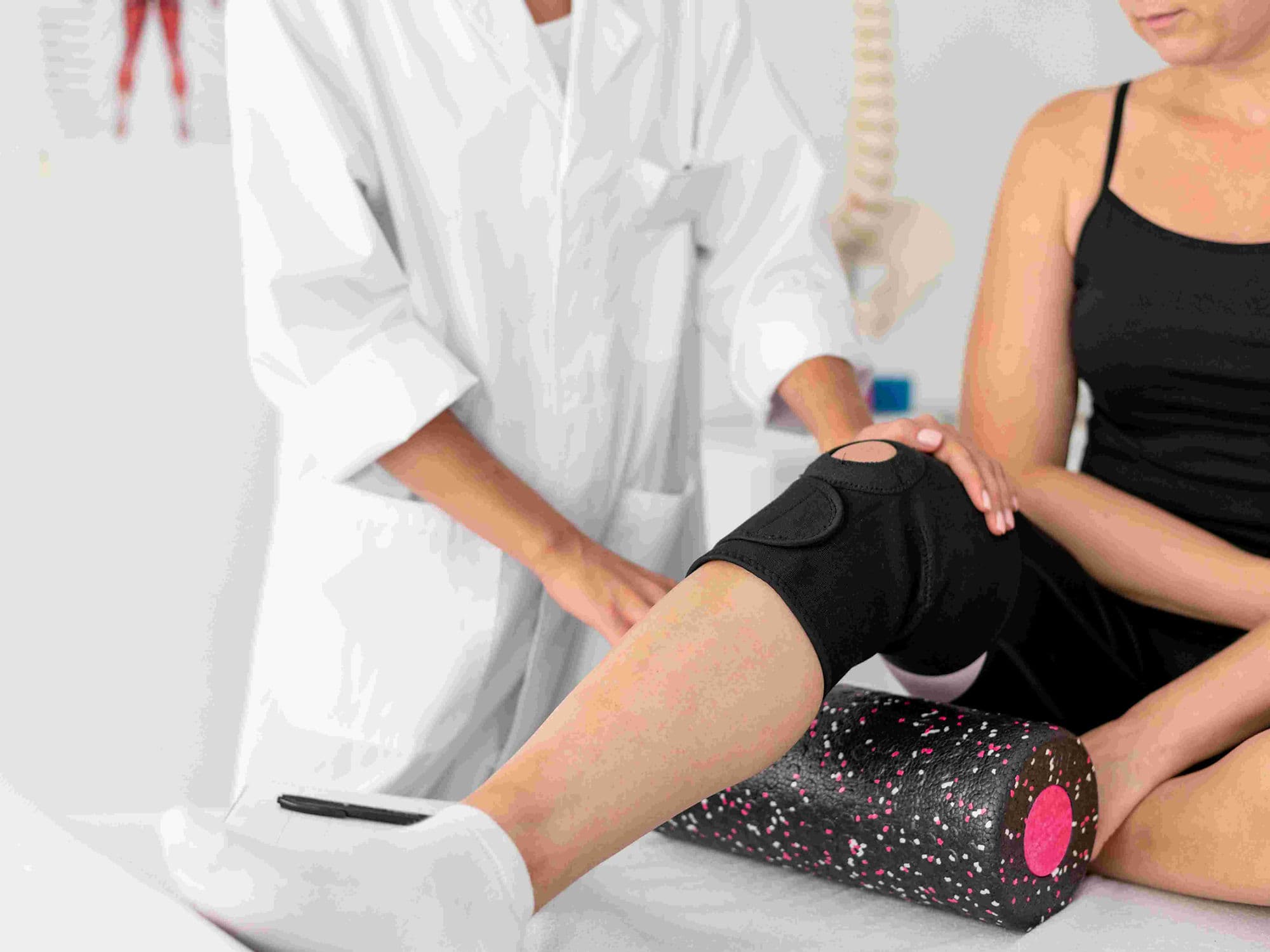
Hospital stays range from same-day discharge to 3 days, followed by 6–12 weeks of intensive physical therapy.
Achievement of 0° extension (straight leg) and at least 120° flexion (bend) is the primary goal of rehabilitation.
Use of blood thinners for 3–6 weeks is required to prevent Deep Vein Thrombosis (DVT).
High-impact sports like running are generally discouraged, but walking, swimming, and cycling are highly recommended.
Benefits of Total Knee Replacement
Significant pain relief and improved joint mobility in over 90% of patients.
Correction of knee deformities and restoration of proper leg alignment.
High durability, with modern implants lasting 15 to 20 years in the vast majority of cases.
Substantial improvement in the ability to perform daily tasks and overall quality of life.