Ventricular Septal Rupture Repair
Ventricular Septal Rupture (VSR) Repair
Ventricular Septal Rupture (VSR) Repair is a high-stakes, emergency surgical procedure to fix a hole in the septum (the wall dividing the left and right ventricles). This rupture is a rare but catastrophic complication of a massive heart attack, occurring when a lack of blood flow causes heart muscle to die and physically tear. Surgical intervention remains the "gold standard," as the condition is almost always fatal without mechanical closure.
When You Should Consider VSR Repair
Acute Heart Failure: When the septum tears, oxygen-rich blood surges into the right side of the heart, causing the heart to lose its ability to pump to the rest of the body.
Pulmonary Flooding: Sudden, excessive blood flow into the lungs leads to rapid fluid buildup (edema) and severe breathing difficulty.
Cardiogenic Shock: If blood pressure drops dangerously low and organs begin to fail due to the massive "shunt" of blood within the heart.
Post-Infarction Complication: Typically occurs within the first 24 hours or 3–5 days following a major heart attack.
High-Risk Stabilization: If a patient is currently on life support (ECMO) or a balloon pump (IABP) specifically to bridge them to a definitive surgical repair.
Surgical Techniques
Infarct Exclusion: The modern standard where a large synthetic patch is "wallpapered" over the hole and anchored to healthy, firm heart muscle away from the fragile tear.
Triple Patch Technique: A newer method using three layers of bovine pericardium and surgical glue to ensure a leak-proof seal and minimize the risk of the hole reopening.
Extended Sandwich Patch: Using two large Dacron patches to "sandwich" the septum from both the left and right sides, often used for complex or posterior ruptures.
Hybrid Repair: A two-stage approach where surgery is followed by a transcatheter "plug" if a small residual leak (shunt) remains after the initial operation.
Concomitant CABG: Since a blocked artery caused the rupture, surgeons almost always perform a heart bypass during the same procedure to protect the remaining muscle.
How Is Performed
Access: A midline incision is made through the breastbone (sternotomy) for the most direct access to the complex rupture site.
Bypass: The patient is connected to a heart-lung machine; the heart is stopped to allow the surgeon to operate on the delicate, damaged tissue.
Ventriculotomy: The surgeon opens the scarred area of the left ventricle (the chamber with the highest pressure) to inspect the tear.
Debridement: Any "mushy" or dead tissue at the edges of the hole is cleared away to reach firmer muscle that can hold sutures.
Patching & Gluing: The synthetic or tissue patch is meticulously secured. Specialized surgical glues are often used to reinforce the suture lines on fragile tissue.
Restarting: The heart is carefully restarted, and a transesophageal echo (TEE) is performed immediately to check for any residual leaks.
[Image showing a synthetic patch being sutured over a ventricular septal defect]
Pre-Procedure Preparation
Emergency Stabilization: Hemodynamic stabilization is the priority; many patients receive an Intra-aortic Balloon Pump (IABP) to reduce the heart's workload.
Fasting: Required, though most patients are already under emergency care and receiving fluids intravenously.
Blood Cross-matching: Extensive cross-matching is performed, as these surgeries carry a high risk of bleeding and often require blood transfusions.
Tissue Friability Review: Surgeons may delay surgery for 3–7 days if the patient is stable enough to let the heart muscle toughen, which increases suture success.
Emergency Consent: Consent is often obtained from family members, as the patient is typically too ill or sedated to provide it themselves.
Tests Before VSR Repair
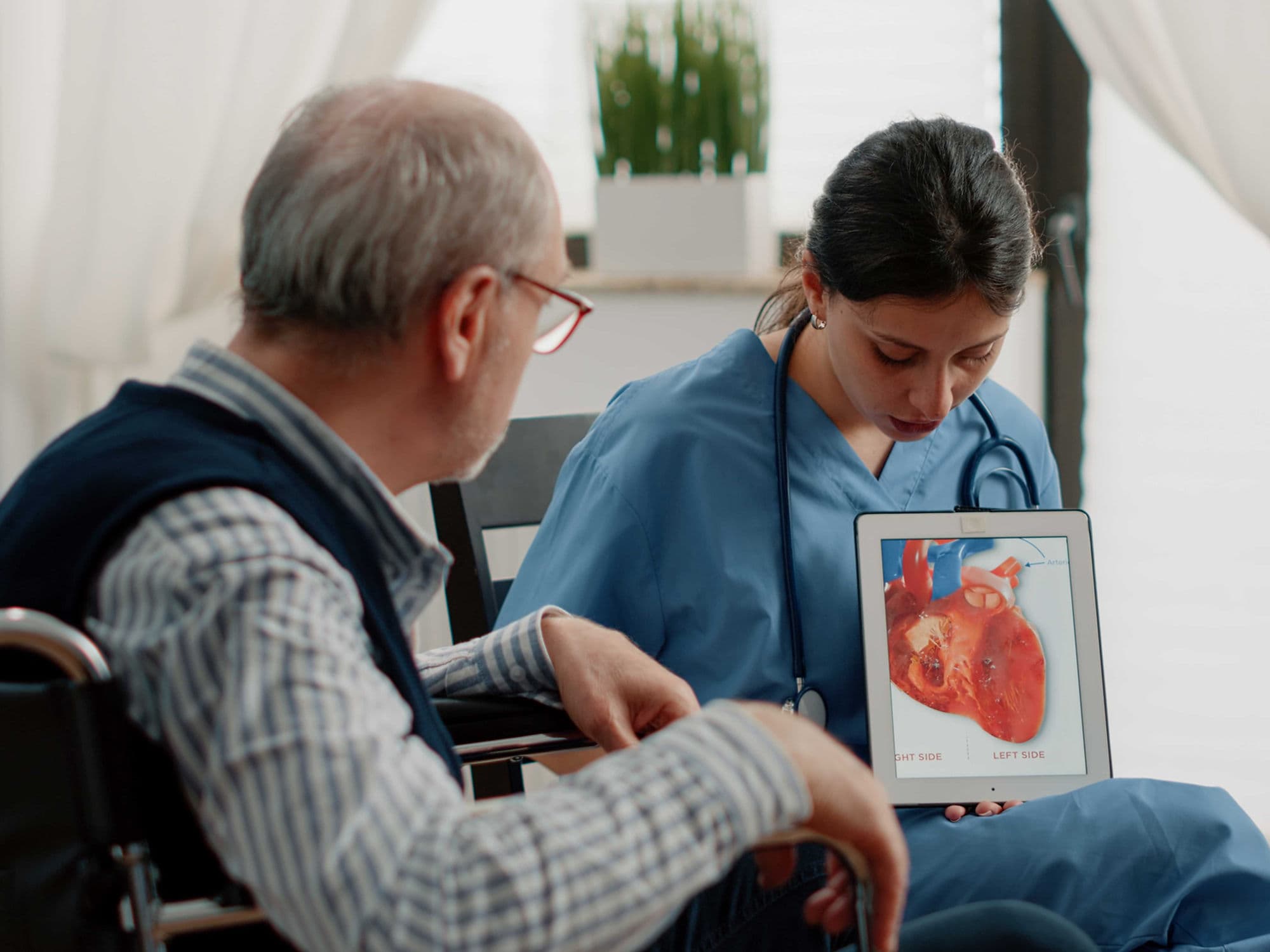
Echocardiogram (TTE/TEE): The essential test to confirm the location and size of the rupture and quantify the "shunt" volume.
Coronary Angiogram: Necessary to identify the blocked artery that caused the heart attack and plan the necessary bypass grafts.
Cardiac CT Scan: Sometimes used to assess the anatomy of the rupture, especially if it is in a difficult-to-reach posterior location.
Swan-Ganz Catheterization: To measure the pressures in the lungs and the degree of oxygen-rich blood mixing in the right side of the heart.
Blood Gas Analysis: To monitor how well the lungs are coping with the sudden influx of extra blood.
Life After VSR Repair
ICU Stay: Patients typically require 3 to 7 days in the ICU on a ventilator with multiple medications to support blood pressure.
Hospital Stay: Total recovery in the hospital usually lasts 2 to 3 weeks due to the severity of the initial heart attack.
Long-term Management: Lifelong heart failure medications (such as Beta-blockers and ARNI therapy) are essential to help the heart recover.
Residual Shunt Monitoring: 10–20% of cases may have a tiny remaining leak; these are monitored via regular echocardiograms and only repaired if they cause symptoms.
Rehabilitation: A slow, medically supervised cardiac rehab program is vital to rebuild strength after such a massive physiological trauma.
Benefits of VSR Repair
Life-Saving Intervention: Without surgery, the mortality rate is nearly 90% within weeks; repair offers the only realistic chance for survival.
Stops Pulmonary Flooding: Immediately halts the surge of blood into the lungs, allowing for easier breathing and recovery from edema.
Restores Systemic Pressure: By closing the hole, the heart can once again send oxygenated blood to the brain, kidneys, and liver.
Improved Outcomes: While high-risk, 30-day survival rates in specialized cardiac centers have improved significantly for stable patients.
Future Heart Health: For those who survive the initial recovery, long-term heart function can improve significantly with proper care.