TMVI/TMVR (Transcatheter Mitral Valve Replacement)
TMVI/TMVR (Transcatheter Mitral Valve Replacement)
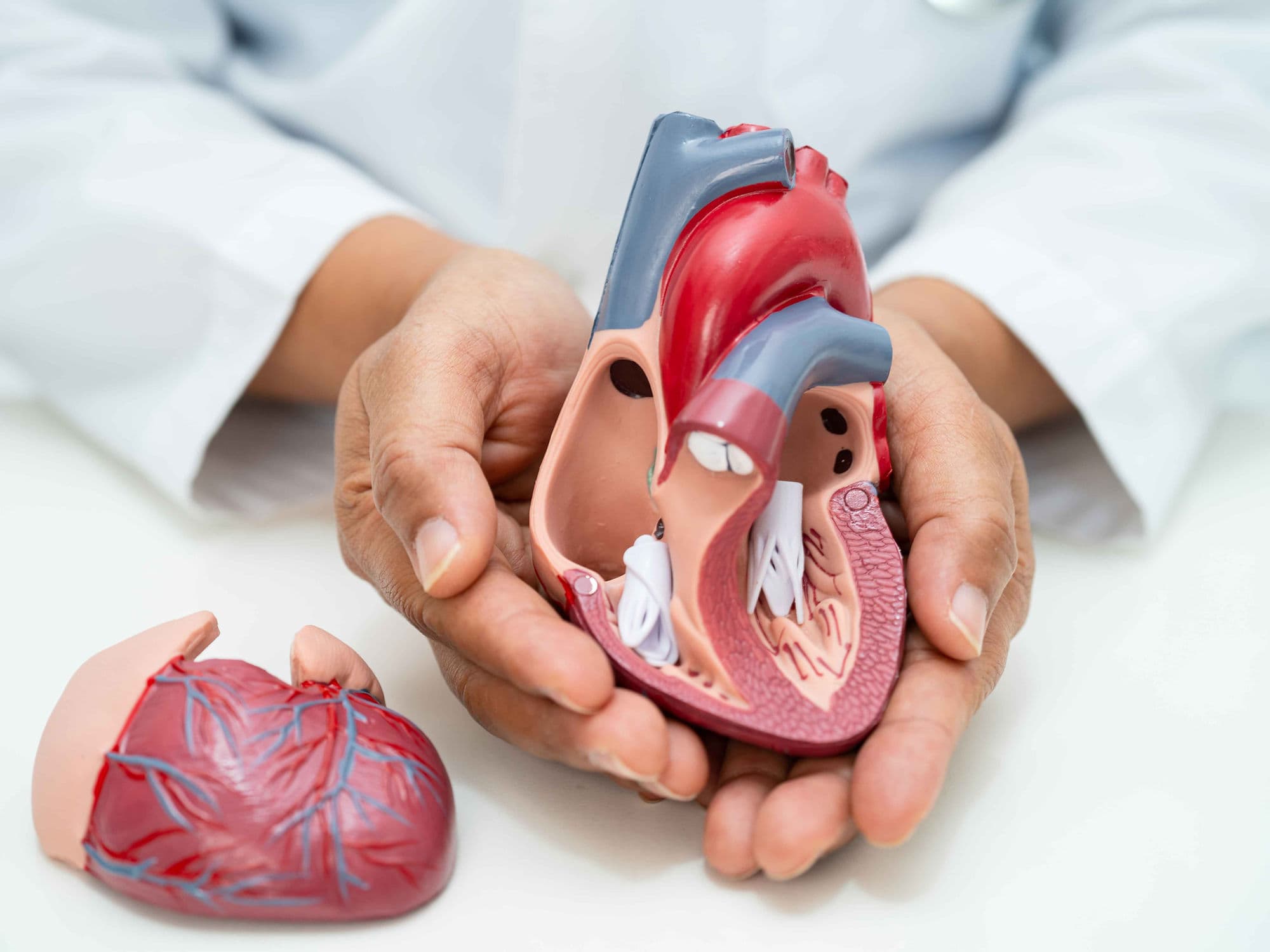
TMVI (Transcatheter Mitral Valve Implantation) and TMVR (Transcatheter Mitral Valve Replacement) are minimally invasive procedures used to replace a diseased mitral valve without the need for traditional open-heart surgery. These procedures are typically reserved for high-risk patients with severe Mitral Regurgitation (a leaking valve) or Mitral Stenosis (a narrowed valve) who may not tolerate a standard sternotomy.
When You Should Consider TMVI / TMVR
Severe Mitral Regurgitation: When the mitral valve does not close tightly, causing blood to flow backward into the lungs.
Mitral Stenosis: When the valve leaflets become thick or stiff, restricting blood flow from the left atrium to the left ventricle.
High Surgical Risk: For patients whose age or underlying health conditions (like lung or kidney disease) make traditional surgery too dangerous.
Failed Previous Valve: A "Valve-in-Valve" procedure for patients whose previously implanted surgical biological valve has begun to wear out.
Functional Mitral Disease: When heart failure has caused the heart to enlarge, pulling the mitral valve leaflets apart and causing a massive leak.
How TMVI / TMVR Is Performed
3D Guidance: The surgical team uses a combination of real-time X-ray (fluoroscopy) and Transesophageal Echocardiography (TEE) to see the heart in three dimensions.
Access Routes: * Transseptal: The most common approach; a catheter is guided from the groin vein, through the wall of the heart (septum), and into the mitral position.
Transapical: A small incision is made between the ribs to access the valve directly through the tip (apex) of the heart.Valve Positioning: A collapsed artificial valve—constructed from biological tissue on a metal frame—is steered precisely into the center of the diseased native valve.
Deployment: The new valve is expanded, either by a balloon or a self-expanding mechanism. This pushes the old valve leaflets aside and anchors the new valve firmly in place.
Leak Check: Before finalizing the placement, the team checks for "paravalvular leaks" to ensure blood cannot escape around the edges of the new device.
Pre-Procedure Preparation
Cardiac CT Scan: A specialized high-resolution scan is mandatory to measure the "neo-LVOT"—ensuring the new valve frame won't block the heart's main exit path.
Transesophageal Echocardiogram (TEE): An ultrasound probe passed down the esophagus to provide the clearest possible images of the valve structure.
Heart Team Evaluation: A collaborative review by interventional cardiologists and cardiac surgeons to confirm this is the safest treatment path.
Dental Clearance: To minimize the risk of bacteria entering the bloodstream and infecting the new heart valve (endocarditis).
Fasting (NPO): No food or drink for at least 8 hours prior to the procedure, as it is performed under general anesthesia.
Tests Before TMVI / TMVR
3D Cardiac CT: Essential for sizing the valve and mapping the internal dimensions of the left ventricle.
Diagnostic Catheterization: To check for blockages in the coronary arteries that might need treatment at the same time.
Blood Panels: To assess kidney function and ensure the blood's clotting ability is within a safe range for the procedure.
Lung Function Tests: To evaluate the patient's overall respiratory health for anesthesia planning.
Life After TMVI / TMVR
Hospital Stay: Usually 2 to 5 days, which is significantly shorter than the recovery for open-heart surgery.
Medication Adherence: Patients must take anticoagulants (blood thinners) for at least 3 to 6 months—and often indefinitely—to prevent clots from forming on the metal frame.
Immediate Improvement: Most patients notice a dramatic reduction in shortness of breath and fatigue almost immediately after the procedure.
Activity Restrictions: Heavy lifting and strenuous exercise are restricted for 2 to 4 weeks while the access site in the groin or chest heals.
Long-Term Follow-up: Regular echocardiograms are required (at 30 days, 6 months, and annually) to ensure the valve remains functional and secure.
Benefits of TMVI / TMVR
No Sternotomy: Avoids the need to open the chest bone, resulting in significantly less pain and a lower risk of wound infection.
Faster Mobilization: Patients are usually up and walking within a day of the procedure.
Effective Symptom Relief: Successfully stops the "back-pressure" on the lungs, allowing for better breathing and increased energy levels.
High Success Rate: Modern devices are highly effective at reducing or eliminating mitral leaks, even in the most complex heart geometries.