Mediastinal Lymph Node Dissection (Cancer)
Mediastinal Lymph Node Dissection
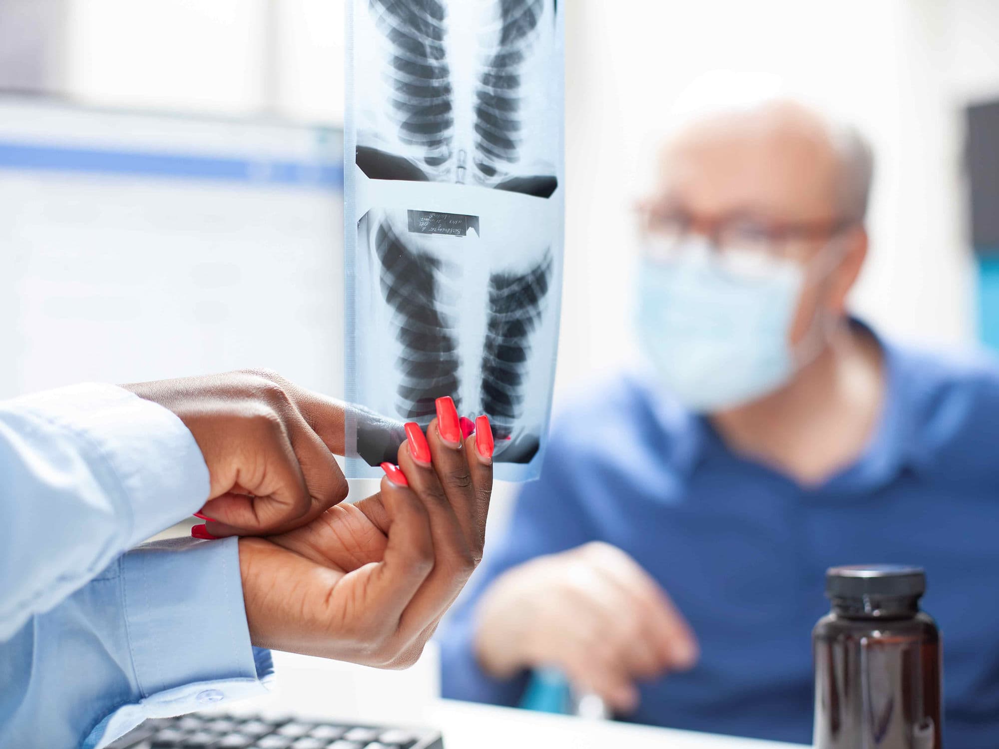
Mediastinal Lymph Node Dissection (MLND) is a surgical procedure to remove the lymph nodes located in the mediastinum—the central area of the chest between the lungs. It is a critical component of lung cancer surgery. Rather than just taking a sample, the surgeon removes all the lymph nodes and surrounding fat within specific "stations" to ensure any microscopic cancer spread is captured. This procedure is the gold standard for accurate pathologic staging, which dictates whether a patient needs further treatment like immunotherapy or chemotherapy.
When You Should Consider MLND
Lung Cancer Surgery: Performed as a mandatory part of a lobectomy or pneumonectomy for Non-Small Cell Lung Cancer (NSCLC).
Staging Accuracy: When imaging (PET-CT) suggests nodes might be involved, or even if they look normal but the primary tumor is large.
Thymic Tumors: For patients with thymoma or thymic carcinoma to check for regional spread.
Esophageal Cancer: Often included in an esophagectomy to clear the lymphatic drainage path of the esophagus.
Diagnostic Uncertainty: When non-surgical biopsies (like EBUS) are inconclusive but suspicion of nodal involvement remains high.
Methods Of MLND
Robotic-Assisted (RATS) Dissection: The preferred modern tool for MLND. Its 3D magnification allows surgeons to see tiny nerves and vessels clearly, making it safer to remove nodes deep in the chest.
Video-Assisted Thoracoscopic (VATS) Dissection: A minimally invasive approach using a camera and specialized instruments through small "keyhole" incisions.
Open Thoracotomy Dissection: Usually performed through the same large incision used for an open lung resection, allowing for direct manual access to the mediastinum.
Mediastinoscopy: A separate, smaller surgical procedure where a scope is inserted through a small notch at the base of the neck to reach the upper nodal stations.
Systematic Nodal Sampling: A less extensive version where only representative nodes are taken, though full dissection (MLND) is preferred for more accurate staging.
How Is Performed
Surgical Access: The surgeon enters the chest cavity using the same approach selected for the primary lung or esophageal resection.
Anatomical Exposure: The surgeon opens the thin lining (pleura) over the mediastinum to expose the fat pads containing the lymph nodes near the trachea, esophagus, and heart.
Systematic Clearance: All lymphoid tissue and surrounding fat within the targeted "stations" are meticulously removed.
Nerve Preservation: Great care is taken to identify and protect the Phrenic nerve (for breathing) and the Recurrent Laryngeal nerve (for the voice) that run through the mediastinum.
Hemostasis: Using advanced energy devices like ultrasonic scalpels, the surgeon seals small lymphatic channels and blood vessels to prevent fluid buildup or "oozing."
Pathology Review: The removed nodes are labeled by their specific station number and sent to a lab where a pathologist examines them under a microscope for cancer cells.
Pre-Procedure Preparation
PET-CT Scan: To identify which nodal stations show "metabolic activity," helping the surgeon prioritize specific areas for thorough dissection.
EBUS-TBNA: Many patients undergo an Endobronchial Ultrasound biopsy before surgery to "pre-stage" the nodes and plan the extent of the dissection.
Cardiovascular Review: Since the surgery occurs near the heart and great vessels, ensuring stable heart function is vital for a safe procedure.
Anticoagulation Management: Stopping blood thinners is critical, as MLND involves working around highly vascular structures where bleeding must be strictly controlled.
Incentive Spirometry: Strengthening the lungs before the procedure to ensure you can cough effectively and clear your airway post-operatively.
Tests Before MLND
High-Resolution Chest CT: To map the anatomy of the lymph nodes in relation to the laryngeal nerve and the superior vena cava.
Endobronchial Ultrasound (EBUS): To provide a preliminary assessment of the nodes through the airway before the definitive surgical removal.
Chest MRI: Sometimes used if nodes are near the spine or major nerves to evaluate if the tumor has invaded those structures.
Blood Coagulation Profile: To ensure the body can effectively stop minor oozing from the lymphatic channels after the nodes are removed.
Baseline Vocal Assessment: Since nerves controlling the voice box are located in the mediastinum, a baseline check of the voice is often performed for comparison after surgery.
Life After MLND
Chest Tube Management: You will have a chest tube for a few days to drain any fluid or air; it is removed once the drainage levels from the dissection site are safe.
Vocal Cord Monitoring: A temporary hoarse voice can occur if the laryngeal nerve is irritated during the dissection; most cases recover with time and specialized therapy.
Dietary Adjustments: In rare cases of "Chylothorax" (lymphatic fluid leak), a specific low-fat diet may be required for a short period to allow the duct to heal.
Pain Management: Dissection near the ribs and spine can cause localized "aching" or soreness; this is managed with nerve blocks and oral medications.
Follow-up Treatment: The final "nodal status" (Pathology Report) typically takes 5–7 days and is the most important factor in determining if you need follow-up chemotherapy or immunotherapy.
Benefits Of MLND
Definitive Staging: MLND provides the most accurate "N" (Nodal) stage, which is far more precise than a PET-CT or EBUS biopsy alone.
Reduced Recurrence: Removing all nodes in a station (rather than just sampling) significantly lowers the chance of the cancer returning in the center of the chest.
Adjuvant Guidance: Knowing exactly which nodes are involved allows oncologists to prescribe targeted therapies or immunotherapies that can significantly improve survival rates.
Minimal Impact on Recovery: When performed robotically or thoracoscopically, adding MLND to a lung resection adds very little time to the hospital stay but provides invaluable data.
Comprehensive Clearance: Ensures that any microscopic clusters of cancer cells in the regional lymph system are physically removed from the body.