Tricuspid Valve Repair
Tricuspid Valve Repair
Tricuspid Valve Repair is a surgical or minimally invasive procedure to fix a leaking (regurgitation) or narrowed (stenosis) tricuspid valve, which sits between the right atrium and right ventricle. Repair is increasingly preferred over valve replacement because it preserves the heart's natural anatomy and avoids the need for lifelong, heavy-duty blood thinners. It is a vital intervention for maintaining proper blood flow from the body into the lungs.
When You Should Consider Tricuspid Valve Repair
Secondary (Functional) Regurgitation: When the valve leaks because the right side of the heart has stretched (common in patients with left-sided heart disease).
Concomitant Repair: When you are already undergoing surgery for a mitral or aortic valve; repairing the tricuspid valve at the same time prevents future heart failure.
Severe Right-Sided Symptoms: Such as significant swelling in the legs, abdominal bloating, or unexplained fatigue.
Direct Valve Damage: Caused by infection (endocarditis), rheumatic fever, or blunt chest trauma.
Pulmonary Hypertension: When high pressure in the lungs forces the tricuspid valve to leak, requiring a surgical "tightening" of the valve base.
Surgical Techniques
Annuloplasty (The Ring): The "gold standard" where a cloth-covered medical ring is sewn around the base of the valve to pull the leaflets together for a tight seal.
Leaflet Repair: Techniques like "bicuspidization" (tucking a leaflet) or patching holes with a piece of the heart's own sac (pericardium).
Neochords: Attaching artificial GORE-TEX strings to support drooping or "flail" leaflets that no longer close properly.
Edge-to-Edge Repair (TriClip): A leading-edge, minimally invasive option where a clip is guided through a leg vein to "pin" leaking leaflets together.
Minimally Invasive Surgery: Performing the repair through a small incision between the ribs (thoracotomy) rather than opening the breastbone.
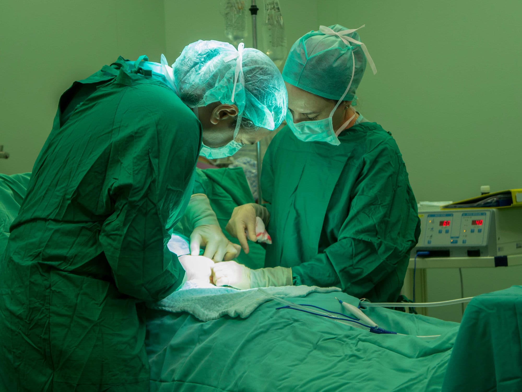
How Is Performed
Access: Performed via a midline incision (sternotomy) or a minimally invasive side incision.
Bypass: The patient is connected to a heart-lung machine, which takes over the work of the heart and lungs during the repair.
Inspection: The surgeon opens the right atrium to inspect the valve leaflets and the supporting "annulus" ring.
Implantation: The annuloplasty ring or neochords are meticulously sewn into place to restore the valve's shape.
Testing: Saline is injected into the ventricle to confirm the valve is leak-proof before the heart is closed and restarted.
Pre-Procedure Preparation
Fasting: Required for 8–12 hours before surgery, as it is performed under general anesthesia.
Extensive Blood Tests: Including liver and kidney function panels, as these organs are often affected by tricuspid issues.
Dental Check-up: To ensure no oral bacteria could cause a post-surgical heart infection.
Medication Adjustment: Specifically regarding blood thinners, as directed by your surgical team.
Sanitization: Shaving and antiseptic cleaning of the chest and any potential graft sites.
Tests Before Tricuspid Valve Repair
Echocardiogram (TTE/TEE): The primary tool used to grade the severity of the leak and measure the size of the heart chambers.
Cardiac Catheterization: To check the pressures in the heart and lungs (pulmonary hypertension) and look for coronary artery blockages.
Cardiac MRI: To get a high-definition 3D view of the right ventricle's function and volume.
Liver Function Tests: To see if the "back-pressure" from the leaky valve has caused liver congestion.
Chest X-ray: To evaluate the size of the heart silhouette and the condition of the lungs.
Life After Tricuspid Valve Repair
Hospital Stay: Usually lasts 5 to 7 days, with the first 24–48 hours spent in the ICU for close monitoring.
Initial Recovery: Most patients are encouraged to sit up and begin walking within 24 hours of surgery.
Sternal Precautions: If a sternotomy was performed, no lifting over 3 kg for 6 to 8 weeks to allow the bone to heal.
Medication: Most patients take a mild blood thinner (like aspirin) for 3–6 months; lifelong Warfarin is typically not required for a repair.
Follow-up: Regular echocardiograms will be scheduled to ensure the repair remains stable and the heart size is shrinking back to normal.
Benefits of Tricuspid Valve Repair
High Durability: Over 90% of repairs are successful and significantly reduce leakage for many years.
Prevents Heart Failure: Directly reduces the risk of right-sided heart failure and associated liver congestion.
Improved Energy: Patients often notice a dramatic reduction in swelling and a significant increase in exercise capacity.
Preserves Heart Function: Keeping your natural valve (rather than a replacement) helps the right ventricle maintain its strength.
High Success Rates: Elective repairs in specialized centers have low complication rates (1% to 3%) and excellent long-term survival.