Medivisor India Treatment Logo
+91 83682 47758
info@medivisorhealth.com
  • Home
    • About Us
    • Our Services
    • Our Team
    • Our Safety Measures
    • Loading treatments...
    • Loading hospitals...
    • Loading doctors...
    • Treatment Cost
    • Treatment Process
    • Visa Process
    • Travel Guide
    • FAQs
    • Why Medivisor
    • Success Stories
    • Patient Activities
    • Social Activities
    • News Coverage
    • Blog
  • Become a Partner

Medivisor India Treatment is a trusted New Delhi–based medical travel company assisting international patients with advanced, affordable, and seamless treatment in India.

  • Medivisor House 359, Sector 1, Vaishali, Ghaziabad, (Delhi/NCR) India
  • +91 8368 247 758
  • info@medivisorhealth.com

About Us

  • About Us
  • Our Services
  • Our Team
  • Our Medical Advisors
  • Our Safety Measures
  • Join Our Network
  • Contact Us

India Treatment

  • Hospitals
  • Treatments
  • Treatment Cost
  • Treatment Process
  • Visa Process
  • Travel Guide
  • FAQs
  • Why Medivisor

Gallery

  • Patient Testimonials
  • Patient Activities
  • News Coverage
  • Blog

Get in Touch

© 2026 Medivisor India Treatment. All rights reserved.

Dr Sandeep Attawar

Chair & Director of Thoracic Organ Transplants

Cardiothoracic & Vascular Surgeon (CTVS)Lung Transplant SurgeonHeart Transplant SurgeonPediatric Cardiac Surgeon

33+ years experience
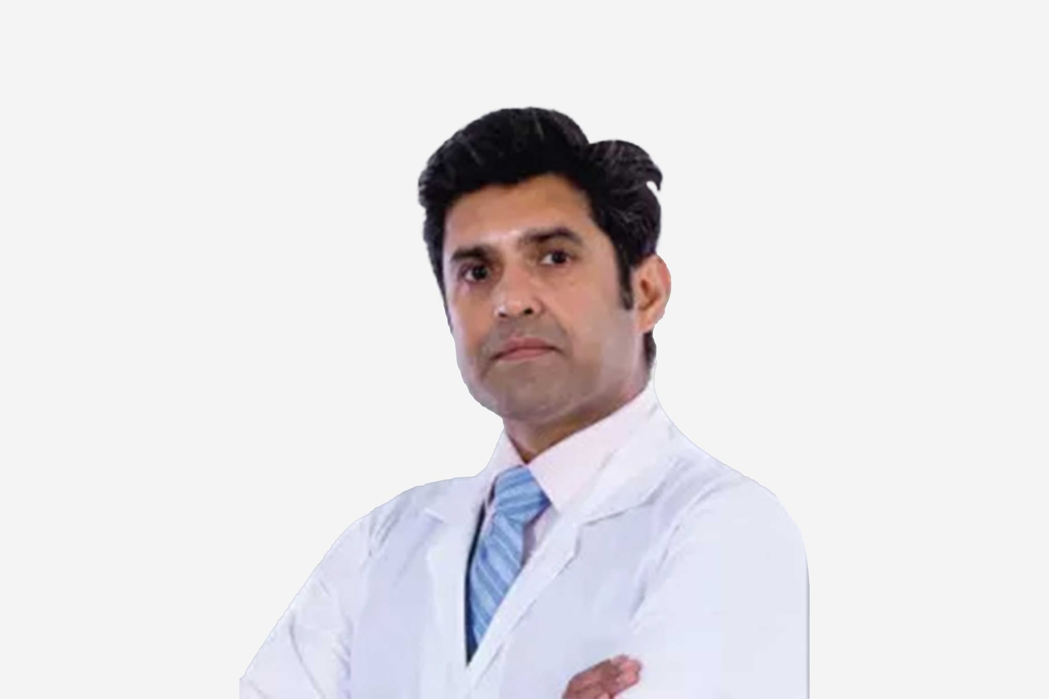
Dr Sandeep Attawar, Chair & Director of Thoracic Organ Transplants
  1. Home
  2. Doctors
  3. Dr Sandeep Attawar

About Dr Sandeep Attawar

Dr. Attawar is a highly distinguished specialist in cardiothoracic surgery and advanced organ transplantation, recognized for his technical mastery in complex cardiovascular and pulmonary interventions. He specializes in utilizing precision-guided surgical platforms to restore cardiopulmonary function and optimize outcomes for patients across all age demographics, from neonates to geriatric populations.

Mastery in Heart and Lung Transplantation

He specializes in the comprehensive management of end-stage heart and lung failure. His clinical practice focuses heavily on executing high-precision thoracic organ transplants, with a documented track record that includes hundreds of complex procedures such as double lung transplants, isolated heart transplants, and combined heart-lung transplantations.

Innovation in Mechanical Circulatory Support and Reconstructive Surgeries

Dr. Attawar possesses profound expertise in treating advanced cardiac failure through the utilization of sophisticated mechanical circulatory support systems, including the implantation of Left Ventricular Assist Devices (LVADs). His structural surgical capabilities encompass a wide spectrum of intricate interventions, including coronary artery bypass grafting, complex valve repair and replacement, and the closure of congenital septal defects.

Pioneering Post-Viral Pulmonary Reconstructions

A primary focus of his surgical practice is the implementation of advanced transplantation pathways to address irreversible, end-stage organ damage. He and his team have pioneered specialized thoracic interventions for severe post-viral complications, executing a globally recognized series of successful double lung transplants for patients with terminal lung damage resulting from COVID-19.

Global Clinical Governance and Institutional Leadership

Throughout his extensive career, Dr. Attawar has integrated modern cardiothoracic innovations with rigorous clinical protocols to lead major healthcare initiatives, playing a foundational role in establishing multiple premier heart centers globally. His commitment to evidence-based medicine is further demonstrated by his appointment to an elite international panel of experts tasked with authoring the consensus guidelines for lung transplantation followed by hundreds of medical institutions worldwide.

Dr. Sandeep Attawar at a Glance

  • Specialist in Cardiothoracic Surgery, Advanced Heart/Lung Transplantation, and Mechanical Circulatory Support.

  • Expert in a comprehensive range of interventions from neonatal pediatric heart surgeries to complex adult bypass operations.

  • Extensive procedural experience encompassing over 450 thoracic organ transplants and dozens of LVAD implants.

  • Pioneer in executing successful double lung transplantations for end-stage COVID-19 pulmonary damage.

  • International guidelines panelist helping shape standard thoracic transplantation protocols used globally.

  • Dedicated to institutional excellence, having established three major heart centers in India and abroad.

MBBS
MS - Surgery
M.CH - CTVS
Board Certified in Cardiothoracic & Vascular Surgeon (CTVS)
Current president of Indian society of heart
lung transplant(INSHLT)
Active member of INSHLT, Society of thoracic surgeons(STS)
the European association of cardio-thoracic surgery(EACTS).
Contributor for the textbook of “Tranplantation & Mechanical circulatory support for end stage lung
heart disease” .
Pioneer in India’s first breathing lung transplant (XVIVO perfusion).
Founder of the institute of lung bioengineering at KIMS.
Scientific advisor for reliant heart
is involved in advancing LVAD.
Proctor for heart mate II & III, JARVIK HEART for India
south east asia.
May we help you?

Enter digits only (6-15 digits). We'll add your country code.

Related Treatments

Aortic Aneurysm Repair (Open)

Open Aortic Aneurysm Repair is a major surgical procedure used to treat a life-threatening bulge in the aorta, the body's main artery. Unlike minimally invasive endovascular repair (EVAR), this traditional "open" approach involves a large incision to directly access the aorta, remove the diseased section, and replace it with a synthetic graft. It remains the "gold standard" for its durability and for treating complex aneurysms that are not suitable for stenting.

When You Should Consider Open Aortic Aneurysm Repair

  • Abdominal Aortic Aneurysm (AAA): When a bulge in the abdominal portion of the aorta reaches a critical size (typically 5.0–5.5 cm) or shows rapid growth.

  • Thoracic Aortic Aneurysm (TAA): For aneurysms located in the chest cavity that carry a high risk of rupture or dissection.

  • Complex Anatomy: When the shape or location of the aneurysm is too close to vital branching vessels, such as the renal (kidney) arteries, making a stent unfeasible.

  • Younger, Fit Patients: Due to the graft's long-term durability, younger patients with a longer life expectancy often benefit from a one-time permanent repair.

  • Ruptured Aneurysm: Open surgery remains a primary life-saving intervention for patients experiencing active internal bleeding from a burst aorta.

Methods Of Open Aortic Aneurysm Repair

  • Transperitoneal Approach: A long vertical incision made from the breastbone to below the belly button to access the abdominal aorta.

  • Retroperitoneal Approach: A side incision often used for patients with previous abdominal surgeries or specific anatomical needs to reach the aorta from behind.

  • Dacron Graft Interposition: The standard method of sewing a durable, woven polyester tube into the healthy parts of the aorta to replace the weakened section.

  • Bifurcated Grafting: A specialized "Y-shaped" graft used when the aneurysm extends down into the iliac arteries that lead to the legs.

  • Thoracoabdominal Repair: An extensive procedure involving both the chest and abdomen for aneurysms that span across the diaphragm.

How Is Performed

  • Surgical Access: Under general anesthesia, the surgeon makes a large incision (chest or abdomen) to provide direct visualization of the diseased aorta.

  • Aortic Clamping: To stop blood flow during the repair, the surgeon places specialized clamps on the aorta above and below the aneurysm site.

  • Organ Protection: During the clamping phase, techniques like mild hypothermia or selective perfusion are used to protect the kidneys and intestines from a lack of oxygen.

  • Graft Insertion: The surgeon cuts open the weakened aortic wall and sews a synthetic tube (the graft) into the healthy tissue above and below the bulge.

  • Aortic Wrap: The original, weakened aortic wall is often wrapped around the new synthetic graft to provide an extra layer of protection and support.

  • Restoring Flow: The clamps are carefully removed to allow blood to flow through the new synthetic lining, and the surgeon checks all suture lines for leaks.

Pre-Procedure Preparation

  • Cardiac Clearance: Extensive heart testing, such as a stress test or echocardiogram, is mandatory to ensure the heart can handle the stress of aortic clamping.

  • Advanced Imaging: High-resolution CT Angiography (CTA) is used to create a precise 3D map of the aneurysm and the branching arteries.

  • Kidney Function Check: Blood tests to evaluate renal health, as the kidneys are temporarily affected by the change in blood flow during surgery.

  • Smoking Cessation: Stopping smoking at least 4 weeks prior is critical to reduce the risk of postoperative lung complications and promote graft healing.

  • Fasting (NPO): No food or drink for 8–12 hours prior to the procedure to ensure safety under general anesthesia.

Tests Before Open Aortic Aneurysm Repair

  • CT Angiogram (CTA): The primary tool for measuring the aneurysm's diameter and identifying its relationship to the renal and mesenteric arteries.

  • Electrocardiogram (EKG): To check baseline heart rhythm and rule out underlying conditions before the major operation.

  • Complete Blood Count (CBC): To ensure adequate hemoglobin levels and check for any signs of infection.

  • Coagulation Profile: To confirm the blood's ability to clot normally, as this procedure carries a risk of significant blood loss.

Life After Open Aortic Aneurysm Repair

  • Hospital Stay: Expect to spend 5 to 10 days in the hospital, typically starting with the first 24–48 hours in the Intensive Care Unit (ICU).

  • Pain Management: Significant abdominal or chest wall soreness is expected; patients are managed with IV pain relief initially, transitioning to oral medications.

  • Incentive Spirometry: Deep breathing exercises are essential to prevent pneumonia, especially after a large abdominal or chest incision.

  • Activity Restrictions: Walking is encouraged within 24 hours to prevent blood clots, but heavy lifting (over 10 lbs) is restricted for 6 to 12 weeks.

  • Long-term Monitoring: Unlike EVAR, which requires annual scans, open repair usually requires less frequent follow-up imaging (often every 5 years) once the graft is secure.

Benefits Of Open Aortic Aneurysm Repair

  • Proven Durability: The synthetic graft is permanently sewn into place and is designed to last for the remainder of the patient's life.

  • Lower Re-intervention Rate: Patients who undergo open repair are much less likely to need follow-up "fix-it" procedures compared to those with stents.

  • Total Removal of Risk: By replacing the diseased section, the threat of a future rupture at that specific site is virtually eliminated.

  • Versatility: Can treat complex aneurysms that are too tortuous or involve too many branching vessels for minimally invasive technology.

  • Peace of Mind: Provides long-term security with a significantly lower requirement for frequent, life-long radiation-heavy CT surveillance.

Aortic Valve Replacement AVR

Aortic Valve Replacement (AVR) is an advanced cardiac procedure that replaces a damaged, stiff, or leaking aortic valve with a new mechanical or tissue valve. This restores healthy blood flow, improves heart pumping capacity, reduces symptoms, and prevents long-term heart failure or life-threatening complications.

When You Should Consider AVR

  • Severe or persistent shortness of breath that limits walking, climbing stairs, or daily activity.

  • Chest pain, pressure, or heaviness due to the heart struggling to push blood through a narrowed valve.

  • Extreme tiredness or low energy even during simple tasks.

  • Dizziness or fainting episodes, especially during exertion.

  • Irregular heartbeat or noticeable palpitations, indicating the heart is under stress.

  • Swelling in the feet, legs, or ankles, a sign of poor blood circulation or early heart failure.

Conditions That Require AVR

  • Severe Aortic Stenosis – the valve becomes narrowed and heavily calcified, restricting blood flow.

  • Severe Aortic Regurgitation – the valve leaks and allows blood to flow backward into the heart.

  • Congenital valve abnormalities, including bicuspid valves.

  • Infection-related valve damage (endocarditis) that weakens or destroys the valve.

  • Aged, stiff, or heavily calcified aortic valve due to long-term wear and tear.

How Aortic Valve Replacement Is Performed

  • General anesthesia is given to ensure a pain-free and comfortable procedure.

  • The surgeon makes either a full chest incision or a minimally invasive cut depending on your case.

  • The damaged aortic valve is carefully removed.

  • A new mechanical or biological valve is implanted to restore proper blood flow.

  • The heart is restarted, and valve function is tested to ensure smooth operation.

  • You are shifted to the ICU for continuous monitoring and early recovery.

Types of Aortic Valve Replacement

  • Mechanical Valve Replacement
    Long-lasting artificial valve; ideal for younger patients. Requires lifelong blood thinners to prevent clots.

  • Biological (Tissue) Valve Replacement
    Made from natural tissue. Offers natural blood flow and usually requires minimal blood thinner use.

  • Minimally Invasive AVR
    Smaller incisions, less pain, reduced blood loss, and faster healing.

  • Robotic AVR
    Performed using robotic precision tools for high accuracy, minimal scars, and quicker recovery.

  • TAVR (Transcatheter Aortic Valve Replacement)
    A non-surgical, catheter-based procedure performed through the groin. Ideal for elderly or high-risk patients.

Pre-Surgery Preparation

  • Quit smoking at least 2–3 weeks before surgery for better lung function.

  • Keep blood pressure, diabetes, and heart rate well controlled.

  • Follow fasting instructions before the procedure.

  • Stop blood thinners only if your cardiologist advises.

  • Complete all required heart and blood tests before the surgery date.

Pre-Surgery Tests

  • ECG to check heart rhythm.

  • 2D Echocardiography to evaluate valve structure and pumping strength.

  • CT scan or MRI for detailed imaging when needed.

  • Coronary Angiography to detect any artery blockages.

  • Chest X-ray to assess lung health.

  • Routine blood tests including CBC, kidney/liver function, and clotting profile.

Why AVR Is Highly Effective

  • Restores normal forward blood flow from the heart.

  • Reduces breathlessness and chest discomfort.

  • Prevents the heart from becoming enlarged or weak.

  • Improves daily stamina, energy levels, and activity tolerance.

  • Provides long-lasting results with modern valve technology.

Recovery After AVR

  • ICU stay: Usually 1–2 days for close monitoring.

  • Early walking begins within 24 hours.

  • Tubes and drains are removed in 48–72 hours.

  • Home recovery: Typically 4–8 weeks depending on the surgery type.

  • Return to work: Usually within 6–10 weeks.

Life After AVR

  • Avoid smoking permanently to protect the new valve.

  • Follow a heart-healthy, low-salt diet for lifelong cardiac wellness.

  • Exercise daily with light walking, avoid heavy lifting initially.

  • Take medications regularly, especially blood thinners if you have a mechanical valve.

  • Join a cardiac rehabilitation program for guided recovery and long-term heart strength.

Mitral Valve Replacement (MVR)

Mitral Valve Replacement (MVR) is a specialized heart procedure that restores healthy blood flow by replacing a diseased mitral valve with a mechanical or biological valve. This improves heart efficiency, reduces symptoms like breathlessness and fatigue, and prevents long-term complications such as heart failure.

When You Should Consider MVR

  • Shortness of breath during daily activities or while lying down.

  • Chest discomfort or pressure caused by poor blood flow through the heart.

  • Fatigue or low energy during simple tasks.

  • Irregular heartbeat or palpitations due to valve dysfunction.

  • Swelling in feet, legs, or ankles from fluid retention.

  • Fainting or dizziness, especially during physical activity.

Conditions That Require MVR

  • Severe Mitral Stenosis – narrowing of the mitral valve restricting blood flow.

  • Severe Mitral Regurgitation – leaking mitral valve causing backward blood flow.

  • Congenital mitral valve defects present from birth.

  • Valve damage from infection (endocarditis).

  • Calcified or thickened mitral valve leading to poor heart function.

How Mitral Valve Replacement Is Performed

  • General anesthesia is administered for a safe, painless procedure.

  • A chest or minimally invasive incision is made based on patient suitability.

  • The damaged mitral valve is carefully removed.

  • A mechanical or biological replacement valve is implanted.

  • Heart function is tested before closing the incision.

  • Patient is moved to the ICU for monitored recovery.

Types of Mitral Valve Replacement

  • Mechanical Valve Replacement
    Long-lasting artificial valve; requires lifelong blood thinners.

  • Biological (Tissue) Valve Replacement
    Natural tissue valve; usually requires minimal blood thinner use.

  • Minimally Invasive MVR
    Smaller incisions, less pain, quicker healing, and reduced scarring.

  • Robotic MVR
    Uses robotic precision for high accuracy, minimal scarring, and faster recovery.

  • Transcatheter Mitral Valve Replacement (TMVR)
    Non-surgical, catheter-based procedure for high-risk or elderly patients.

Pre-Surgery Preparation

  • Stop smoking 2–3 weeks before surgery.

  • Maintain blood pressure, diabetes, and heart rate within target range.

  • Follow fasting instructions as advised.

  • Pause blood thinners only if instructed by your cardiologist.

  • Complete all cardiac and routine blood tests prior to surgery.

Pre-Surgery Tests

  • ECG to check heart rhythm.

  • Echocardiography (2D/3D) to evaluate mitral valve function.

  • CT or MRI scans for detailed imaging if required.

  • Coronary angiography to detect any blocked arteries.

  • Chest X-ray to assess lung and heart health.

  • Routine blood tests including CBC, kidney/liver function, and clotting profile.

Why MVR Is Highly Effective

  • Restores normal blood flow through the heart.

  • Reduces shortness of breath, fatigue, and chest discomfort.

  • Prevents heart enlargement and failure.

  • Improves daily activity tolerance and quality of life.

  • Provides long-lasting results with modern valve options.

Recovery After MVR

  • ICU stay: 1–2 days for close monitoring.

  • Walking usually begins within 24 hours.

  • Tubes and drains are removed in 48–72 hours.

  • Home recovery: 4–8 weeks depending on the procedure type.

  • Return to work: Typically 6–10 weeks, gradually increasing activity.

Life After MVR

  • Avoid smoking permanently.

  • Follow a heart-healthy, low-salt diet.

  • Engage in daily light exercise; avoid heavy lifting initially.

  • Take prescribed medications regularly, especially blood thinners for mechanical valves.

  • Join a cardiac rehabilitation program for optimal long-term recovery.

TAVI/TAVR (Transcatheter Aortic Valve Replacement)

Transcatheter Aortic Valve Implantation (TAVI), also known as TAVR, is a minimally invasive procedure used to treat severe aortic stenosis. As of 2026, it has become a standard of care for patients across all surgical risk categories—from high-risk to low-risk—offering an alternative to traditional open-heart surgery.

When You Should Consider TAVI/TAVR

  • Diagnosis of severe aortic stenosis causing restricted blood flow

  • Chest pain (angina) or tightness during physical activity

  • Frequent shortness of breath or feeling easily winded

  • Dizziness, lightheadedness, or fainting spells

  • Symptoms of heart failure, such as swelling in the ankles or feet

Key Benefits of TAVI/TAVR

  • Minimally invasive approach with no need for a large chest incision

  • Avoids the use of a heart-lung bypass machine in most cases

  • Significantly shorter recovery time compared to open-heart surgery

  • Faster improvement in breathing and energy levels

  • Lower risk of certain complications like major bleeding or infection

How the Procedure Is Performed

  • Access: Usually performed through a tiny incision in the groin (transfemoral approach).

  • Catheterization: A thin tube carries the collapsed replacement valve to the heart.

  • Deployment: The new valve is expanded, pushing the old valve leaflets aside.

  • Immediate Function: The new valve starts working instantly to restore blood flow.

  • Anesthesia: Most procedures use conscious sedation rather than general anesthesia.
2026 Innovations in TAVI Care

  • Universal Risk Application: Now available for low-risk patients as well as high-risk.

  • Advanced Valve Materials: 2026 bioprosthetic valves are designed for greater durability.

  • Conscious Sedation: Improved protocols allow for faster wake-up and recovery times.

  • Cerebral Protection: Specialized filters are used during deployment to reduce stroke risk.

  • Precision Imaging: 3D mapping ensures perfect valve placement and fit.

Recovery and Expectations

  • Hospital Stay: Most patients are ready to go home within 1 to 2 days.

  • Post-Op Activity: Walking is encouraged almost immediately after the procedure.

  • Incision Care: The small groin incision heals quickly with minimal scarring.

  • Follow-up: Regular check-ups include an echocardiogram to monitor valve function.

  • Return to Life: Most patients return to normal daily activities within a week.

Living with Your New Valve

  • Heart-Healthy Lifestyle: Balanced diet and light exercise support long-term success.

  • Medication Management: Patients typically take blood-thinning medications for a short period.

  • Infection Prevention: Always inform dentists and doctors about your valve before procedures.

  • Regular Monitoring: Periodic imaging ensures the valve remains seated and functional.

  • Immediate Relief: Most patients report a dramatic reduction in symptoms right away.

TMVI/TMVR (Transcatheter Mitral Valve Replacement)

TMVI (Transcatheter Mitral Valve Implantation) and TMVR (Transcatheter Mitral Valve Replacement) are minimally invasive procedures used to replace a diseased mitral valve without the need for traditional open-heart surgery. These procedures are typically reserved for high-risk patients with severe Mitral Regurgitation (a leaking valve) or Mitral Stenosis (a narrowed valve) who may not tolerate a standard sternotomy.

When You Should Consider TMVI / TMVR

  • Severe Mitral Regurgitation: When the mitral valve does not close tightly, causing blood to flow backward into the lungs.

  • Mitral Stenosis: When the valve leaflets become thick or stiff, restricting blood flow from the left atrium to the left ventricle.

  • High Surgical Risk: For patients whose age or underlying health conditions (like lung or kidney disease) make traditional surgery too dangerous.

  • Failed Previous Valve: A "Valve-in-Valve" procedure for patients whose previously implanted surgical biological valve has begun to wear out.

  • Functional Mitral Disease: When heart failure has caused the heart to enlarge, pulling the mitral valve leaflets apart and causing a massive leak.

How TMVI / TMVR Is Performed

  • 3D Guidance: The surgical team uses a combination of real-time X-ray (fluoroscopy) and Transesophageal Echocardiography (TEE) to see the heart in three dimensions.

  • Access Routes: * Transseptal: The most common approach; a catheter is guided from the groin vein, through the wall of the heart (septum), and into the mitral position.
    Transapical: A small incision is made between the ribs to access the valve directly through the tip (apex) of the heart.

  • Valve Positioning: A collapsed artificial valve—constructed from biological tissue on a metal frame—is steered precisely into the center of the diseased native valve.

  • Deployment: The new valve is expanded, either by a balloon or a self-expanding mechanism. This pushes the old valve leaflets aside and anchors the new valve firmly in place.

  • Leak Check: Before finalizing the placement, the team checks for "paravalvular leaks" to ensure blood cannot escape around the edges of the new device.

Pre-Procedure Preparation

  • Cardiac CT Scan: A specialized high-resolution scan is mandatory to measure the "neo-LVOT"—ensuring the new valve frame won't block the heart's main exit path.

  • Transesophageal Echocardiogram (TEE): An ultrasound probe passed down the esophagus to provide the clearest possible images of the valve structure.

  • Heart Team Evaluation: A collaborative review by interventional cardiologists and cardiac surgeons to confirm this is the safest treatment path.

  • Dental Clearance: To minimize the risk of bacteria entering the bloodstream and infecting the new heart valve (endocarditis).

  • Fasting (NPO): No food or drink for at least 8 hours prior to the procedure, as it is performed under general anesthesia.

Tests Before TMVI / TMVR

  • 3D Cardiac CT: Essential for sizing the valve and mapping the internal dimensions of the left ventricle.
  • Diagnostic Catheterization: To check for blockages in the coronary arteries that might need treatment at the same time.

  • Blood Panels: To assess kidney function and ensure the blood's clotting ability is within a safe range for the procedure.

  • Lung Function Tests: To evaluate the patient's overall respiratory health for anesthesia planning.

Life After TMVI / TMVR

  • Hospital Stay: Usually 2 to 5 days, which is significantly shorter than the recovery for open-heart surgery.

  • Medication Adherence: Patients must take anticoagulants (blood thinners) for at least 3 to 6 months—and often indefinitely—to prevent clots from forming on the metal frame.

  • Immediate Improvement: Most patients notice a dramatic reduction in shortness of breath and fatigue almost immediately after the procedure.

  • Activity Restrictions: Heavy lifting and strenuous exercise are restricted for 2 to 4 weeks while the access site in the groin or chest heals.

  • Long-Term Follow-up: Regular echocardiograms are required (at 30 days, 6 months, and annually) to ensure the valve remains functional and secure.

Benefits of TMVI / TMVR

  • No Sternotomy: Avoids the need to open the chest bone, resulting in significantly less pain and a lower risk of wound infection.

  • Faster Mobilization: Patients are usually up and walking within a day of the procedure.

  • Effective Symptom Relief: Successfully stops the "back-pressure" on the lungs, allowing for better breathing and increased energy levels.

  • High Success Rate: Modern devices are highly effective at reducing or eliminating mitral leaks, even in the most complex heart geometries.

Lung Biopsy (Surgical)

A Surgical Lung Biopsy is an invasive procedure used to remove a sample of lung tissue for laboratory analysis, typically when less invasive methods—such as needle biopsies—cannot provide a definitive diagnosis. It is a critical diagnostic tool used to identify lung cancer, interstitial lung diseases, or chronic infections. By obtaining a larger tissue sample, pathologists can more accurately determine the exact nature of a lung abnormality and guide a specific treatment plan.

When You Should Consider A Surgical Lung Biopsy

  • Inconclusive Needle Biopsy: When previous, less invasive tests have failed to provide a clear diagnosis of a lung mass or nodule.

  • Interstitial Lung Disease (ILD): To identify the specific pattern of scarring or inflammation in the lung tissue to determine the best course of medication.

  • Complex Lung Infections: When a patient has a persistent infection that has not responded to standard treatments and the specific pathogen remains unknown.

  • Staging Lung Cancer: To confirm if a known cancer has spread to different areas of the lung or to evaluate the characteristics of a secondary nodule.

  • Unexplained Lung Nodules: For a suspicious spot on an X-ray or CT scan that is located in an area difficult to reach with a traditional biopsy needle.

Methods Of A Surgical Lung Biopsy

  • VATS (Video-Assisted Thoracoscopic Surgery): The preferred, minimally invasive method where a surgeon makes 1–3 small "keyhole" incisions to insert a camera (thoracoscope) and surgical tools.

  • Open Lung Biopsy (Limited Thoracotomy): A traditional approach involving a larger incision between the ribs to access the lung directly; this is typically reserved for complex cases where VATS is not feasible.

  • Robotic-Assisted Thoracoscopic Biopsy: A modern variation of VATS that uses robotic precision to navigate tight spaces within the chest cavity.

  • Frozen Section Analysis: A technique where the removed tissue is immediately frozen and examined by a pathologist while the patient is still in surgery to guide the next surgical steps.

  • Transbronchial Cryobiopsy: A specialized method using a bronchoscope and freezing probe; while less invasive than surgery, it is sometimes used in conjunction with surgical planning.

How Is Performed

  • Accessing the Chest: Under general anesthesia, the surgeon creates the necessary incisions (either keyhole for VATS or a single larger opening for an open biopsy).

  • Lung Deflation: A specialized breathing tube is used to temporarily deflate the lung being biopsied, allowing the surgeon a clear view of the tissue.

  • Tissue Resection: Using specialized surgical staplers or instruments, the surgeon removes a small, wedge-shaped piece of lung tissue containing the abnormality.

  • Site Inspection: The surgeon checks the remaining lung tissue for bleeding or air leaks before the procedure is finalized.

  • Chest Tube Placement: A plastic drainage tube is almost always inserted through the chest wall to drain air, blood, or fluid and help the lung re-expand.

  • Incision Closure: The surgical incisions are closed with sutures or surgical staples, and a protective dressing is applied to the site.

[Image showing the placement of a chest tube following lung surgery]

Pre-Procedure Preparation

  • Medication Adjustment: Blood thinners (such as Warfarin, Plavix, or Eliquis) must be stopped several days prior as instructed to minimize the risk of bleeding.

  • Pulmonary Evaluation: Reviewing previous CT scans and X-rays to map the exact location of the tissue sample needed.

  • Physical Assessment: A thorough exam and blood tests to ensure the patient is a safe candidate for general anesthesia.

  • Smoking Cessation: Patients are strongly encouraged to stop smoking at least 4 weeks prior to surgery to reduce the risk of postoperative pneumonia.

  • Fasting (NPO): Patients must typically fast for at least 8 hours before the procedure to ensure safety during anesthesia.

Tests Before A Surgical Lung Biopsy

  • High-Resolution CT Scan: To provide the surgeon with a detailed 3D map of the lung nodules or areas of interstitial disease.

  • Pulmonary Function Tests (PFTs): To measure baseline lung capacity and ensure the patient can tolerate the temporary lung deflation during surgery.

  • Electrocardiogram (EKG): To check heart health and ensure there are no underlying cardiac issues before undergoing a major procedure.

  • Basic Metabolic Panel (BMP): Routine blood work to check kidney function and electrolyte levels.

Life After A Surgical Lung Biopsy

  • Hospital Stay: Patients usually remain hospitalized for 1 to 3 days to monitor lung expansion and manage the chest tube.

  • Chest Tube Management: The drainage tube is typically removed once the surgeon confirms there are no air leaks and the lung remains fully inflated.

  • Respiratory Care: Deep breathing exercises and the use of an incentive spirometer are essential to keep the lungs clear and prevent infection.

  • Pain Management: Discomfort at the incision site and referred shoulder pain are common; these are managed with oral medications or IV drips.

  • Activity Resumption: Patients are encouraged to walk within 24 hours of surgery, but strenuous activity and heavy lifting must be avoided for several weeks.

Benefits Of A Surgical Lung Biopsy

  • Definitive Diagnosis: Provides a much larger and more representative tissue sample than a needle biopsy, significantly increasing diagnostic accuracy.

  • Guides Targeted Treatment: Allows doctors to identify the specific type of lung disease, ensuring the most effective medications or therapies are used.

  • Immediate Surgical Decisions: If a "frozen section" confirms cancer, the surgeon can sometimes proceed immediately with a curative procedure like a lobectomy.

  • Identifies Rare Conditions: Is often the only way to accurately diagnose complex interstitial lung diseases or rare fungal infections.

  • Long-Term Peace of Mind: Resolves the uncertainty of suspicious lung findings that could not be identified through other means.

Mediastinal Lymph Node Dissection (Cancer)

Mediastinal Lymph Node Dissection (MLND) is a surgical procedure to remove the lymph nodes located in the mediastinum—the central area of the chest between the lungs. It is a critical component of lung cancer surgery. Rather than just taking a sample, the surgeon removes all the lymph nodes and surrounding fat within specific "stations" to ensure any microscopic cancer spread is captured. This procedure is the gold standard for accurate pathologic staging, which dictates whether a patient needs further treatment like immunotherapy or chemotherapy.

When You Should Consider MLND

  • Lung Cancer Surgery: Performed as a mandatory part of a lobectomy or pneumonectomy for Non-Small Cell Lung Cancer (NSCLC).

  • Staging Accuracy: When imaging (PET-CT) suggests nodes might be involved, or even if they look normal but the primary tumor is large.

  • Thymic Tumors: For patients with thymoma or thymic carcinoma to check for regional spread.

  • Esophageal Cancer: Often included in an esophagectomy to clear the lymphatic drainage path of the esophagus.

  • Diagnostic Uncertainty: When non-surgical biopsies (like EBUS) are inconclusive but suspicion of nodal involvement remains high.

Methods Of MLND

  • Robotic-Assisted (RATS) Dissection: The preferred modern tool for MLND. Its 3D magnification allows surgeons to see tiny nerves and vessels clearly, making it safer to remove nodes deep in the chest.

  • Video-Assisted Thoracoscopic (VATS) Dissection: A minimally invasive approach using a camera and specialized instruments through small "keyhole" incisions.

  • Open Thoracotomy Dissection: Usually performed through the same large incision used for an open lung resection, allowing for direct manual access to the mediastinum.

  • Mediastinoscopy: A separate, smaller surgical procedure where a scope is inserted through a small notch at the base of the neck to reach the upper nodal stations.

  • Systematic Nodal Sampling: A less extensive version where only representative nodes are taken, though full dissection (MLND) is preferred for more accurate staging.

How Is Performed

  • Surgical Access: The surgeon enters the chest cavity using the same approach selected for the primary lung or esophageal resection.

  • Anatomical Exposure: The surgeon opens the thin lining (pleura) over the mediastinum to expose the fat pads containing the lymph nodes near the trachea, esophagus, and heart.

  • Systematic Clearance: All lymphoid tissue and surrounding fat within the targeted "stations" are meticulously removed.

  • Nerve Preservation: Great care is taken to identify and protect the Phrenic nerve (for breathing) and the Recurrent Laryngeal nerve (for the voice) that run through the mediastinum.

  • Hemostasis: Using advanced energy devices like ultrasonic scalpels, the surgeon seals small lymphatic channels and blood vessels to prevent fluid buildup or "oozing."

  • Pathology Review: The removed nodes are labeled by their specific station number and sent to a lab where a pathologist examines them under a microscope for cancer cells.

Pre-Procedure Preparation

  • PET-CT Scan: To identify which nodal stations show "metabolic activity," helping the surgeon prioritize specific areas for thorough dissection.

  • EBUS-TBNA: Many patients undergo an Endobronchial Ultrasound biopsy before surgery to "pre-stage" the nodes and plan the extent of the dissection.

  • Cardiovascular Review: Since the surgery occurs near the heart and great vessels, ensuring stable heart function is vital for a safe procedure.

  • Anticoagulation Management: Stopping blood thinners is critical, as MLND involves working around highly vascular structures where bleeding must be strictly controlled.

  • Incentive Spirometry: Strengthening the lungs before the procedure to ensure you can cough effectively and clear your airway post-operatively.

Tests Before MLND

  • High-Resolution Chest CT: To map the anatomy of the lymph nodes in relation to the laryngeal nerve and the superior vena cava.

  • Endobronchial Ultrasound (EBUS): To provide a preliminary assessment of the nodes through the airway before the definitive surgical removal.

  • Chest MRI: Sometimes used if nodes are near the spine or major nerves to evaluate if the tumor has invaded those structures.

  • Blood Coagulation Profile: To ensure the body can effectively stop minor oozing from the lymphatic channels after the nodes are removed.

  • Baseline Vocal Assessment: Since nerves controlling the voice box are located in the mediastinum, a baseline check of the voice is often performed for comparison after surgery.

Life After MLND

  • Chest Tube Management: You will have a chest tube for a few days to drain any fluid or air; it is removed once the drainage levels from the dissection site are safe.

  • Vocal Cord Monitoring: A temporary hoarse voice can occur if the laryngeal nerve is irritated during the dissection; most cases recover with time and specialized therapy.

  • Dietary Adjustments: In rare cases of "Chylothorax" (lymphatic fluid leak), a specific low-fat diet may be required for a short period to allow the duct to heal.

  • Pain Management: Dissection near the ribs and spine can cause localized "aching" or soreness; this is managed with nerve blocks and oral medications.

  • Follow-up Treatment: The final "nodal status" (Pathology Report) typically takes 5–7 days and is the most important factor in determining if you need follow-up chemotherapy or immunotherapy.

Benefits Of MLND

  • Definitive Staging: MLND provides the most accurate "N" (Nodal) stage, which is far more precise than a PET-CT or EBUS biopsy alone.

  • Reduced Recurrence: Removing all nodes in a station (rather than just sampling) significantly lowers the chance of the cancer returning in the center of the chest.

  • Adjuvant Guidance: Knowing exactly which nodes are involved allows oncologists to prescribe targeted therapies or immunotherapies that can significantly improve survival rates.

  • Minimal Impact on Recovery: When performed robotically or thoracoscopically, adding MLND to a lung resection adds very little time to the hospital stay but provides invaluable data.

  • Comprehensive Clearance: Ensures that any microscopic clusters of cancer cells in the regional lymph system are physically removed from the body.

Pleurectomy / Decortication

Pleurectomy and Decortication are major thoracic surgeries often performed together to treat diseases of the pleura (the lining of the lungs). While a pleurectomy involves the surgical removal of the diseased lining, decortication focuses on "peeling" off a thick layer of inflammatory or scar tissue—often called a "rind"—that is trapping the lung and preventing it from expanding. Together, these procedures aim to restore lung function and alleviate the chronic "heaviness" or shortness of breath caused by pleural disease.

When You Should Consider Pleurectomy and Decortication

  • Malignant Pleural Mesothelioma: Used as a lung-sparing surgical option to remove as much cancer as possible from the chest lining.

  • Chronic Empyema: When a long-term infection or pus buildup has created a thick, restrictive layer of scar tissue around the lung.

  • Persistent Pleural Effusions: For patients with recurring fluid buildup that has led to a "trapped lung" that can no longer expand on its own.

  • Fibrothorax: When the lung is encased in a rigid layer of fibrous tissue following a previous injury, infection, or inflammatory condition.

  • Chronic Hemothorax: To remove old, clotted blood and the resulting scar tissue that has formed after a traumatic chest injury.

Methods Of Pleurectomy and Decortication

  • Open Thoracotomy: The traditional and most common approach, involving a 6–10 inch incision on the side of the chest to provide the surgeon with maximum access for the meticulous "peeling" process.

  • Video-Assisted Thoracoscopic Surgery (VATS): A minimally invasive method used in earlier stages of infection or cancer, utilizing small "keyhole" incisions and a camera.

  • HIPE (Hyperthermic Intrathoracic Chemotherapy): An advanced technique where heated chemotherapy is circulated within the chest cavity during surgery to target remaining cancer cells.

  • Extended Pleurectomy/Decortication: A more radical version that may include removing the diaphragm or the sac around the heart (pericardium) if the disease has spread to those areas.

  • Robotic-Assisted Decortication: A modern variation of the minimally invasive approach that offers enhanced precision for separating delicate scar tissue from the lung surface.

How Is Performed

  • Surgical Access: Under general anesthesia, the surgeon enters the chest cavity—usually through a thoracotomy—and deflates the lung on the affected side.

  • Pleurectomy: The surgeon meticulously strips away the parietal pleura (the lining attached to the ribs and chest wall), systematically removing the source of disease.

  • Decortication: In this highly delicate stage, the surgeon "peels" the thick, restrictive fibrous rind off the surface of the lung (the visceral pleura).

  • Lung Re-expansion: The surgeon gently inflates the lung to ensure it can now fill the chest cavity and that the fibrous "trap" has been successfully removed.

  • Hemostasis and Air Leak Check: The lung surface is carefully inspected for tiny holes or bleeding points, which are sealed using surgical glues, staples, or sutures.

  • Chest Tube Placement: Two or three large drainage tubes are placed in the chest to remove air, blood, and fluid, ensuring the lung remains expanded during the healing process.

[Image showing a thoracotomy incision and the removal of the pleural lining]

Pre-Procedure Preparation

  • Imaging and Mapping: High-resolution CT scans or MRIs are mandatory to assess the thickness of the rind, while a PET scan may be used to evaluate cancer activity.

  • Pulmonary Function Tests (PFTs): Essential tests to measure baseline lung capacity and ensure the patient can tolerate the temporary deflation of the lung during surgery.

  • Smoking Cessation: Patients must stop smoking at least 4 weeks prior to the procedure to significantly reduce the risk of postoperative pneumonia.

  • Nutritional Support: Because this is an extensive surgery, optimizing protein and calorie intake is vital to support complex tissue healing.

  • Fasting (NPO): No food or drink for 8–12 hours before the surgery to ensure safety under general anesthesia.

Tests Before Pleurectomy and Decortication

  • Chest CT with Contrast: The primary tool used to visualize the "pleural peel" and plan the surgical approach.

  • Quantitative V/Q Scan: Occasionally performed to predict exactly how much each lung is contributing to the patient’s overall breathing.

  • Electrocardiogram (EKG): To ensure heart health, as the procedure involves working near the heart and major blood vessels.

  • Complete Blood Count (CBC): To check for underlying infection (high white blood cell count) or anemia before a procedure where blood loss can be significant.

Life After Pleurectomy and Decortication

  • Hospital Stay: Typically 7 to 14 days; the stay depends heavily on how long it takes for the "air leaks" on the lung surface to seal and for the chest tubes to be removed.

  • Pain Management: This is considered one of the most painful surgical recoveries; patients often receive an epidural or specialized nerve blocks for the first few days.

  • Intensive Respiratory Therapy: Frequent use of an incentive spirometer and deep coughing exercises are mandatory to keep the lung expanded and prevent infection.

  • Early Mobilization: Patients are encouraged to sit up and walk within 24 hours of surgery to improve circulation and prevent blood clots (DVT).

  • Long-Term Recovery: It typically takes 8 to 12 weeks to return to normal energy levels, with dramatic improvements in breathing often felt once the chest wall has healed.

Benefits Of Pleurectomy and Decortication

  • Restores Lung Capacity: By removing the restrictive rind, the lung can once again expand and provide oxygen, significantly improving quality of life.

  • Cytoreduction in Cancer: Effectively removes the vast majority of visible tumor in mesothelioma cases, allowing follow-up treatments to work more effectively.

  • Clears Chronic Infection: Provides a definitive cure for trapped infections (empyema) that cannot be drained by simple needles or tubes.

  • Reduces Chest Heaviness: Alleviates the chronic, "tight" sensation and pain associated with a thickened and scarred pleural lining.

  • Lung-Sparing Approach: Unlike a pneumonectomy, this procedure preserves the lung tissue itself, maintaining a higher level of long-term respiratory function.

Pneumonectomy (Cancer)

A pneumonectomy is the surgical removal of an entire lung. It is a major thoracic operation reserved for cases where a tumor is so centrally located or extensive that removing only a portion of the lung (like a lobectomy) would leave cancer cells behind. While it significantly impacts breathing capacity, many patients successfully adapt to living with one healthy lung through specialized pulmonary rehabilitation.

When You Should Consider a Pneumonectomy

  • Central Tumors: When the cancer is located in the main bronchus (airway) or involves the main pulmonary artery or vein.

  • Multi-Lobar Involvement: When the tumor crosses the anatomical fissures and involves all lobes of a single lung.

  • Locally Advanced NSCLC: For Stage II or III Non-Small Cell Lung Cancer that cannot be cleared by a "sleeve" resection.

  • Malignant Mesothelioma: An Extrapleural Pneumonectomy may be performed to remove the lung, the lining (pleura), part of the diaphragm, and the heart sac (pericardium).

  • Recurrent Cancer: When cancer returns in a lung that has previously undergone a partial removal (Completion Pneumonectomy).

Types of Pneumonectomy

  • Traditional Pneumonectomy: Removal of the entire left or right lung.

  • Extrapleural Pneumonectomy (EPP): A radical version often used for mesothelioma, removing the lung along with surrounding membranes and a portion of the diaphragm.

  • Completion Pneumonectomy: The removal of the remaining part of a lung after a previous surgery has already been performed.

  • Carinal Pneumonectomy: A highly complex procedure where the lung is removed along with the "fork" of the windpipe (carina), followed by reconstruction of the airway.

How Is Performed

  • One-Lung Ventilation: Performed under general anesthesia using a special tube that allows the surgeon to deflate the lung being removed while the other lung is safely ventilated.

  • Thoracotomy Access: Usually requires an incision around the side to the back (posterolateral thoracotomy) to provide the best view of the major heart and lung vessels.

  • Vascular Ligation: The main pulmonary artery and pulmonary veins are carefully tied off and divided using surgical staplers.

  • Bronchial Stump Closure: The main airway is cut close to the windpipe and sealed. Surgeons often reinforce this "stump" with a flap of nearby tissue to prevent air leaks.

  • The "Empty" Cavity: Unlike other lung surgeries, a chest tube is often not used for suction afterward. The empty space naturally fills with fluid over time, which eventually turns into a gel-like substance to prevent the heart from shifting too far.

Pre-Procedure Preparation

  • Extensive PFTs: Comprehensive Pulmonary Function Tests to calculate exactly how much breathing capacity you will have left with just one lung.

  • Cardiac Stress Testing: Because removing a lung puts extra pressure on the heart, an Echocardiogram or Stress Test is mandatory to ensure the heart is strong enough.

  • Nutritional Optimization: A high-protein, calorie-dense diet is started weeks before to ensure the body can handle the significant healing required.

  • Pre-habilitation: Specialized exercises to strengthen the "good" lung and the muscles used for breathing before the surgery begins.

  • Smoking Cessation: Total cessation is required at least 4–8 weeks prior to reduce the high risk of post-operative pneumonia.

Tests Before Pneumonectomy

  • PET-CT and Brain MRI: To confirm that the cancer has not spread outside of the lung being removed.

  • EBUS / Mediastinoscopy: Biopsies of the lymph nodes in the center of the chest to ensure the cancer is still "resectable."

  • V/Q Scan: A quantitative Ventilation/Perfusion scan to determine the percentage of lung function contributed by each lung.

  • Baseline ABG: An Arterial Blood Gas test to measure the current oxygen and carbon dioxide levels in your blood.

  • Blood Type & Cross-match: Due to the risk of bleeding from major vessels, blood is held in reserve for the procedure.

Life After a Pneumonectomy (Recovery & Risks)

  • ICU Stay: Most patients spend the first 24–48 hours in the Surgical Intensive Care Unit for close monitoring of heart rhythm and oxygen levels.

  • Hospital Timeline: Expect a stay of 7 to 10 days. Recovery at home typically takes 2 to 4 months.

  • Atrial Fibrillation (AFib): Common (up to 30%) as the heart adjusts to new pressures in the chest; it is usually temporary and managed with medication.

  • Shortness of Breath: You will likely feel breathless with heavy exertion, but most patients can perform daily activities without supplemental oxygen.

  • Post-Pneumonectomy Syndrome: A rare late complication where the heart shifts too far into the empty space; modern techniques use tissue flaps or fillers to prevent this.

Why Specialized Treatment Is Highly Effective

  • Definitive Local Control: It is the most aggressive way to ensure a "clean margin" when a tumor is large or centrally located.

  • Lung Adaptation: The remaining lung undergoes "compensatory hyperinflation," expanding slightly and becoming more efficient at gas exchange over time.

  • Integrated 2026 Care: Combined with modern neoadjuvant immunotherapy, a pneumonectomy can provide long-term survival for cases previously considered inoperable.

  • Pulmonary Rehab: Supervised rehabilitation programs significantly improve "one-lung" quality of life, helping patients return to travel and hobbies.

Thoracoabdominal Aneurysm Repair

Thoracoabdominal Aortic Aneurysm (TAAA) Repair is one of the most extensive and technically demanding operations in vascular surgery. It involves repairing an aneurysm that spans both the thorax (chest) and the abdomen, affecting the critical segment of the aorta that supplies blood to the spinal cord, kidneys, liver, and intestines. Because this surgery involves the "vital zone" of the aorta, it requires sophisticated organ protection strategies to prevent permanent damage to these life-sustaining systems.

When You Should Consider TAAA Repair

  • Critical Aneurysm Size: When the diameter of the thoracoabdominal aorta exceeds 5.5–6.0 cm, where the risk of rupture outweighs the risks of surgery.

  • Rapid Expansion: If serial CT scans show the aneurysm is growing by more than 0.5 cm within a six-month period.

  • Symptomatic Aneurysms: For patients experiencing new-onset back, chest, or abdominal pain, which may indicate an impending rupture.

  • Connective Tissue Disorders: Patients with Marfan Syndrome or Loeys-Dietz Syndrome often require earlier intervention due to a higher risk of aortic dissection.

  • Acute Aortic Dissection: When a tear in the aortic wall extends from the chest into the abdomen, compromising blood flow to the kidneys or gut.

Methods Of TAAA Repair

  • Open Surgical Repair: The traditional "gold standard" involving a large incision and direct replacement of the aorta with a synthetic Dacron graft.

  • Fenestrated Endovascular Repair (FEVAR): A minimally invasive approach using a custom stent-graft with "windows" precisely aligned to the renal and visceral arteries.

  • Branched Endovascular Repair (BEVAR): Utilizing a stent-graft with small internal or external "cuffs" that connect to the branch arteries via smaller covered stents.

  • Hybrid Repair: A combination of "de-branching" surgery (moving the organ arteries) followed by a standard endovascular stent-graft.

  • Left Heart Bypass: A specialized circulation technique used during open surgery to maintain blood flow to the lower body while the aorta is clamped.

How Is Performed

  • Surgical Access: Under general anesthesia, a large thoracoabdominal incision is made, extending from the side of the chest, across the ribs, and down into the abdomen.

  • Organ Protection Setup: Surgeons place a spinal drain (CSF drainage) to protect the spinal cord and prepare chilled fluid (cold perfusion) for the kidneys.

  • Aortic Clamping: The aorta is clamped above and below the diseased segment. Distal perfusion or bypass is often started to protect the lower organs and legs.

  • Graft Interposition: The aneurysm is opened, and a large synthetic fabric tube (Dacron) is sewn into the healthy parts of the aorta.

  • Visceral Re-attachment: The most critical step; the surgeon meticulously re-sews the individual arteries for the liver, stomach, gut, and kidneys into the side of the new graft.

  • Restoring Circulation: Clamps are gradually removed, and the surgeon confirms that all vital organs are receiving robust blood flow before closing the chest and abdomen.

Pre-Procedure Preparation

  • High-Resolution CT Angiography: Mandatory 3D mapping of the entire aorta to identify the exact location of the renal, celiac, and mesenteric arteries.

  • Cardiovascular Optimization: Extensive heart and lung testing (PFTs and Stress Echo) to ensure the patient can survive the significant physiological stress of the procedure.

  • CSF Drain Placement: For open repairs, a small catheter is placed in the lower back the morning of surgery to regulate spinal fluid pressure and prevent paralysis.

  • Nutritional Loading: High-protein supplementation is often started weeks before surgery to assist with the massive metabolic demands of recovery.

  • Fasting (NPO): No food or drink for at least 8–12 hours prior to the procedure to ensure safety under general anesthesia.

Tests Before TAAA Repair

  • CT Angiogram (CTA): The primary tool for Crawford Classification and determining if the patient is a candidate for endovascular (stent) options.

  • Pulmonary Function Test (PFT): To evaluate the risk of respiratory failure, as the chest incision and lung deflation significantly impact breathing.

  • Carotid Ultrasound: To ensure there are no major blockages in the neck arteries that could lead to a stroke during the period of aortic clamping.

  • Creatinine & GFR: Blood tests to establish a baseline for kidney function, which is at high risk during this specific surgery.

Life After TAAA Repair

  • Hospital Stay: Usually 10 to 14 days, with the first 3–5 days spent in the Intensive Care Unit (ICU) for high-level neurological and organ monitoring.

  • Post-Op Drains: Patients wake up with several temporary tubes (chest tube, abdominal drain, and spinal drain) that are removed as the body stabilizes.

  • Pain Management: Due to the large incision, an epidural or specialized nerve block is typically used for the first week, followed by oral medications.

  • Physical Rehabilitation: Walking is required within 48 hours to prevent blood clots, but it takes 6 to 12 weeks to regain basic daily strength.

  • Long-term Energy: It is common for patients to feel fatigued for 6 months to a year as the body recovers from such a large-scale reconstruction.

Benefits Of TAAA Repair

  • Permanent Fixation: In open surgery, the graft is sewn directly to healthy tissue, providing a highly durable, lifelong solution for the aneurysm.

  • Prevention of Catastrophic Rupture: Successfully treating a TAAA eliminates the high risk of sudden death associated with a burst thoracoabdominal aorta.

  • Comprehensive Treatment: Unlike smaller repairs, TAAA surgery addresses the entire "vital zone" of the aorta in a single, definitive operation.

  • Improved Survival in High-Risk Patients: For those with suitable anatomy, modern endovascular (FEVAR/BEVAR) options offer a life-saving alternative without a large incision.

  • Restores Systemic Stability: Eliminates the "ticking time bomb" of a large aneurysm, allowing patients to return to a normal lifestyle after the recovery period.

Thymectomy
Thymectomy

Thymectomy is the surgical removal of the thymus gland, located in the upper chest directly behind the breastbone (sternum). This procedure is primarily performed to treat Myasthenia Gravis (MG), an autoimmune disorder, or to remove tumors of the thymus known as thymomas. While the thymus is critical for immune development in childhood, it often shrinks and becomes less active in adults, allowing for its safe removal when medically necessary.

When You Should Consider Thymectomy

  • Myasthenia Gravis (MG): For patients with generalized MG, removal of the thymus often improves muscle weakness, reduces the need for heavy medications, and can lead to long-term remission.

  • Thymoma: The discovery of a tumor within the thymus gland, which requires removal to prevent the growth or spread of potentially cancerous cells.

  • Thymic Carcinoma: A more aggressive form of thymic cancer that necessitates a complete surgical resection of the gland and surrounding tissue.

  • Thymic Hyperplasia: When the thymus gland is abnormally enlarged and contributing to autoimmune symptoms.

  • Ocular Myasthenia: In specific cases where eye-related muscle weakness does not respond to standard medical therapies.

Methods Of Thymectomy

  • Robotic-Assisted Thymectomy: A modern, minimally invasive approach that uses robotic arms for extreme precision in the tight space between the heart and the breastbone.

  • Video-Assisted Thoracoscopic Surgery (VATS): A minimally invasive technique using 3 small incisions on the side of the chest and a camera to visualize and remove the gland.

  • Transsternal (Open) Thymectomy: The traditional method where the surgeon splits the breastbone (sternum) to provide a wide, direct view of the entire mediastinum.

  • Transcervical Thymectomy: A less common approach where the gland is removed through a small incision in the lower neck, typically used for non-cancerous cases.

  • Extended Thymectomy: A more thorough removal that includes the thymus and all surrounding fat in the chest to ensure no microscopic thymic tissue remains.

How Is Performed

  • Surgical Access: Depending on the method, the surgeon either splits the sternum or makes small "keyhole" incisions between the ribs to reach the thymus.

  • Gland Isolation: The surgeon carefully separates the thymus from the pericardium (the sac around the heart) and the large blood vessels in the chest.

  • Nerve Identification: Critical care is taken to identify and protect the phrenic nerves, which run along both sides of the thymus and control the diaphragm for breathing.

  • Vessel Ligation: The small veins and arteries supplying the thymus are sealed and cut using specialized surgical clips or energy devices.

  • Complete Resection: The entire gland is removed, often along with the surrounding fatty tissue, to ensure a complete treatment for MG or cancer.

  • Chest Tube Placement: A temporary drainage tube is often placed in the chest cavity to remove any air or fluid and ensure the lungs re-expand properly after surgery.

Pre-Procedure Preparation

  • Diagnostic Imaging: A CT scan or MRI of the chest is mandatory to visualize the gland’s size and its relationship to the heart, lungs, and major vessels.

  • Medical Optimization: For MG patients, symptoms must be strictly controlled with medications like pyridostigmine or treatments like plasmapheresis to prevent a post-operative breathing crisis.

  • Pulmonary Evaluation: Breathing tests (spirometry) to ensure the respiratory muscles are strong enough to handle the recovery period.

  • Smoking Cessation: Stopping smoking at least 4 weeks prior to surgery is essential to reduce the risk of pneumonia and support wound healing.

  • Fasting (NPO): No food or drink for 8–12 hours before the procedure to ensure safety under general anesthesia.

Tests Before Thymectomy

  • Chest CT with Contrast: The primary test used to map the anatomy of the thymus and check for any signs of tumor invasion into nearby structures.

  • Acetylcholine Receptor (AChR) Antibody Test: A blood test used to confirm the diagnosis of Myasthenia Gravis and monitor the severity of the autoimmune response.

  • Electrocardiogram (EKG): To ensure heart health before undergoing a procedure that occurs in close proximity to the heart and great vessels.

  • Basic Metabolic Panel: Routine blood work to check electrolyte levels and kidney function before general anesthesia.

Life After Thymectomy

  • Hospital Stay: Patients who undergo minimally invasive surgery typically stay 1 to 2 days, while open surgery patients may require 3 to 5 days for the breastbone to stabilize.

  • Pain Management: Significant chest wall soreness is expected; patients are managed with oral medications and occasionally nerve blocks for the first few days.

  • Respiratory Care: Using an incentive spirometer and performing deep breathing exercises every hour is critical to prevent lung collapse and infection.

  • Activity Restrictions: If the sternum was split, heavy lifting and driving are restricted for 4 to 6 weeks to allow the bone to heal (similar to a broken arm).

  • Long-Term Monitoring: Improvement in MG symptoms is not immediate and can take 6 months to 2 years; cancer patients will require regular CT scans to check for recurrence.

Benefits Of Thymectomy

  • High Remission Rates: For many MG patients, surgery offers the best chance at achieving a medication-free life or significantly reducing symptom severity.

  • Prevents Cancer Spread: Early removal of a thymoma prevents the tumor from growing into the lungs, heart, or lining of the chest.

  • Minimally Invasive Options: Modern robotic and VATS techniques allow for a much faster recovery and less scarring than traditional open chest surgery.

  • Stabilizes Immune Function: By removing the source of abnormal antibodies in MG, the surgery helps the body return to a more balanced immune state.

  • Curative for Thymoma: Complete surgical resection remains the most effective cure for localized tumors of the thymus gland.

VATS (Video-Assisted Thoracoscopic Surgery)

Video-Assisted Thoracoscopic Surgery (VATS) is a minimally invasive surgical technique used to diagnose and treat conditions within the chest (thorax). Instead of a large open incision (thoracotomy), the surgeon utilizes a small camera called a thoracoscope and specialized long-handled instruments inserted through several "keyhole" incisions. This modern approach allows for complex thoracic procedures to be performed with significantly less trauma to the chest wall, leading to faster recovery times and reduced postoperative pain.

When You Should Consider VATS

  • Lung Cancer Diagnosis: When a suspicious nodule or mass is found on a CT scan and requires a precise tissue biopsy for staging.

  • Early-Stage Lung Cancer Treatment: For the removal of a lung lobe (lobectomy) or a smaller segment (wedge resection) when the tumor is localized.

  • Recurrent Collapsed Lung (Pneumothorax): To repair leaks on the lung surface and perform pleurodesis to prevent the lung from collapsing again.

  • Pleural Effusion: To drain persistent fluid buildup around the lungs and biopsy the chest lining to find the underlying cause.

  • Mediastinal Tumors: For the removal of the thymus gland (thymectomy) or other growths located in the center of the chest.

  • Hyperhidrosis: To perform a sympathectomy, which involves cutting specific nerves to treat excessive hand sweating.

Methods Of VATS

  • VATS Lobectomy: The most common major VATS procedure, involving the removal of an entire lobe of the lung through small incisions.

  • VATS Wedge Resection: Removing a small, triangle-shaped slice of the lung to excise a localized tumor or perform a biopsy.

  • VATS Pleurodesis: A procedure where the lung is intentionally adhered to the chest wall to prevent fluid or air from accumulating in the pleural space.

  • VATS Decortication: Using thoracoscopic tools to "peel" a restrictive layer of infected or fibrous tissue off the lung surface.

  • VATS Sympathectomy: A specialized nerve-interruption procedure performed through the chest to treat severe sweating or certain vascular conditions.

  • Uniportal VATS: An advanced variation where the entire surgery is performed through a single small incision rather than three.

How Is Performed

  • Double-Lumen Intubation: Under general anesthesia, a specialized breathing tube is used to deflate the lung on the operative side, providing the surgeon with a clear space to work.

  • Keyhole Access: The surgeon makes 2 to 3 small incisions (approximately 1–3 cm each) between the ribs, avoiding the need to spread or cut the ribs themselves.

  • High-Definition Visualization: The thoracoscope is inserted, transmitting magnified, high-definition images of the lungs and pleura to a video monitor in the operating room.

  • Instrument Navigation: Using specialized long-handled surgical tools, the surgeon performs the dissection, suturing, or stapling required for the specific procedure.

  • Specimen Removal: If a piece of tissue or a lobe is removed, it is placed in a small surgical bag and pulled through one of the keyhole incisions.

  • Chest Tube Placement: At the end of the procedure, a temporary drainage tube is placed through one of the incisions to help the lung re-expand and drain any residual fluid.

[Image showing the internal view of a lung via a thoracoscope during VATS]

Pre-Procedure Preparation

  • Diagnostic Mapping: Reviewing recent CT scans or PET scans to precisely locate the area of interest within the chest.

  • Pulmonary Function Test (PFT): Mandatory testing to ensure the patient's breathing capacity is sufficient for surgery and temporary lung deflation.

  • Cardiac Clearance: Ensuring the heart is healthy enough for general anesthesia, often involving an EKG or stress test.

  • Medication Management: Patients must stop blood-thinning medications several days before the procedure as directed by their surgical team.

  • Fasting (NPO): No food or drink for 8–12 hours prior to the procedure to ensure patient safety during anesthesia.

Tests Before VATS

  • Chest X-ray and CT Scan: To provide a visual roadmap of the lungs, ribs, and major blood vessels before the incisions are made.

  • Complete Blood Count (CBC): To check for signs of infection or anemia that could affect surgical outcomes.

  • Coagulation Profile: To confirm the blood's ability to clot properly, minimizing the risk of bleeding during the minimally invasive dissection.

  • Basic Metabolic Panel: To assess kidney function and electrolyte balance before receiving anesthesia.

Life After VATS

  • Hospital Stay: Patients typically remain in the hospital for 2–4 days, which is significantly shorter than the stay required for traditional open surgery.

  • Chest Tube Removal: The drainage tube is usually removed within 24–72 hours once the surgeon confirms the lung is fully expanded and there are no air leaks.

  • Pain Management: Postoperative discomfort is generally well-managed with oral medications and occasionally a local nerve block near the incision sites.

  • Incentive Spirometry: Regular use of a breathing device is required to help the lungs re-expand and prevent postoperative pneumonia.

  • Activity Resumption: Most patients can return to light daily activities and work within 2 to 4 weeks, though heavy lifting should be avoided for a month.

Benefits Of VATS

  • Significantly Less Pain: Because the ribs are not spread with a metal retractor, there is far less trauma to the chest wall and intercostal nerves.

  • Reduced Risk of Infection: Smaller incisions result in a lower rate of wound complications and less overall stress on the immune system.

  • Faster Return to Normalcy: Patients experience a much quicker recovery of their physical strength and lung function compared to open thoracotomy.

  • Minimal Scarring: The "keyhole" incisions heal with very small, often barely visible scars compared to the large incision of traditional surgery.

  • Shorter Hospitalization: Most patients return to the comfort of their own homes days sooner, reducing the risk of hospital-acquired complications.

Heart Bypass Surgery (CABG)

Coronary Artery Bypass Grafting (CABG), commonly called "heart bypass surgery," is a major surgical procedure used to treat severe coronary artery disease. It creates new pathways for blood to flow to the heart muscle by bypassing clogged or narrowed sections of the coronary arteries. By using healthy blood vessels from elsewhere in the body to "reroute" blood, CABG restores vital oxygen supply to the heart muscle and reduces the risk of a heart attack.

When You Should Consider CABG

  • Left Main Disease: A severe blockage in the main artery supplying the left side of the heart, which is considered high-risk.

  • Triple Vessel Disease: Significant blockages in all three major coronary arteries.

  • Diabetes: Patients with diabetes and multi-vessel disease often have better long-term outcomes with surgery than with stenting.

  • Complex Anatomy: Blockages that are too long, heavily calcified (hardened), or located in areas where a stent cannot be safely placed.

  • Failed Angioplasty: When previous attempts to open arteries with balloons or stents have not been successful or the artery has narrowed again.

Surgical Techniques

  • On-Pump CABG: The traditional method where a heart-lung bypass machine takes over the work of the heart and lungs, allowing the surgeon to operate on a still, non-beating heart.

  • Off-Pump (Beating Heart) CABG: The surgeon uses specialized stabilizers to operate while the heart continues to beat, avoiding the bypass machine. This is often preferred for patients at high risk for stroke.

  • Minimally Invasive (MIDCAB): Small incisions are made between the ribs rather than through the breastbone. This is typically used for bypassing one or two arteries on the front of the heart.

  • Endoscopic Vessel Harvesting (EVH): A 2026 standard where grafts from the leg or arm are removed through tiny incisions using a camera, reducing scarring and pain.

How CABG Is Performed

  • Incision: A midline incision is made, and the breastbone (sternum) is divided to access the heart.

  • Harvesting: Simultaneously, healthy vessels are harvested: the Internal Mammary Artery (chest), Saphenous Vein (leg), or Radial Artery (arm).

  • Bypass: One end of the graft is attached to the aorta (the main artery) and the other end below the blockage, creating a permanent "detour."

  • Restarting: Once the connections are tested for leaks, the heart is restarted (if it was stopped), and the bypass machine is disconnected.

  • Closing: The sternum is secured with permanent stainless steel wires, and the skin is closed with stitches or staples.

Pre-Procedure Preparation

  • Fasting for at least 8–12 hours before surgery, as it is performed under general anesthesia.

  • Extensive blood work, chest X-rays, and an ECG to ensure you are fit for major surgery.

  • Dental clearance is often required to ensure no hidden infections could travel to the heart.

  • Stopping or adjusting certain medications, especially blood thinners like Clopidogrel or Aspirin, as directed.

  • Shaving and surgical scrubbing of the chest, legs, and arms to prevent infection.

Tests Before CABG

  • Coronary Angiogram: The "roadmap" that shows exactly where the blockages are located.

  • Echocardiogram: To assess the heart's pumping strength (Ejection Fraction) and valve function.

  • Carotid Doppler: To check for blockages in the neck arteries that might increase the risk of stroke during surgery.

  • Pulmonary Function Test (PFT): To ensure the lungs are strong enough to handle anesthesia and recovery.

  • Vein Mapping: Ultrasound of the legs or arms to ensure the vessels are healthy enough to be used as grafts.

Life After CABG

  • ICU Stay: Expect to spend the first 24 hours in the Intensive Care Unit for close monitoring of heart rhythm and blood pressure.

  • Hospital Stay: Total recovery in the hospital usually lasts 5 to 7 days.

  • Sternal Precautions: For the first 6 weeks, you must avoid lifting anything heavier than 2–3 kg to allow the breastbone to heal properly.

  • Cardiac Rehabilitation: Starting around week 6, supervised exercise programs are highly recommended to rebuild strength.

  • Long-term Meds: You will likely remain on Aspirin and cholesterol-lowering medications (statins) indefinitely to keep the new grafts clear.

Benefits of CABG

  • Superior Longevity: Provides a long-term solution for complex multi-vessel disease, often outlasting stents.

  • Symptom Relief: Significant reduction or total elimination of chest pain (angina) and shortness of breath.

  • Reduced Heart Attack Risk: By restoring blood flow to large areas of the heart, the risk of a future major cardiac event is lowered.

  • Improved Quality of Life: Most patients return to an active lifestyle and can exercise more effectively than before surgery.

  • 2026 Success Rates: Elective CABG has a high survival rate (approx. 98–99%) due to advanced surgical and anesthesia protocols.

Tricuspid Valve Repair

Tricuspid Valve Repair is a surgical or minimally invasive procedure to fix a leaking (regurgitation) or narrowed (stenosis) tricuspid valve, which sits between the right atrium and right ventricle. Repair is increasingly preferred over valve replacement because it preserves the heart's natural anatomy and avoids the need for lifelong, heavy-duty blood thinners. It is a vital intervention for maintaining proper blood flow from the body into the lungs.

When You Should Consider Tricuspid Valve Repair

  • Secondary (Functional) Regurgitation: When the valve leaks because the right side of the heart has stretched (common in patients with left-sided heart disease).

  • Concomitant Repair: When you are already undergoing surgery for a mitral or aortic valve; repairing the tricuspid valve at the same time prevents future heart failure.

  • Severe Right-Sided Symptoms: Such as significant swelling in the legs, abdominal bloating, or unexplained fatigue.

  • Direct Valve Damage: Caused by infection (endocarditis), rheumatic fever, or blunt chest trauma.

  • Pulmonary Hypertension: When high pressure in the lungs forces the tricuspid valve to leak, requiring a surgical "tightening" of the valve base.

Surgical Techniques

  • Annuloplasty (The Ring): The "gold standard" where a cloth-covered medical ring is sewn around the base of the valve to pull the leaflets together for a tight seal.

  • Leaflet Repair: Techniques like "bicuspidization" (tucking a leaflet) or patching holes with a piece of the heart's own sac (pericardium).

  • Neochords: Attaching artificial GORE-TEX strings to support drooping or "flail" leaflets that no longer close properly.

  • Edge-to-Edge Repair (TriClip): A leading-edge, minimally invasive option where a clip is guided through a leg vein to "pin" leaking leaflets together.

  • Minimally Invasive Surgery: Performing the repair through a small incision between the ribs (thoracotomy) rather than opening the breastbone.

How Is Performed

  • Access: Performed via a midline incision (sternotomy) or a minimally invasive side incision.

  • Bypass: The patient is connected to a heart-lung machine, which takes over the work of the heart and lungs during the repair.

  • Inspection: The surgeon opens the right atrium to inspect the valve leaflets and the supporting "annulus" ring.

  • Implantation: The annuloplasty ring or neochords are meticulously sewn into place to restore the valve's shape.

  • Testing: Saline is injected into the ventricle to confirm the valve is leak-proof before the heart is closed and restarted.

Pre-Procedure Preparation

  • Fasting: Required for 8–12 hours before surgery, as it is performed under general anesthesia.

  • Extensive Blood Tests: Including liver and kidney function panels, as these organs are often affected by tricuspid issues.

  • Dental Check-up: To ensure no oral bacteria could cause a post-surgical heart infection.

  • Medication Adjustment: Specifically regarding blood thinners, as directed by your surgical team.

  • Sanitization: Shaving and antiseptic cleaning of the chest and any potential graft sites.

Tests Before Tricuspid Valve Repair

  • Echocardiogram (TTE/TEE): The primary tool used to grade the severity of the leak and measure the size of the heart chambers.

  • Cardiac Catheterization: To check the pressures in the heart and lungs (pulmonary hypertension) and look for coronary artery blockages.

  • Cardiac MRI: To get a high-definition 3D view of the right ventricle's function and volume.

  • Liver Function Tests: To see if the "back-pressure" from the leaky valve has caused liver congestion.

  • Chest X-ray: To evaluate the size of the heart silhouette and the condition of the lungs.

Life After Tricuspid Valve Repair

  • Hospital Stay: Usually lasts 5 to 7 days, with the first 24–48 hours spent in the ICU for close monitoring.

  • Initial Recovery: Most patients are encouraged to sit up and begin walking within 24 hours of surgery.

  • Sternal Precautions: If a sternotomy was performed, no lifting over 3 kg for 6 to 8 weeks to allow the bone to heal.

  • Medication: Most patients take a mild blood thinner (like aspirin) for 3–6 months; lifelong Warfarin is typically not required for a repair.

  • Follow-up: Regular echocardiograms will be scheduled to ensure the repair remains stable and the heart size is shrinking back to normal.

Benefits of Tricuspid Valve Repair

  • High Durability: Over 90% of repairs are successful and significantly reduce leakage for many years.

  • Prevents Heart Failure: Directly reduces the risk of right-sided heart failure and associated liver congestion.

  • Improved Energy: Patients often notice a dramatic reduction in swelling and a significant increase in exercise capacity.

  • Preserves Heart Function: Keeping your natural valve (rather than a replacement) helps the right ventricle maintain its strength.

  • High Success Rates: Elective repairs in specialized centers have low complication rates (1% to 3%) and excellent long-term survival.

Off-Pump Bypass (Beating Heart Surgery)

Off-Pump Coronary Artery Bypass (OPCAB), also known as "Beating Heart Surgery," is a specialized technique where the surgeon performs the bypass while the heart continues to beat. Unlike traditional CABG, it does not use a heart-lung bypass machine to stop the heart and take over its function. This approach is highly valued for reducing systemic inflammation and protecting vital organs, particularly in high-risk patients.

When You Should Consider OPCAB

  • Elderly Patients (70+ years): Those who may be more vulnerable to the systemic physiological stress of a heart-lung machine.

  • History of Stroke: Patients with a "porcelain" (heavily calcified) aorta where clamping the vessel during traditional surgery increases the risk of a stroke.

  • Chronic Kidney Disease: Maintaining natural blood pressure and pulsatile flow during surgery is generally safer for renal function.

  • Liver Disease or Blood Disorders: Patients who may face higher complications from the intense blood-thinning required for "on-pump" machines.

  • Lung Issues: Those with respiratory compromise who benefit from being taken off a ventilator as quickly as possible following the procedure.

The Core Technology: How It Works

  • Suction Stabilizers: Small, mechanical arms that "grip" a tiny area (1–2 cm) of the heart surface, keeping that specific spot perfectly still while the rest of the heart continues to pump.

  • Intracoronary Shunts: Tiny plastic tubes inserted into the artery during the stitching process to ensure blood continues to flow to the heart muscle while the surgeon sews the graft.

  • Heart Positioners: Suction devices used to gently lift and rotate the beating heart, allowing the surgeon to reach blockages on the side or back walls.

  • Transit Time Flow Measurement (TTFM): A clinical standard used during surgery to verify that blood flow through the new graft is perfect before closing the chest.

  • Deep Pericardial Stay Sutures: Specialized internal stitches that allow the surgeon to maneuver the heart safely into the necessary positions without stopping it.

How Is Performed

  • Surgical Access: Under general anesthesia, a standard midline incision is made through the breastbone (sternotomy) to reach the heart.

  • Graft Harvesting: Healthy vessels are prepared from the chest (internal mammary artery), leg (saphenous vein), or arm (radial artery) to be used as the new bypass routes.

  • Heart Positioning: The surgeon carefully maneuvers the beating heart using positioners to expose the specific blocked coronary arteries.

  • The Bypass: The stabilizer is applied to the target site, and the surgeon meticulously sews the graft onto the artery using ultra-fine sutures.

  • Verification & Closing: After confirming flow with TTFM, the stabilizer is removed, and the breastbone is secured with permanent stainless steel wires.

Pre-Procedure Preparation

  • Fasting (NPO): No food or drink for at least 8–12 hours before surgery to ensure safety during general anesthesia.

  • Baseline Diagnostics: Extensive blood tests, chest X-rays, and an ECG to assess overall surgical readiness and organ function.

  • Dental Clearance: A check to rule out any active oral infections that could travel through the bloodstream and compromise the heart surgery.

  • Medication Adjustment: Reviewing all prescriptions; anti-platelet drugs or blood thinners may need to be paused or adjusted several days prior.

  • Surgical Scrub: Shaving and antiseptic scrubbing of the chest and any potential graft harvest sites on the legs or arms.

Tests Before OPCAB

  • Coronary Angiogram: The essential "roadmap" that identifies the exact location and severity of blockages for the surgical team.

  • Echocardiogram: An ultrasound to evaluate the heart's pumping strength and identify any underlying valve issues.

  • Carotid Ultrasound: To assess stroke risk by checking the health of the arteries supplying blood to the brain.

  • CT Scan of the Aorta: Specifically used to check for heavy calcification (porcelain aorta) that would favor an off-pump approach.

  • Vein/Artery Mapping: Ultrasound imaging to ensure the quality and size of the blood vessels intended for use as bypass grafts.

Life After OPCAB

  • ICU Recovery: Patients typically spend the first 12 to 24 hours in the Intensive Care Unit for close hemodynamic monitoring.

  • Hospital Discharge: The total stay is usually 4 to 5 days, which is often 1–2 days shorter than traditional "on-pump" bypass surgery.

  • Sternal Precautions: To allow the breastbone to heal, patients must avoid lifting anything heavier than 2–3 kg (about 5 lbs) for 6 to 8 weeks.

  • Gradual Recovery: Most patients return to light daily activity quickly but require 2 to 3 months for a full return to strenuous levels.

  • Cardiac Rehab: Participating in a supervised exercise and education program starting around week 6 is vital for long-term cardiovascular health.

Benefits Of OPCAB

  • Reduced Stroke Risk: Avoiding the clamping of a calcified aorta minimizes the chance of dislodging plaque that could travel to the brain.

  • Organ Protection: Shorter ventilator times and more natural, pulsatile blood flow help protect the sensitive kidney and lung systems.

  • Less Bleeding: Beating heart surgery generally requires fewer blood transfusions than procedures involving a bypass machine.

  • Lower Inflammatory Response: Avoiding the heart-lung machine reduces the "whole-body" inflammation often seen after major cardiac surgery.

  • Faster Return to Normalcy: Many patients experience shorter hospital stays and a quicker initial recovery phase compared to traditional methods.

ASD Device Closure
ASD Device Closure

Atrial Septal Defect (ASD) closure is a specialized cardiac procedure performed to repair a hole in the septum, which is the wall separating the heart's upper chambers. This treatment is essential for restoring normal blood flow, preventing the heart from overworking, and reducing the risk of long term complications such as pulmonary hypertension or stroke.

When You Should Consider ASD Closure

  • Persistent shortness of breath, especially during exercise or physical activity.

  • Frequent respiratory infections or lung issues.

  • Chronic fatigue or low energy levels during simple daily tasks.

  • Heart palpitations or the sensation of a skipped heartbeat.

  • Swelling in the legs, feet, or abdomen caused by fluid buildup.

  • Detection of a heart murmur during a routine physical checkup.

Conditions That Require ASD Closure

  • Secundum ASD which is the most common form located in the middle of the atrial wall.

  • Primum ASD which occurs in the lower part of the septum and may affect heart valves.

  • Sinus Venosus ASD located near the entry points of the large veins into the right atrium.

  • Coronary Sinus ASD which involves a defect in the wall between the coronary sinus and the left atrium.

  • Large defects that cause significant blood shunting and heart chamber enlargement.

How ASD Closure Is Performed

  • General anesthesia is administered to ensure the patient is comfortable and pain free.

  • For transcatheter closure, a thin tube is guided through a vein in the groin to the heart.

  • For surgical repair, a chest incision is made to provide direct access to the heart wall.

  • A specialized mesh device or a surgical patch is placed to permanently seal the hole.

  • The heart function is tested using real time imaging to ensure the defect is fully closed.

  • Patients are moved to a specialized recovery unit for continuous monitoring.

Types of ASD Closure

  • Transcatheter Device Closure A minimally invasive method using a catheter to deliver a permanent sealing device to the heart.

  • Open Heart ASD Repair The traditional surgical approach used for very large or complex defects involving a chest incision.

  • Minimally Invasive ASD Surgery Performed through small incisions between the ribs to minimize scarring and speed up healing.

  • Robotic Assisted Repair Uses advanced robotic systems for high precision closure with the smallest possible incisions.

Pre Surgery Preparation

  • Stop smoking at least two to three weeks before the procedure for better lung recovery.

  • Ensure blood pressure and blood sugar levels are well controlled.

  • Follow specific fasting instructions provided by your Medivisor India Treatment coordinator.

  • Adjust or pause blood thinning medications only as advised by your cardiologist.

  • Complete all required cardiac imaging and blood work before the scheduled surgery date.

Pre Surgery Tests

  • ECG to monitor the electrical activity and rhythm of the heart.

  • 2D or 3D Echocardiography to visualize the size and location of the defect.

  • Transesophageal Echo (TEE) for a more detailed view of the heart structures.

  • Chest X ray to evaluate the size of the heart and the condition of the lungs.

  • Routine blood panels including CBC, liver function, and clotting profiles.

Why ASD Closure Is Highly Effective

  • Restores normal blood circulation and prevents oxygen rich blood from mixing with poor blood.

  • Eliminates symptoms like breathlessness and chronic fatigue within weeks.

  • Prevents the right side of the heart from becoming enlarged or failing.

  • Significantly improves daily stamina and long term quality of life.

  • Provides a permanent solution with high success rates in both children and adults.

Recovery After ASD Closure

  • ICU or recovery room stay for one to two days for close observation.

  • Early mobilization and walking are encouraged within twenty four hours.

  • For transcatheter patients, discharge is often possible within forty eight hours.

  • Surgical patients typically require four to seven days of hospital care.

  • Most patients return to school or work within one to four weeks depending on the method.

Life After ASD Closure

  • Exercise tolerance often improves significantly within two to three months of the repair.

  • Follow a heart healthy diet and stay hydrated to support the healing process.

  • Take daily aspirin or blood thinners for six months as prescribed to prevent clots.

  • Use antibiotics before dental procedures for six months to prevent heart infections.

  • Attend regular follow up appointments with a cardiologist to monitor heart health.

Left Ventricular Aneurysm Repair

Left Ventricular (LV) Aneurysm Repair, often called an "Aneurysmectomy" or the "Dor Procedure," is a major surgical operation to correct a "bulge" in the heart's main pumping chamber. This bulge is typically a patch of thin, scarred, non-functioning muscle that forms after a massive heart attack. The focus of this surgery is "Ventricular Restoration"—reshaping the heart from a balloon-like state back into its natural, efficient oval shape to restore pumping power.

When You Should Consider LV Aneurysm Repair

  • Congestive Heart Failure: When the scarred area "balloons" outward, wasting the heart's energy and causing severe breathlessness and fatigue.

  • Recurrent Blood Clots: When blood pools and stagnates inside the bulge, creating clots that carry a high risk of stroke.

  • Refractory Arrhythmias: Life-threatening fast heartbeats (Ventricular Tachycardia) triggered by the border between healthy muscle and scar tissue.

  • Large Aneurysm Size: Even if symptoms are mild, a very large or expanding aneurysm may require repair to prevent progressive heart stretching.

  • Concomitant Surgery: Often performed if you already need a heart bypass (CABG) or mitral valve repair to fully restore heart efficiency.

Surgical Techniques

  • Linear Repair: For smaller aneurysms, the surgeon removes the scarred tissue and sews the healthy muscle edges back together.

  • The Dor Procedure (Endoventricular Circular Patch Plasty): The modern "gold standard" where a synthetic or tissue patch is placed inside the ventricle to rebuild its internal structure.

  • Hybrid LV Restoration: A 2026 approach combining surgical repair with catheter-based techniques for patients who are too high-risk for traditional surgery.

  • Extracellular Matrix (ECM) Patches: A newer option using biological "scaffolding" that may help the heart tissue integrate better than traditional synthetic materials.

  • Ventricular Reconstruction: Using internal sutures to "exclude" the dead tissue from the pumping chamber without actually cutting it out.

[Image showing a synthetic patch being sutured inside the left ventricle during a Dor Procedure]

How LV Aneurysm Repair Is Performed

  • Access: A midline incision is made through the breastbone (sternotomy) to reach the heart.

  • Bypass: The patient is connected to a heart-lung machine; the heart is stopped to allow the surgeon to safely open the ventricle.

  • Clot Removal: Any old blood clots (thrombi) trapped within the aneurysm are carefully removed to prevent future strokes.

  • Reshaping: The surgeon identifies the "border zone" of healthy muscle and secures the patch or sutures to create a new, smaller, and stronger pumping chamber.

  • Verification: An intraoperative ultrasound (TEE) is performed to ensure the heart's "Stroke Volume" (the amount of blood pumped per beat) has significantly improved.

Pre-Procedure Preparation

  • Fasting for at least 8–12 hours before the surgery, which is performed under general anesthesia.

  • Extensive blood work, including kidney function tests and cross-matching for potential blood transfusions.

  • Dental clearance to eliminate any hidden infections that could compromise the surgical site or the patch.

  • Adjusting medications, specifically heart failure drugs like ACE inhibitors and blood thinners, as directed by the surgeon.

  • Review of a "Viability Study" to confirm that the remaining heart muscle is strong enough to support the repair.

Tests Before LV Aneurysm Repair

  • Cardiac MRI: The best tool for mapping the exact size of the aneurysm and distinguishing between scar tissue and healthy muscle.

  • Echocardiogram (TEE): To measure the Ejection Fraction and check if the mitral valve is leaking due to the aneurysm.

  • Coronary Angiogram: To identify blockages in the arteries that will likely be bypassed during the same operation.

  • Cardiac CT Scan: To assess the proximity of the aneurysm to the chest wall, especially important for "redo" surgeries.

  • EP Study (Electrophysiology): Occasionally done if the patient has had life-threatening arrhythmias to locate the "trigger" points.

Life After LV Aneurysm Repair

  • ICU Stay: Usually 2 to 3 days for intensive monitoring of blood pressure, heart rhythm, and fluid levels.

  • Hospital Stay: Total stay typically ranges from 7 to 12 days, depending on the speed of recovery.

  • Mechanical Support: Some patients may briefly require a temporary pump (like an IABP) to help the reshaped heart work in the first 48 hours.

  • Sternal Precautions: No lifting anything heavier than 3 kg for 8 to 12 weeks to ensure the breastbone heals.

  • Long-term Meds: Lifelong use of beta-blockers and blood thinners is often necessary to protect the repair and prevent new clots.

Benefits of LV Aneurysm Repair

  • Improved Pumping Efficiency: Reshaping the heart significantly increases the Ejection Fraction and overall cardiac output.

  • Dramatic Symptom Relief: Most patients report a major decrease in shortness of breath and a return of energy within 4–8 weeks.

  • Reduced Stroke Risk: By removing the "pocket" where blood stagnates, the primary source of heart-related strokes is eliminated.

  • Rhythm Stability: Repairing the "border zone" often resolves or simplifies the management of dangerous heart arrhythmias.

  • 2026 Success Rates: In specialized Indian centers, the success rate for the Dor Procedure is approximately 90–95% for elective cases.

Ventricular Septal Rupture Repair

Ventricular Septal Rupture (VSR) Repair is a high-stakes, emergency surgical procedure to fix a hole in the septum (the wall dividing the left and right ventricles). This rupture is a rare but catastrophic complication of a massive heart attack, occurring when a lack of blood flow causes heart muscle to die and physically tear. Surgical intervention remains the "gold standard," as the condition is almost always fatal without mechanical closure.

When You Should Consider VSR Repair

  • Acute Heart Failure: When the septum tears, oxygen-rich blood surges into the right side of the heart, causing the heart to lose its ability to pump to the rest of the body.

  • Pulmonary Flooding: Sudden, excessive blood flow into the lungs leads to rapid fluid buildup (edema) and severe breathing difficulty.

  • Cardiogenic Shock: If blood pressure drops dangerously low and organs begin to fail due to the massive "shunt" of blood within the heart.

  • Post-Infarction Complication: Typically occurs within the first 24 hours or 3–5 days following a major heart attack.

  • High-Risk Stabilization: If a patient is currently on life support (ECMO) or a balloon pump (IABP) specifically to bridge them to a definitive surgical repair.

Surgical Techniques

  • Infarct Exclusion: The modern standard where a large synthetic patch is "wallpapered" over the hole and anchored to healthy, firm heart muscle away from the fragile tear.

  • Triple Patch Technique: A newer method using three layers of bovine pericardium and surgical glue to ensure a leak-proof seal and minimize the risk of the hole reopening.

  • Extended Sandwich Patch: Using two large Dacron patches to "sandwich" the septum from both the left and right sides, often used for complex or posterior ruptures.

  • Hybrid Repair: A two-stage approach where surgery is followed by a transcatheter "plug" if a small residual leak (shunt) remains after the initial operation.

  • Concomitant CABG: Since a blocked artery caused the rupture, surgeons almost always perform a heart bypass during the same procedure to protect the remaining muscle.

How Is Performed

  • Access: A midline incision is made through the breastbone (sternotomy) for the most direct access to the complex rupture site.

  • Bypass: The patient is connected to a heart-lung machine; the heart is stopped to allow the surgeon to operate on the delicate, damaged tissue.

  • Ventriculotomy: The surgeon opens the scarred area of the left ventricle (the chamber with the highest pressure) to inspect the tear.

  • Debridement: Any "mushy" or dead tissue at the edges of the hole is cleared away to reach firmer muscle that can hold sutures.

  • Patching & Gluing: The synthetic or tissue patch is meticulously secured. Specialized surgical glues are often used to reinforce the suture lines on fragile tissue.

  • Restarting: The heart is carefully restarted, and a transesophageal echo (TEE) is performed immediately to check for any residual leaks.

[Image showing a synthetic patch being sutured over a ventricular septal defect]

Pre-Procedure Preparation

  • Emergency Stabilization: Hemodynamic stabilization is the priority; many patients receive an Intra-aortic Balloon Pump (IABP) to reduce the heart's workload.

  • Fasting: Required, though most patients are already under emergency care and receiving fluids intravenously.

  • Blood Cross-matching: Extensive cross-matching is performed, as these surgeries carry a high risk of bleeding and often require blood transfusions.

  • Tissue Friability Review: Surgeons may delay surgery for 3–7 days if the patient is stable enough to let the heart muscle toughen, which increases suture success.

  • Emergency Consent: Consent is often obtained from family members, as the patient is typically too ill or sedated to provide it themselves.

Tests Before VSR Repair

  • Echocardiogram (TTE/TEE): The essential test to confirm the location and size of the rupture and quantify the "shunt" volume.

  • Coronary Angiogram: Necessary to identify the blocked artery that caused the heart attack and plan the necessary bypass grafts.

  • Cardiac CT Scan: Sometimes used to assess the anatomy of the rupture, especially if it is in a difficult-to-reach posterior location.

  • Swan-Ganz Catheterization: To measure the pressures in the lungs and the degree of oxygen-rich blood mixing in the right side of the heart.

  • Blood Gas Analysis: To monitor how well the lungs are coping with the sudden influx of extra blood.

Life After VSR Repair

  • ICU Stay: Patients typically require 3 to 7 days in the ICU on a ventilator with multiple medications to support blood pressure.

  • Hospital Stay: Total recovery in the hospital usually lasts 2 to 3 weeks due to the severity of the initial heart attack.

  • Long-term Management: Lifelong heart failure medications (such as Beta-blockers and ARNI therapy) are essential to help the heart recover.

  • Residual Shunt Monitoring: 10–20% of cases may have a tiny remaining leak; these are monitored via regular echocardiograms and only repaired if they cause symptoms.

  • Rehabilitation: A slow, medically supervised cardiac rehab program is vital to rebuild strength after such a massive physiological trauma.

Benefits of VSR Repair

  • Life-Saving Intervention: Without surgery, the mortality rate is nearly 90% within weeks; repair offers the only realistic chance for survival.

  • Stops Pulmonary Flooding: Immediately halts the surge of blood into the lungs, allowing for easier breathing and recovery from edema.

  • Restores Systemic Pressure: By closing the hole, the heart can once again send oxygenated blood to the brain, kidneys, and liver.

  • Improved Outcomes: While high-risk, 30-day survival rates in specialized cardiac centers have improved significantly for stable patients.

  • Future Heart Health: For those who survive the initial recovery, long-term heart function can improve significantly with proper care.

LVAD Implantation
LVAD Implantation

Left Ventricular Assist Device (LVAD) Implantation is a major surgical procedure to install a mechanical pump that assists a weakened heart in circulating blood throughout the body. Unlike a total artificial heart, an LVAD works with your existing heart to take over the pumping work of the left ventricle—the heart's main pumping chamber. These devices are increasingly used as "Destination Therapy" for those who may not be eligible for a heart transplant, serving as a long-term life-support system.

When You Should Consider an LVAD

  • End-Stage Heart Failure: When medications and other treatments no longer help and the heart is too weak to support the body's metabolic needs.

  • Bridge to Transplant (BTT): To keep a patient stable and healthy enough to undergo a heart transplant while waiting for a suitable donor organ.

  • Destination Therapy (DT): As a permanent solution to improve quality of life for patients ineligible for a transplant due to age or other medical conditions.

  • Bridge to Recovery (BTR): In cases where heart failure is expected to be temporary (such as viral myocarditis), supporting the heart until it can pump on its own.

  • Severe Symptom Burden: When life is severely limited by extreme fatigue, shortness of breath even at rest, and frequent emergency hospitalizations.

Core Components Of The LVAD System

  • The Internal Pump: Surgically implanted at the apex (tip) of the left ventricle to pull blood out and push it directly into the aorta.

  • The Driveline: A thin, reinforced cable that passes from the internal pump through the skin of the abdomen to connect to the external computer.

  • External Controller: A small computer worn on a belt or harness that monitors the pump's function and provides vital alerts or alarms.

  • Power Source: Uses rechargeable lithium-ion batteries for mobile use or a power cord that plugs into a standard electrical outlet while sleeping.

  • Mobile Monitoring: Modern controllers often sync with smartphone apps to allow caregivers and medical teams to monitor pump flow and battery life remotely.

How Is Performed

  • Surgical Access: The surgeon makes an incision down the center of the chest and separates the breastbone (sternotomy) to reach the heart.

  • Heart-Lung Bypass: A bypass machine takes over heart and lung functions so the surgeon can safely work on a still heart.

  • Implantation: The inflow end of the pump is sewn into the left ventricle, and the outflow graft is meticulously attached to the aorta.

  • Driveline Tunneling: The power cable is carefully tunneled through the abdominal wall to exit the skin at a specific "exit site" on the abdomen.

  • Activation: Once the device is tested and circulating blood, the bypass machine is disconnected and the chest is secured with surgical wires.

Pre-Procedure Preparation

  • Fasting (NPO): No food or drink for 8–12 hours before surgery, as the procedure is performed under general anesthesia.

  • Multidisciplinary Evaluation: Extensive review by a "Heart Failure Team," including cardiologists, surgeons, social workers, and nutritionists.

  • Organ Function Screens: Blood tests to ensure the liver and kidneys are healthy enough to withstand the surgery and the new circulatory demands.

  • Caregiver Training: Both the patient and a designated "caregiver" must learn how to manage the device, change batteries, and handle emergency alarms.

  • Infection Prevention: Dental clearance is required to ensure no oral bacteria could lead to an infection of the mechanical pump components.

Tests Before LVAD Implantation

  • Echocardiogram: To assess the strength of the right ventricle; if the right side of the heart is too weak, a standard LVAD may not be effective.

  • Right Heart Catheterization: To measure the pressures in the heart and lungs to ensure the body can handle the pump's mechanical flow.

  • Cardiac CT Scan: To map the anatomy of the chest and identify the best surgical placement for the pump and the outflow graft.

  • Pulmonary Function Tests: To ensure the lungs are strong enough for the patient to be successfully taken off a ventilator after the procedure.

  • Psychosocial Assessment: To ensure the patient has the necessary support system and cognitive ability to manage the device daily.

Life After LVAD Implantation

  • ICU Recovery: Patients spend the first few days in the Intensive Care Unit for close monitoring of the pump's speeds and blood flow parameters.

  • Hospital Education: Total recovery in the hospital typically lasts 2 to 3 weeks as the patient and family learn to live with the device.

  • Anticoagulation Therapy: Lifelong use of blood thinners (typically Warfarin) is required to prevent blood from clotting inside the mechanical pump.

  • Daily Maintenance: The driveline exit site requires meticulous daily cleaning and sterile dressing changes to prevent serious infections.

  • Activity Restrictions: While most return to an active life, swimming and soaking in baths are prohibited to keep the exit site completely dry.

Benefits Of LVAD Implantation

  • Significant Longevity: One-year survival is approximately 80% to 85%, offering years of life to those with otherwise terminal heart failure.

  • Improved Quality of Life: Most patients see a dramatic reduction in shortness of breath and can return to activities like walking, gardening, and traveling.

  • Organ Protection: By improving systemic blood flow, the LVAD helps protect the kidneys and liver from damage caused by chronic congestion.

  • Advanced Technology: Newer "fully levitated" centrifugal pumps have significantly reduced the risk of stroke and mechanical pump malfunctions.

  • Bridge to Transplant: Successfully keeps patients in peak physical condition so they are ready when a donor heart becomes available.

ECMO Cannulation
ECMO Cannulation

ECMO (Extracorporeal Membrane Oxygenation) Cannulation is a critical surgical or percutaneous procedure where large-bore tubes (cannulas) are inserted into major blood vessels to connect a patient to an ECMO machine. This "heart-lung" bypass technology acts as a temporary life-support system by taking over the work of the heart and/or lungs, allowing these organs to rest and heal. Advances in portable platforms and AI-driven monitoring have expanded the use of this therapy from the ICU to emergency field transport.

[Image comparing VV-ECMO (venous return) and VA-ECMO (arterial return) setups]

When You Should Consider ECMO Support

  • Severe ARDS: When the lungs are so damaged (e.g., from pneumonia) that a ventilator can no longer maintain oxygen levels.

  • Cardiogenic Shock: When the heart is unable to pump enough blood to support the body’s vital organs, often after a massive heart attack.

  • Bridge to Transplant: To keep patients alive and stable while they wait for a donor heart or lung.

  • E-CPR (Extracorporeal CPR): Used during active cardiac arrest in specialized trauma centers to restore circulation when traditional CPR fails.

  • Post-Surgical Recovery: When a patient’s heart or lungs are "stunned" and unable to function independently after complex cardiac surgery.

Major Cannulation Strategies

  • Veno-Venous (VV) ECMO (Lung Support): Blood is drained from a large vein, oxygenated by the machine, and returned to the venous system. It supports the lungs only.

  • Veno-Arterial (VA) ECMO (Heart & Lung Support): Blood is drained from a vein and returned to an artery, bypassing both the heart and lungs to provide full circulatory support.

  • Veno-Arterio-Venous (VAV) ECMO: A hybrid configuration used when a patient needs both the cardiac support of VA and additional oxygenation for the lungs.

  • Dual-Lumen Cannulation: Using a single, specialized tube inserted in the neck that both drains and returns blood, allowing for earlier patient movement.

  • Distal Perfusion Cannula: In leg-based VA ECMO, a smaller third cannula is often added to ensure blood flow reaches the lower leg and prevent limb injury.

How Is Performed

  • Preparation: The procedure is done in an emergency setting or OR; the patient is heavily sedated and given blood thinners (Heparin) to prevent clots in the machine.

  • Percutaneous Access: Using the "Seldinger Technique" where needles and wires guide the cannulas through the skin into the femoral (groin) or jugular (neck) vessels.

  • Surgical Cut-down: If vessels are too small or damaged, a surgeon makes an incision to directly see and enter the artery or vein.

  • Imaging Guidance: Real-time Ultrasound and Transesophageal Echo (TEE) are used to ensure the cannula tips are perfectly positioned near the heart.

  • Connection: Once the tubes are secured, they are connected to the "primed" ECMO circuit, and the machine gradually takes over organ function.

Pre-Procedure Preparation

  • Emergency Nature: As an emergency life-support measure, formal preparation time is often zero; the medical team acts immediately once the decision is made.

  • Hemodynamic Stabilization: Medications (vasopressors) are used to keep blood pressure high enough to allow for safe cannula insertion.

  • Rapid Blood Cross-matching: The procedure involves moving large volumes of blood outside the body, so blood products must be ready.

  • Anticoagulation Baseline: Checking the patient's clotting status to calibrate the blood-thinning medication required for the ECMO circuit.

  • Consent: If the patient is unconscious, emergency consent is obtained from the next of kin.

Tests Before ECMO Cannulation

  • Point-of-Care Ultrasound (POCUS): To check the size and health of the femoral and jugular vessels for the largest possible cannula fit.

  • Arterial Blood Gas (ABG): To confirm that oxygen levels are critically low despite maximum ventilator support.

  • Echocardiogram: To evaluate right and left heart function, which determines whether VV or VA ECMO is needed.

  • Chest X-ray: To assess the severity of lung "white-out" or damage before the procedure begins.

  • Coagulation Profile: Testing PT/INR and platelet counts to assess the risk of bleeding during the invasive insertion.

Life After ECMO Recovery

  • ICU Monitoring: Patients are usually kept in a medically induced coma initially, though modern protocols emphasize "Awake ECMO" where possible to keep muscles strong.

  • Decannulation: Once the heart or lungs show signs of healing (verified by "trialing off" the machine), the cannulas are surgically removed.

  • Physical Rehabilitation: Because patients are bedbound for days or weeks, intensive physical therapy is required to regain the ability to walk.

  • Long-term Follow-up: Survivors may experience "Post-ICU Syndrome," requiring respiratory therapy and psychological support.

  • Organ Monitoring: Regular checks on kidney and liver function are necessary, as these organs can be stressed during the period of support.

Benefits of ECMO Cannulation

  • The "Ultimate" Life Support: Provides a critical window of time—days to weeks—for the heart and lungs to heal from otherwise fatal injuries.

  • Restores Oxygen Levels: Immediately corrects life-threatening hypoxia that would otherwise lead to brain death.

  • Reduces Ventilator Injury: Allows doctors to turn down the pressure on ventilators, preventing further scarring of the lungs (barotrauma).

  • High Survival Rates: Modern survival rates for neonatal respiratory failure on ECMO are as high as 75%.

  • Bridge to Permanent Solutions: Acts as a vital safety net for patients waiting for a heart transplant or a long-term LVAD pump.

Realted Specialist

Dr Anvay Mulay
Dr Anvay Mulay
Cardiothoracic & Vascular Surgeon (CTVS), Lung Transplant Surgeon, Heart Transplant Surgeon, Cardiac Surgeon
Sir H. N. Reliance Foundation Hospital and Research Centre
30+years experience
View Profile
Dr Chandrashekhar Kulkarni
Cardiothoracic & Vascular Surgeon (CTVS), Lung Transplant Surgeon, Heart Transplant Surgeon
Nanavati-Max Super Specialty Hospital, Mumbai
24+years experience
View Profile
Dr Devananda N S
Dr Devananda N S
Cardiothoracic & Vascular Surgeon (CTVS), Lung Transplant Surgeon, Heart Transplant Surgeon
Manipal Hospital, Old Airport Road
35+years experience
View Profile
Dr Govini Balasubramaniam
Cardiothoracic & Vascular Surgeon (CTVS), Lung Transplant Surgeon, Heart Transplant Surgeon
BGS Gleneagles Hospital, Kengeri, Bengaluru
18+years experience
View Profile
Dr K Madhan Kumar
Dr K Madhan Kumar
Cardiothoracic & Vascular Surgeon (CTVS), Lung Transplant Surgeon, Heart Transplant Surgeon
Kauvery Hospital, Chennai
29+years experience
View Profile
Dr Sanjeev Jadhav
Dr Sanjeev Jadhav
Cardiothoracic & Vascular Surgeon (CTVS), Lung Transplant Surgeon, Heart Transplant Surgeon
Fortis Hiranandani Hospital, Vashi, Navi Mumbai
23+years experience
View Profile
Dr Surendra Nath Khanna
Cardiothoracic & Vascular Surgeon (CTVS), Lung Transplant Surgeon, Heart Transplant Surgeon, Cardiac Surgeon
Artemis Hospital, Gurugram
40+years experience
View Profile
Dr Vishal Khullar
Dr Vishal Khullar
Cardiothoracic & Vascular Surgeon (CTVS), Lung Transplant Surgeon, Heart Transplant Surgeon
Nanavati-Max Super Specialty Hospital, Mumbai
32+years experience
View Profile
Dr Ajay Kaul
Dr Ajay Kaul
Cardiothoracic & Vascular Surgeon (CTVS), Heart Transplant Surgeon, Cardiac Surgeon
Fortis Hospital, Noida
38+years experience
View Profile
Dr Devi Prasad Shetty
Cardiothoracic & Vascular Surgeon (CTVS), Pediatric Cardiac Surgeon, Cardiac Surgeon
Narayana Institute of Cardiac Sciences, Bengaluru
43+years experience
View Profile
Dr Dhaval Naik
Dr Dhaval Naik
Cardiothoracic & Vascular Surgeon (CTVS), Heart Transplant Surgeon
Marengo CIMS Hospital, Ahmedabad
23+years experience
View Profile
Dr K R Balakrishnan
Dr K R Balakrishnan
Cardiothoracic & Vascular Surgeon (CTVS), Lung Transplant Surgeon
MGM Healthcare, Chennai
48+years experience
View Profile
Dr Naresh Kumar P V
Dr Naresh Kumar P V
Cardiothoracic & Vascular Surgeon (CTVS), Lung Transplant Surgeon
Apollo Health City, Jubilee Hills
25+years experience
View Profile
Dr V V Bashi
Dr V V Bashi
Cardiothoracic & Vascular Surgeon (CTVS), Heart Transplant Surgeon
SIMS Hospital, Chennai
43+years experience
View Profile
Dr Vishal Khante
Dr Vishal Khante
Cardiothoracic & Vascular Surgeon (CTVS), Heart Transplant Surgeon, Interventional Cardiologist
Yashoda Hospital, Secunderabad, Hyderabad
11+years experience
View Profile
Dr Vivek Jawali
Dr Vivek Jawali
Cardiothoracic & Vascular Surgeon (CTVS), Heart Transplant Surgeon, Cardiac Surgeon
Fortis Hospital, BG Road, Bengaluru
39+years experience
View Profile
Dr Vijay Shankar C S
Cardiothoracic & Vascular Surgeon (CTVS)
Apollo Hospital, Greams Road, Chennai
30+years experience
View Profile
Dr Yugal Kishore Mishra
Cardiothoracic & Vascular Surgeon (CTVS), Cardiac Surgeon
Manipal Hospital, Dwarka, Delhi
45+years experience
View Profile
Dr Yusuf M M
Dr Yusuf M M
Cardiothoracic & Vascular Surgeon (CTVS)
Apollo Hospital, Greams Road, Chennai
20+years experience
View Profile
Dr Z S Meharwal
Dr Z S Meharwal
Heart Transplant Surgeon, Cardiac Surgeon
Fortis Escorts Heart Institute, New Delhi
35+years experience
View Profile

May we help you?

Enter digits only (6-15 digits). We'll add your country code.